Histology: Cardio, Lymph, Eyes, Ears Flashcards
Which Leukocyte is shown?

Basophil
Which leukocyte is shown?

Neutrophil
Which leukocyte is shown?
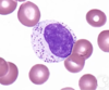
Lymphocyte
Which leukocyte is shown?

Basophil
Which leukocyte is shown?

Neutrophil
Which leukocyte is shown?

Monocyte
Which leukocyte is shown?

Eosinophil
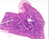
ID Tissue Type and all Structures noted
Lymph Node
- Germinal Center
- Lymph Nodule
- Capsule
- Maybe Trabeculae???
- Sinusoid
- Dr. Jones doesnt know
- Cortex (deep to this is medulla)
ID Tissue and Structures noted
Tonsil
- Germinal Center
- Nodule
- Epithelium
*Crypt not shown, but know it (refer to other tonsil slide)
Imagine a Tonsillar crypt of a Tonsil tissue. Check the back for you answer.
ID tissue and Specific structures labeled
Spleen
- Lymphatic Nodule
- Germinal Center
- Capsule
- Red Pulp
*keep in mind: splenic cord, venous sinus, trabecula
ID tissue and specific strucutures labeled

Spleen
- Splenic cord
- Venous Sinus
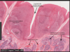
ID Structures shown

- Heart endocardium
- Subendocardial layer
- purkinje fibers shown
- Myocardial Layer
ID different structures shown

- Epicardium
- Mesenchymal outer layer
- Connective tissue
- Autonomic nerves
- Myocardium
ID structure shown at pointer

Lymph Vessel
- Notice how the blood is “tacky”, stuck on the sides. It has a pale red color due to H&E, but there are no clear RBCs because lymph is just plasma and other protein stuff
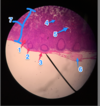
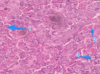
ID structures in the slide

- Capillary
- Various sized arterioles
ID structures shown

Sinusoid capillary with surrounding adipocytes and Hematopoetic cells
*sample from bone marrow i believe*
ID structure shown.
What is at the pointer?

Thymus
Hassal’s Corpuscle
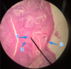
ID structure shown at pointer.
What is the structure above and to the left of the pointer?

- Arteriole
- Vein
ID structure shown at pointer

- Purkinje Fibers
ID the structure shown

- Purkinje fibers
ID structure shown at pointer

- Epicardium
ID structures shown

- Endothelium
- Subendocardium
- Purkinje fibers
- Myocardium
ID structures shown at the pointer and above the pointer to the right

- Vein
- Artery
ID the specific tissue shown
Thymus
ID structure shown.
What peculiar thing do you see toward the upper left of the lumen?

- Vein; valve is open
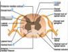
Which organ is shown? Label the Green, Blue and Orange arrows.

Pituitary Gland
- Anterior Pituitary
- Green = Pars Distalis (Anterior Pituitary)
- Blue = Pars Intermedia
- Posterior Pituitary
- Orange = Pars Nervosa (Posterior Pituitary)
ID the tissue shown and ID each arrow

Adenohypophysis
- Dark Blue - Sinusoid
- Light Blue - basophil
- Pink - chromophobe
- Yellow - acidophil
ID tissue shown and Arrows
*Bonus Q: what portion of the tissue is it?

Pituitary Gland
- Red arrows = Herring bodies which are clusters of hormones at the termini of axons
- Blue arrows = Pituicytes which are cells that assist storage and release of hormones
*Pars Nervosa
ID the cells within the Pars Distalis prep shown:
- Beta cell (basophil)
- Alpha cell (acidophil)
- Chromophobe (C) cell
ID the Endocrine tissue shown
What are the scattered purple dots called?

- Pineal Gland
- Pinealocytes
ID the Endocrine tissue shown in the prep.

Adrenal Gland
What is the numbered structure in this Pancreas prep?
What indistinguishable cells are inside this area?

- Pancreatic Islet (islet of Langerhans)
- Alpha and Beta cells
Which Endocrine organ is shown?
What’s a defining feature that could distinguish it from the pineal gland?

- Pancreas
- Pancreatic islets are visible. Pineal glands look very similar, but they do not have islets
ID the Tissue shown and each numbered structure

- Parafollicular cells (light pink stain)
- Colloid
- Follicle
- Follicular Cells (dark nuclei in permiter)
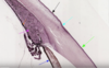
ID the structures shown in this Nerve prep

- Schwann cell nuclei
- Schwann cell
- Endoneurium*
- Perineurium
*double check
ID what the pointer is pointing at; Nerve prep shown

Epineurium
ID the tissue shown and specific structures numbered

Spinal Cord
- Posterior Median Sulcus
- Gray Commissure
- Central Canal
- lined by ependymal cells
- Ventral (anterior) horn
- motor neuron cell bodies
- Anterior Median Fissure
- Dorsal (posterior) Horn
- interneuronal cell body
- Gray Matter
- axons (oligodendrocyte fat makes lighter in color)
- White Matter
- cell bodies + Glial cells
ID the numbered structures
- Posterior Median Sulcus
- Dorsal Horn
- Gray commissure
- Ventral horn
- Central Canal
ID the structures at each arrow [spinal cord]
What is the term for the outer covering formed by these?

- Green arrow = Arachnoid Layer (fibrous and less dense)
- Yellow arrow = Subarachoid Layer
- Red arrow = Pia Mater (thin fibrous layer)
ID the structures noted in this slide

- Neuron cell body
- Glial cell
ID the tissue and the numbered structures

Cochlea
- Scala Vestibuli
- Holds Perilymph
- Basilar Membrane
- Scala Media
- Holds Endolymph
- Scala Tympani
- Holds Perilymph
- Tectorial Membrane of Spiral Organ of corti
- Inside are Hair cells
- Vestibular Membrane
ID the structures in this Peripheral Nervous tissue
- Unipolar Nerve cell (normally round)
- Satellite cells
ID tissue and structures labeled

Cerebellum
- Green arrow = White matter
- Blue arrow = Gray matter
ID the layers of this Cerebellum

Gray matter
- Red = Molecular Layer
- Blue = Purkinje Layer
- Green = Granular Layer
White matter
- Yellow = White matter
ID the tissue and the structures

Thyroid
- Follicular Cells
- Colloid
ID the Tissue and the structures noted
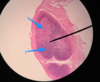
Parathyroid
- Oxophil
- Chief Cell (principal cell; stain dark purple)
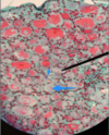
ID the tissue shown and the numbered structures labeled

Adrenal Gland
Adrenal Cortex
- Capsule
- Zona Glomerulus
- Zona Fasciculata
- Zona Reticularis
- Adrenal Medulla
- Chromaffin cells (darker purple)
ID the tissue and structures labeled:
- Blue circle
- Green line
- Red/Yellow lines
What does the Organ of Corti lie on?

- Organ of Corti
- Tectorial Membrane
- Hair Cells
Organ of Corti lies on the basilar membrane
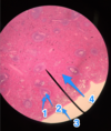
ID the various lines and arrows in this slide prep:

Fibrous Layer
- Green arrow = Cornea (refracts light)
Vascular Layer
- Blue arrows = Iris (control light)
- Black arrow = Ciliary body (support lens, secrete humor)
- Purple arrow = Lens
- Red arrow = Anterior chamber
- Yellow arrow = Posterior chamber
- Pink line = Vitreous space
Id the structures denoted by the arrows (except the light arrow)
Which tissues comprise the Fibrous Tunic?
Which tissues comprise the Vascular Tunic?
Fibrous Tunic
- Black = Sclera
- Green = Cornea
*where the cornea meets the sclera is the baby blue arrow
Vascular Tunic
- Orange = Choroid
- Purple = Ciliary Body
- Dark Blue = Iris
ID the tissues and structures noted

Sensory Tunic
- Nerve Fiber layer
- Ganglionic layer
- Inner nuclear layer
- Outer nuclear layer
- Photoreceptor layer
- Pigmented layer
Vascular Tunic
- Choroid
Fibrous Tunic
- Sclera
