Hand trauma Flashcards

1
Q

What do the thumb collateral injuries include?

A
  • Radial collateral ligament- Rare
  • Ulnar collateral ligament- most common
    • aka Gamekeepers thumb- chronic injury
    • Skiers thumb- acute injury
    • Steiner Lesion= avulsed ligament w/wout bony attachment is displaced ABOVE ADDUCTOR APONEUROSIS
      • won’t heal without surgical repair
2
Q

What is the epidemiology of thumb collateral injuries?

A
  • Ulnar more common than radial
3
Q

What is the aetiology of ulnar collateral injury?

A
  • Hyperabduction or extension at MCPJ
4
Q

Describe the anatomy of the UCL?

A
  • Proper collater ligament
    • Runs MC head to volar aspect of prox phalanx
    • resist valgus load w thumb in Flexion
    • primary contraint in flexion
  • ​Accessory collateral ligament & volar plate
    • Runs palmar to proper ligament inserts into volar plate
    • ​resist valgus load w thumb in EXTENSION
    • Primary contrainst w volar plate in extension
  • _​_Valgus laxity in both flexion and extension = complete UCL rupture
5
Q

What are the signs and symptoms of collateral thumb injury?

A

HX

  • Hyperabduction injury to thumb

Symptoms

  • Pain at ulnar aspect of Thumb at MCPJ

​Signs

  • Mass from torn ligament & possible bony avulsio may be present
  • Stress joint w radial deviation both at NEUTRAL/30o
  • Instability in 30o flexion = injury PROPER UCL
  • Instability in neutral - ACCESSORY UCL injury
  • compare to uninjured thumb!
6
Q

What investigations are useful to dx thumb collateral damage?

A
  • xrays- AP , lateral and oblique of thumb
    • Valgus stress view may aid diagnosis if bony avulsionhas been ruled out
  • MRI - aid dx
7
Q

What is a stener lesion?

A
  • Displacement of the distal end of the completely ruptured UCL such that it comes to lie SUPERFICIAL and PROXIMAL to ADDUCTOR APONEUROSIS
  • Must be operated on!!
8
Q

What is the TX of thumb collateral damage?

A

Non operative

  • Immobilisation 4-6/52
    • partial tears with <20o side to side variation

Operative

  • Ligament repair
    • In acute injury >20o side to side variation
    • >35o of opening
    • Stener lesion
      • ​can use suture/ suture anchor/ small screw to repair ligament
  • Reconstruction of ligament w tendon graft, MCP fusion, adductor advancement
    • chronic injury
9
Q

What is the tx of radial collateral ligmanet injuries?

A
  • rare
  • tx is non operative= **Immobilisation **
  • indicated in most cases
  • Stener lesion - doesn’t occur
10
Q

Describe a repair of UCL?

11
Q

Define digital collateral ligament injury?

A
12
Q

What is the epidemiology of scaphoid fractures?

A
  • Most frequently fractured carpal bone
  • Accounts for up to 15% acute wrist injuries
  • location
    • waist -65%
    • Proximal third- 25%
    • Distal third 10%
    • distal pole is most common location in kids due to ossification sequence
13
Q

Describe the pathoanatomy of scaphoid fractures?

A
  • Axial load across hyper-extended and radially deviated wrist
  • Common in contact sports
  • transverse fractures more stable than vertical/oblique fractures
14
Q

What is the prognosis of Scaphoid fractures?

A
  • Incidence of AVN with fracture location
  • Proximal 5th AVN rate = 100%
  • Proximal 3rd AVN rate= 33%
15
Q

Describe the anatomy of the scaphoid?

A
  • 75% scaphoid covered in CARTILAGE
  • Blood supply
    • major supply- DORSAL CARPAL Branch of radial artery- enters scaphoid in a nonarticular ridge on dorsal surface and supplies proximal 80% scaphoid via a RETROGRADE blood flow
    • minor via SUPERFICIAL PALMAR Arch branch of volar radial artery- enters DIstal TUBERCLE and supplies 20% DISTAL SCAPHOID
  • ​Both intrinsic/extrinisic ligaments attach
  • Scaphoid flexes w wirst flexion & radial deviation adn extends during wrist extension& ulnar deviation
16
Q

Describe the signs and symptoms of scaphoid fracture?

A

O/E

  • Anatomic snuffbox tenderness dorsally
  • Scaphoid tubercle tenderness volarly
  • Pain w reisted pronation
17
Q

What investigations are useful in DDx scaphoid fracture?

A
  • Xrays
    • AP and lateral
    • Scaphoid view- 30o wrist extension, 20o ulnar deviation
    • 45o pronation view
    • no fracture but suspicion rpt 7-14 days
  • Bone scan
    • diagnose occult fracture at 72 hrs
    • specificity 98%, sensitivity 100%, PPV 85-93% within 72hrs
  • MRI
    • Most sensitive method within 24hrs
    • also vasularity of prox pole
  • CT Scan
  • less effective than bone scan/ MRI
  • Useful for location of ffracture and progression of nonunion/union post surgery
18
Q

Describe the tx of scaphoid fracture?

A

Non operative

  • stable non displaced fractures
  • normal xray- cast then immobilise 12-21 days and reexam/rpt xrays
  • Start immobilisation early- non union rates increase w delay of immobilisation > 4 wks post injury
  • no concensus on casting- can use volar cast
  • duration of casting
    • distal fracture- 3/12
    • mid waist- 4/12
    • proximal third -5 /12
  • outcomes fractures with <1mm displacment union 90%

​Operative

ORIF vs Percutaneous screw fixation

19
Q

what are the indications for surgery for scaphoid fractures?

A
  • Unstable fractures
  • Proximal pole fractures
  • Displacment >1mm
  • 15 o humpback deformoty
  • radiolunate angle >15o= DISI
  • Scaphoid frac ass with perilunate disslocation
  • comminuted fractures
  • in undisplaced fractures- to allow decreased time to union, faster return to work/sport, cheaper costs to casting- McQueen et al JBJS Br 2008
20
Q

What are the outcomes of surgery for scaphoid fracture?

A
  • Union rates of 90-95%
21
Q

CAn you describe the technique for fixation of scaphoid fracture?

A

Approach

  • Dorsal approach for Proximal Pole fractures
    • Preserve blood supply when entering dorsal rige by limiting exposure to proximal half of scaphoid
    • Percutaneous higher risk of unrecongnised screw pentetration of sunchondral bone
  • Volar approach- for waist and distal pole fractures w humpback flexion
    • allows exposure of whole scaphoid
    • uses interval between FCR and radial artery
  • Arthroscopic assisted also described
  • Fixation is optimised by long screw down CENTRAL AXIS of SCAPHOID
  • Radial stylectomy preformed if impaction between radial styloid adn scaphoid
22
Q

Describe the complications of scaphoid fixation?

A
  • Non union->
  • SNAC wrist- degenerative changes first in radioscaphoid area followed by pancarpal /midcarpal arthritis
  • Tx with
    • **Interposition (Fisk) bone graft- **open wedge graft, 72-95% unon rates
    • Inlay (Russe) bone graft- minimal deformity, 92% union rates
    • Vascular bone graft from radius- 1-2 intercompartmental supraretinaculuar artery ( branch of radial artery) harvested to provide vascular bone graft from dorsal aspect wrist
  • Outcomes:
    • Punctate bleeding during surgery good prognostic indicator of union
    • 92% w obvious bleeding, 71% questionable bleeding, 0% no bleeding
    • Pt w scaphoid nonuions>5 yrs or proximal pole necrosis have less favourable outcomes
23
Q

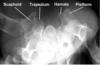
What do hamate fractures include?

A
  • Hook of hamate- most common
  • Hamate body fractures- v rare
24
Q

What is the hx of hamate fractures?

A
  • Hx of blunt trauma to palm of hand
  • often seen racquet sports
    • hockey
    • golf- miss ball hit ground
    • tennis
  • Must distinguish from BIPARTITE HAMATE- Smooth cortical surfaces
25
Describe the symptoms of hamate fractures?
* Hypothenar symptoms- Pain * **Decreased grip strength** * **Parathesisa** in ring and small fingers - ulna N compression in Guyon's canal, numbness in thumb, index, middle and ring finger from median n compression in carpal tunnel * Motor weakness of intrinsics- Ulnar n compression in Guyon's canal
26
What imaging is useful in Hamate fractures?
* Difficult to visualises on AP * Best seen on **CARPAL TUNNEL VIEW** * **CT** best to make diagnosis
27
Describe the TX of hamate fractures?
Non operative * **Immobilise 4-6 weeks** * Acute hook of hamate fractures * body of hamate fractures **Operative** * **Excision of hamate fracture fragment- return to level of sport 7-10 wks** * Chronic hook of hamate fractures * _ORIF is possible but little benefit_
28
What is this?
**Pisiform fracture**
29
Describe the anatomy of the pisiform?
* A **sesmoid bone** * located within **FCU tendon** * **Contributes to stability of ulnar column by preventing TRIQUETRAL subluxation** * Acts as a fulcrum for forces transmitted from forearm to hand
30
What is the epidemiology, aetiology of pisifrom fractures? Name any associated fractures?
* 1-3% of carpal fractures * fall on outstretched hand * 50% pisiform fractures occur with distal radius, hamate or triquetral fractures
31
Describe the signs and symptoms of pisiform fracture?
Symptoms * Ulnar sided wrist pain Signs * Point tenderness
32
What investigations are useful for ddx of pisiform fractures?
Radiographs * **Lateral with 30 degrees of supination** or carpal tunnel view * **CT scan-** best seen
33
Describe the treatment of pisiform fractures?
Non operative * Immobilisation- first line * **Short arm cast with 30o wrist flexion** & ulnar deviation 6 wks * Outcomes- heal without posttraumatic arthritis Operative * **Pisiformectomy-** painful NON union * Outcomes- pisiformectomy reliable way to relieve apin & doesn;t impair wrist function
34
What do base of thumb fractures include?
* **Bennett fracture**- Intra-articular * **Rolando Fracture**- intra-articular * **Extra-articular fractures**
35
What is the epidemiology of bse of thumb fractures?
* 80% thumb fractures involve **METACARPAL BASE** * most common variant is **BENNET Fracture**
36
What is the pathophysiology of base of thumb fractures?
* Most fracture caused by **_AXIAL force_** applied to Thumb * 3 muscles deforming forces at base of thumb * **Abductor pollicis Longus**- PIN * **Extensor pollicis longus**- PIN * **Adductor pollicis** - Ulnar N
37
Describe what a Bennet's fracture is?
* **Inra-articular fracture of base of 1st MC characterised** by * **VOLAR LIP OF Metacarpal base attached to VOLAR OBLIQUE LIGAMENT** * ligament holds this fragment in place * small fragment of 1st MC continues to articulate with trapezium
38
Describe the pathoanatomy of bennett's fracture?
* **Lateral retraction of distal 1st MC shaft** by **APL and Adductor pollicis** * **​Volar- ulnar fragement is held reduced by ant oblique ligament while strong deforming forces pull the reamining metacarpal shaft proximally and dorsally, angulate the shaft ulnarly and supinate it.** * **​Abductor pollicis longus-** inserts MC base- pulls MC shaft proximally & dorsally * **Adductor pollicis**- inserts ulnar base proximal phalanx - angulates MC shaft ulnarly and supinates it * **EPL**- less important inserts base of distal phalanx adn adds to ulnar angulation of distal fragament
39
What is the prognosis of the bennet's fracture?
* Better than Rolando
40
What investigations are helpful in DDX of Bennet's fracture?
* **Hyper-pronated thumb view** * Minimal joint step-off considered best
41
Describe the TX of Bennet's fracture?
Non operative * Closed Reduction & cast Immobilisation * non displaced fractures * reduction with **traction, extension, pronation and adduction** Operative * **Closed reduction and Percutaneous pinning** * Volar fragment too small to hold with screw * Anatomic reduction unstable * can attempt reduction of shft to trapezium to hold reduction * **ORIF** * large fragment * 2mm+ joint displacement
42
What are the complications of a Bennet's fracture?
* **Post traumatic arthritis** * There is no agreement regarding the relationship of post- fixation joint incongruity and post traumatic arthritis.
43
What is a Rolando fracture?
* **Intra-articualar fracture of base of the 1st metacarpal** characteised by * **intra-articular comminution**
44
What is the epidemiology and pathophysiology of Rolando fracture?
* Less common than Bennet's fracture Pathoanatomy * Deforming forces are the same as Bennett's fracture * **Lateral retraction of 1st distal metacarpal shaft by Abductor Pollic Longus and adductor pollicis** * **​Volar- ulnar fragement is held reduced by ant oblique ligament while strong deforming forces pull the reamining metacarpal shaft proximally adn dorsally, angulate the shaft ulnarly and supinate it.** * **Abductor pollicis longus- inserts MC base-** * *pulls MC shaft proximally & dorsally** * **Adductor pollicis- inserts ulnar base proximal phalanx -** * *angulates MC shaft ulnarly and supinates it** * **EPL- less important inserts base of distal phalanx** * *and adds to ulnar angulation of distal fragament** * typically the base spit into volar and dorsal fragment * commonly called a **Y fracture**
45
What is the prognosis of Rolando fracture?
* Worse than a bennet's fracture
46
What is the TX of Rolando fracture?
Non operative * Immobilisation- severe comminuted, stable * start early rom Operative * **External fixation, CRPP** * **​severe comminuation, unstable** * can approximate large fragment w K wires * **​ORIF** * **​MOST common fixed method** * **use T plate or blade plate** * can use K wire if fragments are too small
47
What are the complications of rolando's fracture?
* Post - traumatic arthritis
48
What types can extra-articular fratures be?
* Transverse of Oblique in nature
49
Describe the tx of extr-articular thumb fractures?
Non operative * Spica casting * **if joint reduced & \<30o of angulation** ​​Operative * **CRPP** * If reduction cannot be held to result in less than 30o of angulation * Typically these fractures have the best outcome
50
What can metacapral fractures be divided into?
* Metacarpal head, neck and shaft fractures * Tx based on which MC involved & location of fracture * Acceptable angulation vaires on location * _NO degree of malrotation acceptible_
51
Describe the epidemiology of MC fractures?
* Accounts for **40% o**f all hand injuries * **Men aged 10-29 **most common * **Metacarpal Neck** is most common site * **5TH Metacarpal** is most commonly injured
52
What is the aetiology of MC fractures?
* **Direct blow** to hand or **rotational injury with axial load** * **HIgh energy injuries-\>** multiple injuries
53
Name any associated injuries with MC fractures?
* Wounds= open fractures * Tendon lacerations * Neurovascular injury * compartment syndrome * closed injury mutiple fractures * Crush injuries
54
Describe the anatomy of MC ?
* Concave on palmar side * 1st,4th,5th digits-\>Mobile border * 2nd,3rd digits-\> stiffer central pillar * index MC most firmly fixed, while thumb MC articulated with Trapzium & acts independently from others * **3 Palmer and 4 Dorsal Interosssei** arise from MC shafts * **ECRL/B** - inserts base MC 2, 3- assists with wrist extension/ radial flexion * **ECU**- inserts base 4, extends/flexes wrist when digits being flexed, assist ulnar flexion of wrist * **Abductor pollicis longus**- inserts trapezium and base of MC 1- adducts thumb in frontal plane, extends thumb at CMCJ * **Opponens Pollicis**- inserts MC I, Flexes MC 1 to OPPOSE thumb to fingertips * **Opponens digiti minimi**- inserts medial side of MC V, flexes MC V at CMCJ when little finger is moved into opposition with tip of thumb, deepens palm of hand
55
What are the signs of MC fracture?
Signs * Inspect openw wounds * fight bite over MCP open until proven otherwise * Extensor tendon lacerated and retracted * Dorsal wounds over MC - often open * Deformity * At MC base may indicate CMCj Dislocation * Shortening- cf contralateral side * Malrotation- fingernails in part felxion- see pics * Motor exam- flexor/extensor tendons * NV exam * dorsal wounds = affect dorsal sensory branch of radial/ulnar n * volar wounds - involve digitial nerves
56
What investigations are helpful in DDX MC fracture?
* Xray * Standard AP, Oblique & lateral * **Oblique-** evaluate CMCJ * 30o pronated lateral = 4/5th CMC * 30o Supinated lateral-= 2/3 CMC * Roberts view= thumb CMCJ * Brewerton view= MC head fractures * CT scan * inconculsive CMC fract/dislocations * Multiple CMC dislocations * complex MC head fractures
57
Describe the accepted angulation , shaft shortening and neck angulation for consx tx for MC fractures?
* Index/ Middle ringer * Shaft Angulation 10-20o * shaft shortening 2-5mm * Neck angulation 10-15o * ​Ring Finger * Shaft angulation 30o * Shaft shortening 2-5mm * Neck angulation 30-40o * Little Finger * Shaft angulation 40o * Shaft shortening 2-5mm * Neck angulation 50-60o
58
What are the operative indications fof MC fractures?
* Intra-articular fractures * Rotational malalignment * Significantly displaced fractures * Mutliple MC shaft fractures * Loss inherent stability from border digit during healing process
59
Describe the operative Tx of MC fractures?
* **ORIF** * no degress of articular displacment is acceptable * Majority required surgical fixation- see pic * **External fixation** * Severely comminuted fractures * **MCP Arthroplasty** * Severely comminuted fractrues * **MCPJ fusion** * rarelt done due to limited rom of hand
60
Describe the operative technique for ORIF for MC Head fractures?
* Approach * **dorsal incision** * either **centrally splt extensor apparatus or release and repair sagittal band** * Hardware cannot protrude joitn surface * FIX w multiple small screws in collateral recess , headless screws or K wires * ideal fixation should allow early motion
61
What are the complications of MC head fractures?
* Stiffness * most common * prevent by early motion * CRPS
62
Describe the TX for MC shaft fractures?
Non operative * Immobilisation * Non displaced MC neck fractures * Acceptable angulation * No malrotation * Shortening- aethetic problem only * Immobilise **MCPJ 70-90o** * Cast **4 weeks** Operative * **ORIF vs CRPP** * for open fractures * unacceptable angulation * any malrotation * multiple fractures * **CRPP=** place retrograde thru MC base /antegrade thru collateral recess remove pins 4/52 * **Open reduction w lag screw** * mutliple lags for long spiral fractures- at least 2 * **Open reduction w dorsal plating** * best for transverse fractures * try to cover plate w periosteum to prevent tendon irritation * begin early motion to prevent tendon irritation
63
Describe the TX for MC neck fractures?
Non operative * **Reduction and casting** * Acceptble degrees of apex dorsal angulation * Immobilise MCP 70-90 flexion, leave PIPJ free * Cast 4 weeks * Reduce **Jahss technique** * 90o flexion, dorsal pressure thru proximal phalanx whilst stabilising MC shaft Operative * **Reduction and Fixation** * **​**Unaccetpable angulation * open fracture * any malrotation * intrarticular fractures * **CRPP w MCP flexed** * Antegrade thru MC base * Retrograde thru Collateral recess * **ORIF** * Perform if cannot get reduction with CRPP * Difficult ro place because of limited bone distal fixation
64
What is this?
* Dorsal PIP dislocation
65
What is the epidemiology of dorsal PIP dislocations?
* More common than volar dislocations * leads to **injury to VOLAR PLATE** and at least the **COLLATERAL ligament ** * **Swann neck deformity** will result if left
66
Describe the classification system of PIP dislocations?
* **Simple**- middle phalanx in contact with condyles proximal phalanx * **Complex**- Base of middle phalanx not in contact with condyle of proximal phalanx, Bayonet appearance * volar plate acts as block to reduction with longitudinal traction
67
Describe the TX for dorsal PIP dislocations?
Non operative * Reduce and buddy tape to adjacent finger 3-6 wks * dislocation is reducible * usually preformed by patient * if complex reduce w HYPEREXTENSIONof middke phalanx followed by palmar force * complx- Swan neck deformity ( volar plate injury) Pipj Flexion contracture- pseudoboutonniere Operative * Open reduction & extraction of volar plate * Failed reduction * closed reduction failed often due to **volar plate interposition** * Open reduction failed due to **dislocated FDP tendon** * **dorsal approach w incision between central slip & lateral band**
68
What is this?
* **Dorsal PIP fracture dislocation**
69
Name and describe the classification system of dorsal PIP fracture dislocations?
* **Hastings** * Based on amount of P2 articular surface involvement * **Type 1- Stable** * **\<30%** -= tx dorsal based extension block splint * **Type 2- Tenous** * **30-50%** - if reducible in flexion, dorsal based extension block splint * **Type 3- Unstable** * **\>50%** - ORIF/HAMATE Autograft, Volar plate arthroplasty
70
Describe the tx of dorsal fracture dislocation of PIP?
Non operative * Dorsal extension block splint * if \<40% joint involved & stable * much achieve adequate joint redcution for favourable outcome Operative * **ORIF vs CRPP** * If \>40% joint involved/ unstable * reduction of middle phalanx on condyles of prox phalanx is primary goal * adequate volar exposure of volar plate requires resection of **proximal C2 pulley, Entire A3 pulley and distal C1 pulley** * **​Dynamic Distraction external fixation** * **​**highly comminuted pilon fracture * follw w early mobiliasation * **volar plate arthroplasty/arthrodesis** * chronic injuries
71
What is this?
* **A volar pip dislocation** * Less common than dorsal * leads to injury to **CENTRAL SLIP and at least ONE COLLATERAL Ligament** * failure to tx -\> **Boutonniere deformity**
72
Describe the tx of volar PIP fracture/ dislocation?
Non operative * **Spinting in extension 6-8 wks** * most pip dislocations * If **Fracture \<40%** joint involved & stable Operative * **ORIF vs CRPP** * reduction of middle phalanx on condyles of proximal phalanx is primary goal * i**f joint \>40% involved**
73
What could this be?
* **Rotatory PIP dislocation** * One of phalangeal condyles is **buttonholed between central slip & lateral band**
74
Describe how you would tx rotatory PIP dislocation?
Non operative * Only of reduction successful * reduce by applying traction to finger with **MPJ & PIPJ in 90o Flexion** * Flexion relaxes volar displaced lateral band , allowing to slip back dorsally * Reduction is confirmed w post reduction lat xray Operative * **Open reduction**- most cases
75
What is this?
* Dorsal DIP dislocation with fracture
76
What is the tx of dorsal DIP fracture dislocation?
Non operative * **Closed reduction, immobilisation in slight flexion w dorsal splint for 2 weeks** * ​first line * ​​Tuft fractures no specific tx- temporary splinting Operative * **Open reduction** * 2 failed attempts at reduction * **Volar plate interposition** is most common block to reduction in irreducible **closed** DIP joint dislocation * **FDP** may be blocking if injury **open** * May require percutanoeus pinning to suport nail bed injury * **Amputation** * if highly comminuted injuries with significant soft tissue loss