H&N - Face and Scalp Flashcards

1
Q

Surface marking for parotid gland

A

Starts around tragus

Follows inferior to zygomatic arch

Drops down to angle of mandible and wraps around it

Climbs to inferior part of lobule

2
Q

Parotid duct aka

A

Stensen’s duct

3
Q

Surface marking of Stenson duct

A

Middle third of a line between intertragic line (between tragus and anti tragus) and philtrum

4
Q

How does parotid duct get into mouth

A

After masseter muscle, dives into buccal fat and pierces buccinator muscle

5
Q

End point of parotid duct

A

Near the second upper molar tooth

6
Q

Structures through the parotid gland

A

Facial nerve

External carotid artery (and its branches; the maxillary and superficial temporal)

Retromandibular vein

Auriculotemporal nerve

7
Q

Branches of facial nerve in facial canal

A
  1. greater petrosal nerve
  2. nerve to stapedius
  3. chorda tympani
8
Q

Facial nerve branches through the stylomastoid foramen

A

Posterior auricular

Digastric

Stylohyoid

9
Q

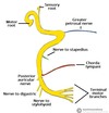
Facial nerve branches after entering parotid gland

A

The Zebra Buggered My Cat

Temporal branch

Zygomatic branch

Buccal branch

Marginal mandibular branch

Cervical branch

10
Q

Functions of facial nerve

A

Face, ear, taste, tear

Taste: anterior 2/3

Tear: PSN to lacrimal and salivary glands

11
Q

Facial nerve end branches motor test

A

Temporal: raise eyebrow

Zygomatic: close eyes tightly

Buccal : blow cheeks

mandibular: show lower teeth
cervical: tense skin under your chin

12
Q

External carotid artery branches

A

Some attendings like freaking out potential medical students

Sup thyroid

Ascending Pharyngeal

Lingual

Facial

Occipital

Post auricular

Maxillary

Sup temporal

13
Q

Tribuaries of retromandibular vein

A

superficial temporal and maxillary veins

14
Q

Arterial supply of parotid

A

External carotid artery

15
Q

Venous supply of parotid

A

Retromandibular vein

16
Q

Lymphatic drainage of parotid

A

deep cervical

17
Q

Innervation of parotid gland

A

Glossopharyngeal -> otic ganglion -> Auriculotemporal

18
Q

Branches of opthalmic nerve

A

Supra-orbital

Supratrochlear

Infratrochlear
Lacrimal

External nasal

19
Q

Branches of maxillary nerve

A

Zygomaticotemporal

zygomaticofacial

Infra-orbital

20
Q

Branches of mandibular nerve

A

Auriculotemporal

Lingual

Inferior alveolar

Nerve to the mylohyoid

Mental

21
Q

Layers of scalp

A

Skin

Connective tissue (dense)

Aponeurotic layer of occipito frontalis (Galear layer)

Loose connective tissue
Pericranium (ie periosteum)

22
Q

Innervation of scalp

A

Anterior to vertex: trigeminal

Posterior to vertex: C2 and C3 (great auricular, lesser and greater occipital, third occipital)

23
Q

In which layer of scalp do blood vessels and nerves lay

A

In dense connective tissue

24
Q

Scalp blood supply

A

Opthalmic artery (internal carotid): supratrochlear and supraorbital

External carotid: Post auricular, superficial temporal, occipital

25
Why scalp lacerations bleed so profusely
Arterial wall firmly attached to fibrous tissue, cant vasospasm/contract so continues to bleed.
26
How to rx scalp laceration and bleed
apply pressure primary closure by suturing
27
Which muscles close the jaw
Medial pterygoid Masseter Temporalis
28
Submandibular gland duct
Whartons duct
29
Which muscle divides the submnadibular gland into two parts
mylohyoid muscle
30
Nerve around whartons duct
lingual
31
What is the acini composition of the three salivary glands
Parotid: mainly serous Sublingual: mainly mucous Submandiblar: mixture of mucous and serous
32
Where do majority of salivary gland stones form
Submandibular: Slow movement of saliva * up ward against gravity * mucous composition * alkaline (deposits calcium crystals)
33
Muscles of mastication
Masseter Temporalis Medial /lateral pterygoid
34
Which muscle opens the jaw
lateral pterygoid muscle
35
Position of lacrimal gland
Superior and lateral to the orbit
36
Angle of the jaw innervation
C2-C3 (greater auricular nerve)
37
Which muscles does mandibular nerve innervate
Muscles of mastication Tensor veli oalatine Mylohyoid Anterior belly of digastric Tensor tympani
38
Blood supply of lacrimal apparatus
Ophthalmic artery
39
Which nerve runs close to parotid duct
Buccal nerve of facial
40
Mx of facial nerve laceration
Lateral to lateral canthus: surgical repair Medial to lateral canthus line: surgery =conservative Always check parotid duct (look for blood at papilla or put a cannula in)
41
Where are scalp flaps taken from
Subgaleal layer (loose areolar connective tissue), fewer vessels More superficial is very vascular and firmly attached Deeper also done sometime but more vascular/attached to suture lines
42
Branches of the maxillary artery
Terminal 6: * Posterior superior alveolar * Pterygoid canal * Palatine (greater- \> hard palate, lesser → soft palate) * Sphenopalatine/nasopalatine * Pharyngeal * Infraorbital
43
Muscles of soft palate
Tensor veli palatini Levator veli palatini Palatoglossus Palatopharyngeus Muscularis uvulae
44
Action of tensor veli palatini
Pulls laterally flattening and tensing palate
45
Action of levator veli palatini
Elevates soft palate, stopping food entering nasopharyinx during eating Also helps tensor veli palatini to open the cartilagenous auditory tube to equalise middle ear pressure
46
Action of palatopharyngeus
Elevates larynx and pharynx Depresses soft palate
47
Action of palatoglossus
inserts into lateral portion of tongue Depresses soft palate
48
Nerve supply to soft palate muscles
All vagus (pharyngeal plexus from CN10 and CN11) Except tensor veli palatini → CN5V3
49
Facial muscles
Frontalis Procerus Orbicularis oculi Nasalis Levator labi superioris alaeque nasi Levator labi superioris Zygomaticofacial major and minor Orbicularis oris Risorius Depressor labii inferioris Depressor anguli oris Mentalis
50
Muscles deep to facial nerve
Mentalis Buccinator levator anguli oris
51
Where are facial muscles cause horizontal and vertical wrinkles in glabella region?
Vertical: Corrugator causes Horizontal: procerus