Abdomen- abdominal wall Flashcards
Branches of coeliac artery
Left gastric
Splenic
Common hepatic

Branches of common hepatic artery
Proper hepatic artery
Gastroduodenal artery

Branches of gastroduodenal artery
Right gastroepiploic artery
Superior anterior pancreatodeodenal artery

Branches of proper hepatic artery
Left proper hepatic artery
Right gastric artery
Right proper hepatic artery -> cystic artery

Lesser sac vs greater sac of abdomen
Space created by lesser and greater omentum

Diaphragm insertions
L3 on the right, L2 on the left
Median crus over aorta, medial over psoas major , lateral over quadratus lumborum

Superficial abdominal fascia layers
Camper’s fascia
Scarpa’s fascia

Camper’s fascia features
Contains large amounts of fat
Continuous over inguinal ligament and perineum
Scarpa’s fascia features
Mainly fibrous tissue (little fat)
Called fascia lata when enters lower limb
Continuous with superficial perineal fascia (Colle’s fascia)
Forms darto’s fascia
Transversalis fascia
deep fascia layer below transversus abdominus
External oblique muscle apponeurosis cover
Most superficial
Ends at inguinal ligament

Ligaments associated with inguinal ligament
Lacunar: inguinal ligament to pecten pubis
Pectineal (cooper’s) ligament

Innervation of flat muscles of anterior abdo wall (eg external/internal oblique/transversus)
T7-T12
Flat muscles of abdo wall
External oblique
Internal oblique
Transversus abdominus

Midline muscles of anterior abdo wall
Rectus abdominis
Pyramidalis

Upper and lower quarters of rectus sheath

Arcuate line
The border between upper 3/4 and lower 1/4 of rectus abdominis

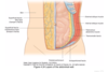
All the layers of abdominal wall
Note the extra-peritoneal fascia

Innervation of anterior abdominal wall
Lateral and anterior cutaneous branches of T7 to T12
Ilio-hypogastric nerve (L1)
Ilio-inguinal nerve (L1)

Blood supply of upper anterior abdominal wall
Internal thoracic: Musculophrenic branch and superior epigastric branch

Arterial supply of inferior abdo wall
Femoral artery: superficial epigastric and superficial iliac branches
External iliac artery: inferior epigastric and deep circumflex iliac branches

Superficial lymph drainage of abdo wall
Above umbilicus: axillary nodes
Below umbilicus: superficial inguinal nodes
Deep lymph drainage of anterior abdo wall
parasternal
lumbar
external iliac
Linea semiluranis
Border of rectus abdominis
Inferior epigastric artery crosses it


