Glomerular Disease (Pathology) Flashcards
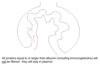
what is this showing?
a normal glomerulus

what can be seen either side of the glomerulus?
Need 2 small arterioles on either side to regulate blood pressure other wise the capillaries will burst

a
Podocyte

b
Red blood cell in capillary loop

c
Endothelial cell lining capillary loop

what is a Podocyte?
Podocytes are cells in the Bowman’s capsule in the kidneys that wrap around capillaries of the glomerulus. Podocyte cells make up the epithelial lining of Bowman’s capsule, the third layer through which filtration of blood takes place
where does blood enter the glomerulus?

Blood is filtered across glomerular membrane, what is not?

From outside podocytes have what?
From outside podocytes have interdigitating ‘fingers’ or foot processes

Filter barrier = membrane = 3 things, what are they?
endothelial cell cytoplasm, basal lamina and podocyte

what is the Mesangium?
The mesangium is a space which is continuous with the smooth muscles of the arterioles. It is outside the capillary lumen, but surrounded by capillaries. It is in the middle (meso) between the capillaries (angis)
Mesangial cells = ‘tree-like’ group of cells which support capillaries

where does filtrate flow?

where do the things that cannot be filtered into bowmens space go?
Blood cells, some fluid and albumin and larger proteins exit via efferent arteriole

what is Glomerulonephritis?
= Disease of glomerulus
Can be inflammatory or non-inflammatory
Glomerulonephritis is inflammation of the tiny filters in your kidneys (glomeruli). Glomeruli remove excess fluid, electrolytes and waste from your bloodstream and pass them into your urine. Glomerulonephritis can come on suddenly (acute) or gradually (chronic)
what is the aetiology of Glomerulonephritis?
Some are due to immunoglobulin deposition
Some are diseases with no immunoglobulin deposition – for example - diabetic glomerular disease
Is Glomerulonephritis just one disease?
- Large range of conditions
- Difficult to cover all variants
what are 4 common presentations of Glomerulonephritis?
- Haematuria (blood in urine)
- Heavy proteinuria (nephrotic syndrome)
- Slowly increasing proteinuria
- Acute renal failure
Case 1:
- 40 year old male
- Discoloured urine
- Dipstick urine – positive for blood
what are the main causes of Haematuria?
- Urinary tract infection
- Urinary tract stone
- Urinary tract tumour
- Glomerulonephritis - not as common of a cause as the ones above
case 1 continued:
- Send off urine culture
- Arrange hospital appointment for ultrasound examination
- Urine culture normal
- Ultrasound of abdomen – normal
- Check his clotting then proceed to renal biopsy: what is seen on biopsy?

Case 1 biopsy - what is seen on immunoflourescence?
(a method in biology that relies on the use of antibodies chemically labeled with fluorescent dyes to visualize molecules under a light microscope)
Immunoglobulin (of IgA type) and complement component C3 in mesangial area of all glomeruli
IgA deposits (yellow arrow) cause increased proliferation of mesangial cells

what is the aetiology of IgA glomerulonephritis?
unknown – Is excess antibody produced?
Excess antibody (IgA) sometimes present in serum, but this is also true of some people who do not have IgA glomerulonephritis
Why is IgA not removed by glomerulus?
Unknown, but probably very important part of cause of disease
Does IgA get filtered into urine?
No, the IgA is ‘stuck’ within the mesangium
Mesangium, not the filter membrane, becomes clogged with antibody
How does this cause red blood cells to escape into urine?
unknown
what does IgA cause to mesangial cells?
IgA – ‘irritates’ mesangial cells and causes them to proliferate and produce more matrix

what is the prognosis of a IgA nephropathy?
- Usually self-limiting, ie return to normal (haematuria goes away)
- Small % go onto chronic renal failure (via continued deposition of matrix)

case 2:
- 50 year old male
- 3 weeks of feeling unwell and swollen legs
- Send of blood biochemistry and haematology tests - Serum albumin is low
- Dipstick proteinuria
- Send into hospital to see nephrologist
- Nephrologist measures protein (albumin) in urine – very heavy loss
what is this and what is it due to?
• Clinical diagnosis of nephrotic syndrome – must be abnormality of glomerular filter
Case 2:
• Check clotting screen then do renal biopsy:
what would be see?

Membranous glomerulonephritis: electron microscopy

In Membranous glomerulonephritis, what happens to IgG?
IgG is stuck in membrane
IgG deposits itself between basal lamina and podocyte but cannot go further and is not filtered into urine

IgG is too big to be filtered into urine, but IgG activates what?
complement (C3), which punches holes in filter
IgG is too big to be filtered into urine, but IgG activates complement (C3), which punches holes in filter
what does this now cause?
Leaky filter now allows albumin to be filtered into urine - nephrotic syndrome

What is the prognosis of Membranous glomerulonephritis?
1/4 in chronic renal failure within 10 years
What is the underlying cause of IgG production and accumulation in membranous glomerulonephrits?
- Unknown but can sometimes have underlying malignancy
- In many patients antigen is phospholipase A2 receptor – why this protein? – as yet unknown.
Case 3:
- 31 year old woman
- Type 1 diabetes since 7 years of age
- Long periods of poor glycaemic control
- Developed retinopathy
- Albumin in urine slowly increasing over last few years. Now has heavy protein leakage into urine
- Check clotting screen then do renal biopsy
what would be seen?
Glycated molecules - Matrix deposition in basal lamina underlying endothelium and in mesangial matrix - thickened but leaky basement membranes + mesangial matrix compresses capillaries (no immune complexes)

what does diabetic nephropathy look like on histology?

in diabetic nephropathy, nodules of mesangial matrix can be seen, what are they?
Kimmelsteil-Wilson lesion = gross excess of mesangial matrix forming nodules

what is the prognosis of diabetic nephropathy?
Inevitable decline if 1. established diabetic nephropathy and if 2. continued poor diabetic control
Case 4:
- Female, 50 years old
- Unwell for 3 weeks
- Cough
- Serum biochemistry – creatinine 500 (was 60 one year before)
what is her diagnosis?
Rapidly rising creatinine = acute renal failure
case 4:
- Ultrasound: no renal tract lesion
- Check clotting then renal biopsy: what is seen?
Early endothelial damage with fibrin deposition
Cellular proliferation and influx of macrophages (= crescent) around glomerular tuft, within Bowman’s space

another picture showing crescent

what is the cause of Crescentic glomerulonephritis?
Many causes of this pattern of injury, for example:
- Granulomatosis with polyangiitis (previously known as Wegener’s granulomatosis)
- Microscopic polyarteritis (a disease very much like granulomatosis with polyangiitis)
- Antiglomerular basement membrane disease
- Other - Many other forms of glomerulonephritis
Our example = Granulomatosis with polyangiitis (previously known as Wegener’s granulomatosis)
what is it?
A form of vasculitis (= inflammation in vessels) which affects vessels in kidneys, nose and lungs
What are further tests for Wegeners?
Serum test shows presence of anti-neutrophil cytoplasmic antibodies (ANCA)
what are Anti-neutrophil cytoplasmic antibodies (ANCA)?
autoantibodies directed against antigens found in the cytoplasmic granules of neutrophils and monocytes. ANCA testing is usually performed to help diagnose or exclude Wegener’s granulomatosis and microscopic polyangiitis
Not deposited in kidney
Antibodies directed against proteinase 3 and myeloperoxidase, 2 enzymes in primary granules of neutrophils

How does Anti-neutrophil cytoplasmic antibodies (ANCA) cause tissue damage?
Antibodies produce tissue damage via interactions with primed neutrophils and endothelial cells
Anti-neutrophil cytoplasmic antibodies (ANCA) – why do they form?
Unknown at present - ? A form of autoimmunity
what is the prognosis of Wegener’s?
- Fatal (mean survival 6 months) if left untreated
- Cyclophosphamide – 75% complete remission