GI Histo Pictures Flashcards
What layer of the GI tract wall are the arrows pointing to?

Submucosa
What layer of the GI tract wall are the arrows pointing to?

Mucosa
What layer of the GI tract wall are the arrows pointing to?

Muscularis Externa
What layer of the GI tract wall are the arrows pointing to?

Serosa or Adventitia
This is a cross-section through what part of the GI tract?

Esophagus
Identify A, B, C, D, & E?

A- Mucosa
B - Submucosa
C - Mucus Gland
D - Muscularis Externa
E - Adventitia
Identify A, B, & C

A- Mucosa
B- Submucosa
C- Muscularis Externa
Where is this cross-section from?

Esophagogastic Junction
Identify A in this cross-section of the stomach

Cardiac glands
Identify A in this cross-section of the stomach

Pyloric Gland
What kind of gland is this?

Fundic gland
Identify A, B, and C in this cross-section of the intestinal wall.

A - Mucosa
B - Submucosa
C - Muscularis
Identify A

Zymogen Granules within Chief Cells
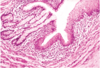
Identify A

Vili
Where do you find the lympathic nodules like the ones pictured here?

Ileum (small intestine) -
•In the ileum, groups of lymphatic nodules are present in the lamina propria and submucosa. These aggregated lymphatic follicles are referred to as Peyer’s patches.
Where do you find this mucosa?

Large intestine
Where is this cross-section from?
Recto-Anal Junction - about 2 cm from anal opening, converging back to skin (non-keratinized stratified squamous epithelium)
What are the arrows pointing to in this cross-section of the pancreas?

Pancreatic Islets, also known as Islets of Langerhans.
Where are these cross-sections from?

Liver
