(gastro) appetite Flashcards

1
Q

how does the body control thirst? (3)

A
  • increase in plasma osmolality
  • reduction in blood volume
  • reduction in blood pressure
2
Q

which is the most potent stimulus for thirst control and how?

A

plasma osmolality increase is the more potent stimulus

= change of 2-3% induces a strong desire to drink

3
Q

how do changes in plasma osmolality compare to changes in blood volume and arterial pressure?

A

plasma osmolality = needs to increase by 2-3% maximum to induce strong thirst

but decreases of 10-15% are required in blood volume or arterial pressure to have the same effect

4
Q

which hormone is responsible for the regulation of osmolality?

A

ADH (anti-diuretic hormone)

(i.e. vasopressin)

5
Q

what is ADH and what is it alternatively known as?

A

anti-diuretic hormone

= alternatively known as vasopressin

6
Q

where does ADH act in the kidney?

A

acts on the basolateral membrane of the renal collecting duct cells

(specifically, the V2 receptors)

7
Q

what is the function of ADH?

A

regulates the volume and osmolality of urine by stimulating water reabsorption from the renal collecting duct, back into the systemic circulation

= concentrating the urine

8
Q

what happens as a result of low plasma ADH?

A

a large volume of urine is excreted (water diuresis)

9
Q

what happens as a result of high plasma ADH?

A

a small volume of urine is excreted (anti-diuresis)

10
Q

where is ADH stored?

A

posterior pituitary gland

11
Q

what is water diuresis and when does it occur?

A

when the plasma ADH levels are low, less water reabsorption occurs so larger volumes of more dilute urine are produced

12
Q

what is anti-diuresis and when does it occur?

A

when the plasma ADH levels are high, more water reabsorption occurs so smaller volumes of more concentrated urine are produced

13
Q

how is the osmolality of the plasma detected?

A

osmoreceptors (sensory receptors) on the surface of the neurones of the hypothalamus

= detect changes in plasma osmolality

14
Q

what are osmoreceptors?

A

sensory receptors that detect changes in plasma osmolality

15
Q

where are osmoreceptors found?

A

found on the surface of specific hypothalamic neurones

(in the OVLT, SFO of the hypothalamus)

16
Q

what are osmoreceptors responsible for?

A

osmoregulation

= altering ADH secretion into the systemic circulation to either increase or decrease water reabsorption

17
Q

which two hypothalamic regions contain osmoreceptors?

A

OVLT (organum vasculosum of the lamina terminalis)

SFO (subfornical organ)

18
Q

which of the two hypothalamic osmoreceptor regions has a greater effect?

A

OVLT (organum vasculosum of the lamina terminalis)

19
Q

explain the process by which osmoreceptors regulate ADH release in a hypertonic environment

A

hypertonic solution (more concentrated than the intracellular fluid)

= cell shrinkage (as water moves out of the cell via osmosis)

= increased proportion of active cation channels

= increase influx of positive charge

= depolarisation

= increased action potential firing frequency

= increased signals to ADH releasing neurones

= increased ADH secretion

= stimulates fluid retention and invokes drinking

20
Q

explain the process by which osmoreceptors regulate ADH release in a hypotonic environment

A

hypotonic solution (less concentrated than the intracellular fluid)

= cell expansion (as water moves into the cell via osmosis)

= reduced proportion of active cation channels

= reduced influx of positive charge

= hyperpolarisation

= reduced action potential firing frequency

= reduced signals to ADH releasing neurones

= reduced ADH secretion

= increased fluid loss

21
Q

how does the neuronal response differ in a hypertonic environment compared to that of a hypotonic environment?

A

in a hypertonic environment = cell shrinkage leads to depolarisation and increases stimulation of ADH secretion

in a hypotonic environment = cell expansion leads to hyperpolarisation and decreases stimulation of ADH secretion

22
Q

when the plasma osmolality is increased, how do the neurones respond?

A

neurones shrink and the proportion of active cation channels in the membrane increases

23
Q

why does the proportion of active cation channels increase when plasma osmolality is high?

A

a hypertonic solution stimulates the movement of water out of the neurone, causing neuronal cell shrinkage

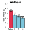
= increases the number of active cation channels in the membrane

24
Q

how does ADH secretion increase when plasma osmolality is high?

A

increased plasma osmolality

= cell shrinkage
= increased number of active cation channels in the neuronal membrane
= depolarisation
= increased firing rate to stimulate increased ADH secretion

25
Q

what does high plasma ADH stimulate?

A

fluid retention and invokes drinking

26
Q

when the plasma osmolality is decreased, how do the body cells respond?

A

neurones expand and the proportion of active cation channels in the membrane decreases

27
Q

what is the impact of cell expansion on the proportion of active cation channels?

A

the proportion of active cation channels in the membrane decreases

28
Q

how does ADH secretion decrease when plasma osmolality is low?

A

reduced plasma osmolality

= cell expansion
= reduced number of active cation channels in the neuronal membrane
= hyperpolarisation
= reduced firing rate to reduce ADH secretion

29
Q

what does low plasma ADH stimulate?

A

fluid loss = diuresis

30
Q

how does a hypotonic environment lead to hyperpolarisation of neurones?

A

hypotonic = cell expansion = reduced number of active cation channels = reduced influx of positive charge = hyperpolarisation

31
Q

what is the impact of hyperpolarisation on ADH secretion?

A

reduced neuronal firing rate

= reduce stimulation of ADH secretion
= reduced plasma ADH

32
Q

what is the impact of depolarisation on ADH secretion?

A

increased neuronal firing rate

= increased stimulation of ADH secretion
= increased plasma ADH

33
Q

how quickly are changes in osmolality corrected by drinking water?

A

not rapidly

= delay in absorption of water in GI tract and subsequent correction of osmolality

34
Q

how is thirst decreased? (2)

A

1) partial decrease in thirst = osmoreceptors in the mouth, pharynx and oesophagus detect water intake and decrease thirst partially
2) complete decrease in thirst = once plasma osmolality returns to normal or blood volume/arterial pressure

35
Q

explain how thirst is partially decreased

A

osmoreceptors in the mouth, pharynx and oesophagus detect water intake and decrease thirst partially

= relief of thirst sensation via these receptors is short-lived

36
Q

explain how thirst is fully decreased

A

only when plasma osmolality returns to normal or blood volume or arterial pressure is corrected, thirst is fully decreased

37
Q

which receptors are involved in the partial decrease of thirst?

A

receptors in mouth, pharynx, oesophagus

= thirst relief from these receptors in short-lived (partial)

38
Q

when and how is thirst fully decreased?

A

thirst is only completely satisfied once plasma osmolality is decreased or blood volume or arterial pressure corrected

39
Q

explain what happens when the blood pressure decreases, in the context of RAAS

A

blood pressure decreased

= renal afferent arteriole hypoperfusion
= increased renin secretion from the juxtaglomerular cells (of renal afferent arteriole)
= increased conversion of angiotensinogen to angiotensin I in the liver
= angiotensin I conversion into angiotensin II via ACE in the lungs

= angiotensin II increases aldosterone secretion, stimulates sympathetic activation and arteriole vasoconstriction among other things

40
Q

which cells respond to the decrease in blood pressure and why?

A

juxtaglomerular cells of renal afferent arteriole

= detect hypotension due to hypoperfusion of the renal afferent arteriole

41
Q

how do the juxtaglomerular cells respond to hypotension?

A

increased renin secretion into the systemic circulation

42
Q

what is renin and what is it alternatively known as?

A

renin is an enzyme secreted by the kidneys and is responsible for converting angiotensinogen into angiotensin I in the liver

(also known as an angiotensinogenase)

43
Q

which cells release renin and why?

A

juxtaglomerular cells of renal afferent arteriole

= response to the renal hypoperfusion

44
Q

what is the function of renin?

A

responsible for converting angiotensinogen into angiotensin I in the liver

45
Q

where is angiotensin I produced?

A

in the liver

46
Q

how is angiotensin I produced?

A

renin cleaves angiotensinogen in the liver to form the physiologically inactive precursor, angiotensin I

47
Q

where is angiotensin II produced?

A

capillary blood vessels/vascular tissue of the lungs

(site of most ACE expression and most angiotensin II production in the body)

48
Q

how is angiotensin II produced?

A

the enzyme ACE in the lung vascular tissue is responsible for converting angiotensin I into angiotensin II in the lung capillaries

49
Q

what converts angiotensinogen into angiotensin I?

A

renin (angiotensinogenase)

50
Q

what converts angiotensin I into angiotensin II?

A

ACE (angiotensin-converting enzyme)

51
Q

where is angiotensinogen found?

A

produced in the liver and is found continuously circulating in the plasma

52
Q

what are the impacts of increased angiotensin II secretion?

A

neurovascular effects

  • increased sympathetic activation
  • increased arteriole vasoconstriction

endocrine effects

  • increased ADH secretion from the PPG
  • increased aldosterone secretion from the zona glomerulosa of the adrenal cortex

renal effects

  • (increased aldosterone) results in increased Na+ retention and K+ excretion
  • increased salt retention results in increased H2O retention
53
Q

how does angiotensin II impact aldosterone secretion?

A

increased aldosterone secretion from the zona glomerulosa of the adrenal cortex

54
Q

where does angiotensin II act in the adrenal cortex and why?

A

acts on the zona glomerulosa of the adrenal cortex

= to increase aldosterone production to then increase Na+ reabsorption and K+ excretion

55
Q

what are the impacts of increased aldosterone secretion?

A

increase Na+, Cl- reabsorption and K+ excretion

= to stimulate H2O retention to increase blood volume

56
Q

what impact does angiotensin II have on the renal collecting duct?

A

increase tubular Na+, Cl- reabsorption and K+ excretion

= to stimulate H2O retention to increase blood volume

57
Q

what impact does angiotensin II have on the posterior pituitary gland?

A

stimulates ADH secretion from the posterior pituitary gland

58
Q

what impact does angiotensin II have on the arterioles and why?

A

stimulates arteriole vasoconstriction

= to increase blood pressure

59
Q

what impact does angiotensin II have on the autonomic nervous system?

A

stimulates sympathetic nervous system activation

60
Q

which two drugs are used to treat hypertension that affect RAAS?

A

direct renin inhibitors and ACE inhibitors

61
Q

how do variations in fat mass affect food intake and energy expenditure?

A

reduction in fat mass increases food intake and reduces energy expenditure

increases in fat mass (i.e. adipose tissue expansion) decreases food intake and increases energy expenditure

62
Q

what happens to food intake and energy expenditure when fat mass reduces?

A

reduction in fat mass increases food intake and reduces energy expenditure

63
Q

what happens to food intake and energy expenditure when adipose tissue expands?

A

adipose tissue expansion reduces food intake and increases energy expenditure

64
Q

how does the body respond to the overfed, weight augmented state?

A

increased sympathetic activation

increased energy expenditure

reduced hunger and food intake

= weight loss

65
Q

how does the body respond to the underfed, weight reduced state?

A

reduced sympathetic activation

reduced energy expenditure

increased hunger and food intake

(reduced thyroid function)

= weight gain

66
Q

which cerebral structure is responsible for appetite regulation?

A

hypothalamus

67
Q

which hormones are responsible for appetite regulation?

A

short-term

  • ghrelin
  • peptide YY (PYY)

long-term
- leptin

68
Q

explain how gut hormones have an effect on higher brain structures

A

ghrelin + PYY

= peripheral stimulation that travels via vagus nerve to the brainstem
= then onto the hypothalamus
= then onto the higher CNS centres of brain e.g. amygdala

69
Q

how does the hypothalamus regulate appetite?

A
  • receives all trigger (ghrelin, PYY, leptin, neural input from the periphery)
  • synthesises response to them by regulation food intake or increasing/decreasing energy expenditure
70
Q

which factors does the hypothalamus alter to regulate appetite?

A

food intake

energy expenditure

71
Q

where is the arcuate nucleus found?

A

located at the base of the hypothalamus on either side of the third ventricle

(in the medial-basal part of the brain)

72
Q

what is the arcuate nucleus?

A

aggregation of neurones in the medial-basal part of the
adjacent to 3rd ventricle

= involved in the regulation of food intake

73
Q

what is the most important terminal field of the arcuate nucleus and why is this important?

A

most important terminal field is the paraventricular nucleus (also adjacent to the third ventricle)

= has neurones that project onto the posterior pituitary gland where ADH is stored

74
Q

what is the paraventricular nucleus and why is this important?

A

adjacent to the third ventricle

many of its neurons project to the posterior pituitary + secrete oxytocin and a smaller amount of ADH

75
Q

where is the paraventricular nucleus found?

A

the hypothalamus, adjacent to the third ventricle

76
Q

what is the orexigenic effect?

A

appetite-increasing

77
Q

what is the anorexigenic effect?

A

appetite-reducing

78
Q

what do posterior pituitary gland neurones release?

A

oxytocin and ADH

79
Q

what are the two divisions of the hypothalamus that are important in appetite regulation?

A

lateral hypothalamus

ventromedial hypothalamus

80
Q

what is the function of the lateral hypothalamus?

A

only produces orexigenic peptides

= appetite-increasing effect

81
Q

what is the function of the ventromedial hypothalamus?

A

associated w satiety

82
Q

what is likely to happen if there is a lesion to the ventromedial hypothalamus?

A

lesions in this region can lead to severe obesity (seen in rats)

83
Q

name some hypothalamic factors implicated in appetite regulation

A

endocannabinoids

AMP-activated protein kinase

protein tyrosine phosphatase

84
Q

how is the arcuate nucleus adapted to interact with peripheral hormones?

A

has an incomplete blood-brain barrier

= allows access to peripheral hormones

85
Q

how many neuronal populations are there in the arcuate nucleus and what are they called?

A

two: one stimulatory population, one inhibitory population

  • stimulatory = NPY/Agrp neurone
  • inhibitory = POMC neurone
86
Q

what is the stimulatory neuronal population in the arcuate nucleus?

A

NPY/Agrp neurone

(neuropeptide Y/Agouti-related peptide)

87
Q

what is the inhibitory neuronal population in the arcuate nucleus?

A

POMC neurone

(pro-opiomelanocortin)

88
Q

how do NPY/Agrp neurones stimulate appetite?

A

make peptides that stimulate food intake

increased NPY signalling and reduced melanocortin signalling
= more stimulation and less inhibition of appetite

89
Q

which two hormones can affect the arcuate nucleus neuronal populations?

A

leptin and insulin

90
Q

how does leptin affect the arcuate nucleus neurones?

A

leptin stimulates the POMC neurones (increased inhibition of appetite)

leptin inhibits the NPY/Agrp neurones (reduced stimulation of appetite)

91
Q

how does leptin affect POMC neurones and what is the effect of this?

A

stimulates the POMC neurones

= increased inhibition of appetite

92
Q

how does leptin affect NPY/Agrp neurones and what is the effect of this?

A

inhibits the NPY/Agrp neurones

= reduced stimulation of appetite

93
Q

which conditions can cause food intake to increase by affecting the arcuate neuronal populations?

A

fasting, uncontrolled diabetes, general leptin deficiency

94
Q

explain the impact of general leptin deficiency

A

leptin deficiency

= less stimulation of POMC (so less inhibition of appetite)
= less inhibition of NPY/Agrp (so more stimulation of appetite)

= collectively act to increase appetite and food intake

95
Q

how do circulating factors control appetite?

A

circulating factors arrive at the arcuate nucleus

can penetrate into the arcuate nucleus via the incomplete blood-brain barrier

  • factors stimulate the POMC neurones (to decrease appetite)
    OR
  • factors stimulate the NPY/Agrp neurones (to increase appetite)
96
Q

how can circulating factors decrease appetite?

A

circulating factors arrive at the arcuate nucleus and penetrate via the incomplete blood-brain barrier

= act on POMC neurones stimulating them to decrease appetite

97
Q

how can circulating factors increase appetite?

A

circulating factors arrive at the arcuate nucleus and penetrate via the incomplete blood-brain barrier

= act on NPY/Agrp neurones stimulating them to increase appetite

98
Q

explain how the melanocortin system works in terms of POMC neurones

A

melanocortins (e.g. aMSH) are products of POMC neurones

melanocortins will bind to MC4R, expressed in the paraventricular

= leads to reduction in appetite, weight and decreased food intake

99
Q

what happens if there is a mutation in the MC4R?

A

fewer melanocortins will bind to the MC4R

= so less stimulation to decrease food intake
= so the opposite happens (i.e. food intake increases = fat mass increases)

100
Q

how do the POMC neurones act on the MC4Rs?

A

POMC neurones release melanocortins such as aMSH that bind to the MC4Rs of the paraventricular nucleus and stimulate a decrease in food intake

101
Q

how do the Agrp neurones act on the MC4Rs?

A

Agrp neurones bind to the MC4Rs of the paraventricular nucleus and inhibit the decrease in food intake (reduced inhibitory signal)

= consequently, food intake increases

102
Q

which CNS mutations are known to affect appetite?

A

no NPY or Agrp mutations associated with appetite have been discovered in humans = so no lack of stimulation of appetite

POMC deficiency and MC4-R mutations cause morbid obesity = lack of inhibition of appetite

103
Q

what is the implication of the following statement?

no NPY or Agrp mutations associated with appetite discovered in humans

A

no lack of/impaired stimulation of appetite

= food intake remains intact

104
Q

what is the implication of the following statement?

POMC deficiency and MC4-R mutations cause morbid obesity

A

lack of inhibition of appetite

= food intake increases hugely

105
Q

why is the amygdala important in appetite?

A

controls rewards and motivation pathways = strong effect on appetite

106
Q

what is the lateral hypothalamus associated with?

A

appetite-stimulant (orexigenic) peptides produced

107
Q

what is the ventromedial hypothalamus associated with?

A

associated with satiety

108
Q

how is neuronal information carried from the GI tract to the brain?

A

neuronal info from GI tract carried to the brainstem via the vagus nerve and then to hypothalamus and then to amygdala

109
Q

what is the adipostat mechanism?

A

keep an individual’s fat mass in a very narrow range despite changes in physical activity and diet

110
Q

explain how the adipostat mechanism works

A
  • adipose tissue produces circulating hormone
  • hypothalamus senses the concentration of circulating hormone present
  • hypothalamus then alters neuropeptides to increase or decrease food intake
111
Q

how do variations in adipose tissue alter the adipostat mechanism?

A

= more adipose tissue
= more hormone being produced
= neuropeptides production altered to (increase or) decrease food intake

112
Q

where is leptin produced?

A

made by adipocytes in white adipose tissue

113
Q

what are the two types of adipose tissue?

A

white adipose tissue
brown adipose tissue

114
Q

what is the function of white adipose tissue?

A

stores energy

115
Q

what is the function of brown adipose tissue?

A

dissipates energy, less common

116
Q

differentiate between white and brown adipose tissue

A

white adipose tissue = stores energy

brown adipose tissue = dissipates energy, less common

117
Q

where is leptin found?

A

in adipose tissue

118
Q

what is the function of leptin?

A

signals to the hypothalamus to control food intake and energy expenditure over the long term

(not from meal to meal = unlike ghrelin and PYY that act short-term)

119
Q

what is the function of leptin?

A

signals to the hypothalamus to decrease food intake and energy expenditure over the long term (+ increase thermogenesis)

(not from meal to meal = unlike ghrelin and PYY that act short-term)

120
Q

explain the pathophysiology of congenital leptin deficiency

A

condition that causes severe obesity beginning in the first few months of life

= reduced leptin levels
= reduced stimulation of the inhibitory POMC neurones + reduced inhibition of the stimulatory NPY/Agrp neurones
= collective effect to increased food intake
= can lead to obesity

(despite adipose tissue expanding, leptin levels do not proportionally increase)

121
Q

how do serum leptin levels vary in obesity?

A

would expect them to be high leptin with a high amount of adipose tissue, as part of the normal physiological mechanisms BUT

= leptin production is high instead because most obese people are insensitive to endogenous leptin production

122
Q

why does the body stop responding to leptin and what is the impact of this?

A

leptin receptor signalling defective

= decreased sensitivity to leptin
(obese people have reduced sensitivity to endogenous leptin production)

results in increased food intake

123
Q

what are normal leptin levels in an individual with low body fat?

A

low body fat = low leptin

124
Q

what are normal leptin levels in an individual with high body fat?

A

high body fat = high leptin

125
Q

how do leptin levels vary with body fat?

A

leptin circulates in plasma in concentrations proportional to fat mass

126
Q

what are gastrointestinal hormones?

A

hormones at are secrete into the GI tract to control various functions of digestive organs

127
Q

where are gastrointestinal hormones produced?

A

secreted by enteroendocrine cells in the stomach, pancreas & small intestine

128
Q

which two gastrointestinal hormones are involved in appetite regulation?

A

ghrelin
peptide YY

129
Q

what is the function of ghrelin?

A

stimulates appetite, increases gastric emptying

130
Q

what is the function of peptide YY?

A

inhibits food intake

131
Q

when are ghrelin levels highest and why?

A

blood levels of ghrelin are highest before meals

= help prepare for food intake by increasing gastric motility and acid secretion

132
Q

how does ghrelin work?

A
  • increases appetite
  • regulates reward, taste sensation, memory, circadian rhythm
133
Q

where does ghrelin act?

A

directly modulates neurons in the arcuate nucleus

  • stimulates NPY/Agrp neurons
  • inhibits POMC neurons.
134
Q

explain the pattern of ghrelin secretion

A

plasma ghrelin levels increase almost twofold before each meal time

preprandial rise and post prandial fall in ghrelin concentration

135
Q

what does the preprandial rise and postprandial fall in ghrelin concentration indicate?

A

physiological role of ghrelin in meal initiation via increasing gastric motility and gastric acid secretion

136
Q

what does the following graph indicate?

A

food intake increases with ghrelin dose

= associated with weight gain and increasing adiposity

137
Q

what does the following graph indicate?

A

increased appetite and energy intake for those given a dose of ghrelin

138
Q

what is PYY?

A

peptide YY (peptide tyrosine tyrosine)

released into the terminal ileum and colon to inhibit food intake (by acting on the hypothalamus)

139
Q

when and where is PYY released?

A

released in the terminal ileum (TI) and colon in response to feeding

140
Q

what is the function of PYY?

A

reduces appetite (can be digested or injected IV)

141
Q

where does PYY act?

A

acts on the arcuate nucleus of the hypothalamus

specifically

  • inhibits NPY/Agrp neurones
  • stimulates POMC neurones

(bu initially released into the terminal ileum and colon)

142
Q

how does ghrelin increase appetite?

A

ghrelin stimulates NPY/Agrp neurones and inhibits POMC neurones

143
Q

how does PYY decrease appetite?

A

PYY inhibits NPY/Agrp and stimulates POMC

144
Q

compare the mechanism of action of ghrelin to that of PYY

A

ghrelin stimulates NPY/Agrp neurones and inhibits POMC neurones whereas PYY does the opposite by inhibiting NPY/Agrp and stimulating POMC

145
Q

what does the following graph indicate?

A

the more PYY released in the GI tract, the greater the reduction in food intake and the more weight reduces

146
Q

what is the relationship between postprandial PYY release and calorie intake?

A

postprandial PYY release is proportional to the calorie intake

(greater calorie intake, greater PYY release postprandially)

147
Q

what is the effect of PYY on food intake and hunger?

A

PYY reduces food/calorie intake

148
Q

name some comorbidities associated with obesity

A
149
Q

what are some conclusions that can be drawn from this graph?

A

healthy environment

  • subjects who are genetically prone to obesity BUT in a healthy environment = very few will develop obesity
  • subjects that are genetically resistant + in a healthy environment = very unlikely to develop obesity

toxic environment

  • subjects that are genetically prone and in a toxic environment = highest risk of severe obesity
  • but those genetically resistant in a toxic environment = likely unaffected

= genes + environment both affect obesity

150
Q

how do ghrelin and peptide YY differ compared to leptin?

A

leptin = long term appetite regulation

ghrelin and peptide YY = immediate appetite regulation