Final Review Flashcards
What are the common disc protrusion sites?
T12-T13, T13-L1, C2-C3, C3-C4
Why is anesthesia recommended for spinal radiographs?
Prevents narrowing of the disc space with muscle spasms
In spinal radiographs, high ___ and low ___ create better contrast
high kVp, low mAs
Use of a ____ increases contrast for spinal radiographs
Grid
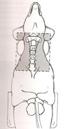
When positioning for a spinal radiograph, the vertebral column must always be _____ with the table top
parallel
When positioning for spinal radiographs, the disc spaces must be _____ with the table top
perpendicular
Common views for spinal radiographs
lateral and VD
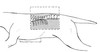
Use of smaller cassettes and multiple views for spinal radiographs allow for what?
Perpendicular view of the disc spaces
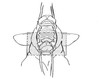
Why are DV spinal views not as accurate?
Not parallel
Object-film distance distortion
How many views may be required for a full spinal study on a dog vs a cat?
Dog - 4-5 views
Cat - 1-2 views
What is the vertebral formula?
C7 T13 L7 S3 Cd20+ (tail)
Vertebral bone with no spinous process and two large lateral wings, articulate via synovial joints allowing greater movement
Atlas (C1)
Vertebral bone with large “blad-like” spinous process, peglike dense at cranial aspect forming atlantoaxial joint. No intervertebral disc between C1 and C2, articulates caudally with C3 via intervertebral disc
Axis (C2)
Vertebral bone with large transverse process (ventral lamina)
C6
What species has 18 pairs of ribs? (One breed does not)
Horses
Species with thoracic vertebrae that have tall spinous processes, large articular facets that form joints with the heads of the ribs
Dogs
In what species are rib heads cranial to respective vertebral body?
Cat
Where would you find the narrowest intervertebral disc space in a dog?
Between T10-T11
Which vertebra is the Anticlinal vertebra?
T11
What species have 4 fused vertebrae making up the sacrum?
pigs and sheep
What species have 5 fused vertebrae making up the sacrum?
Horses, cattle, goats and humans
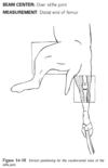
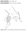
Where should the beam be centered for a lateral view of the cervical spine?
C4
Where should the measurement be taken for a lateral view of the cervical spine?
C6
What should be included in the field of view for cervical spine XRays?
Base of skull to first few thoracic vertebrae
How should the patient be positioned for a flexed cervical spine radiograph?
Lateral recumbency with legs pulled caudally
Where should the beam be centered for a lateral thoracic spinal radiograph?
T7
How should limbs be positioned for a thoracic spine lateral view?
Moderately extended in opposite directions
What is the field of view for a thoracic spine lateral radiograph?
Last cervial to first lumbar vertebrae (shoulder joint to past the origin of the last rib)
Where should the beam be centered for a VD view of the thoracic spine?
Over caudal border of the scapula (T6)
How should the patient be positioned for a VD thoracic spine view?
Dorsal recumbency with legs pulled cranially
In a VD view of the thoracic spine how should the sternum be positioned?
Superimposed over the spine
What is the field of view for a lateral view of the lumbar spine?
T12 to the sacrum and include entire lumbar spine
What view visualizes the “nike swoosh”?
Lumbar spine - Transver spine superimposed at the origins
What is the field of view for lateral lumbosacral vertebrae?
L6 and entire pelvis
How should the patient be positioned for a VD view of the sacrum?
Dorsal recumbency with legs in normal position
How should the tube head be angled for a VD view of the sacrum?
30 degrees
What is the field of view for a VD view of the sacrum?
L6 and entire pelvis
How should the patient be positioned for a caudal spine radiograph?
Lateral recumbency with limbs in a natural position, tail extended caudally
What is the field of view for a caudal spine radiograph?
4-5 vertebrae on either side of area of interest or full tail
How should the cassette be placed for a caudal spine view?
Raise cassette with a foam pad to keep on same plane as the tail
What is measured if sinus cavities need to be radiographed?
Measure just rostral to the thickest part of the cranium to avoid overexposure
What are the views for a routine skull or survey study?
Lateral (affected side down), DV, or VD
Where is the measurement for a lateral skull view?
Over high point of zygomatic arch
Where is the beam centered for a DV view of the skull?
Lateral canthus of the eye
Where is the measurement taken for a DV view of the skull?
Over high point of the cranium
How should the patient be positioned for a DV view of the skull?
Sternal recumbency with head resting on cassette, sand bag on neck and front limbs relaxed or pulled caudally
What is the field of view for a DV view of the skull?
Include top of nose to base of skull
How should the patient be positioned for a VD view of the skull?
Dorsal recumbency, legs pulled caudally, sand bags on either side of the neck, nose parallel with cassette
Where should the beam be centered for a VD view of the skull?
Lateral canthus of the eye
Where should the measurement for a VD view of the skull be taken?
Lateral canthus of the eye
Where should the beam be centered for a rostrocaudal closed mouth view of the frontal sinuses (90 degree)
Through center of frontal sinuses and between the eyes
How should the patient be positioned for a rostrocaudal closed mouth view of frontal sinuses?
Dorsal recumbency with neck flexed, nose pointing upwards, legs pulled caudally alongside body
Where should the beam be centered for a rostrocaudal closed mouth view of the cranium (rostral 30 degree DCdVO)
Mid point between the eyes
Where is the measurement taken for a rostrocaudal closed mouth view of the cranium?
Area of frontal sinuses
How should the patient be positioned for a rostrocaudal closed mouth view of the cranium?
Dorsal recumbency with neck flexed, more flexion than frontal sinus view
What is the patient positioning for VD open mouth view of the nasal cavity?
Dorsal recumbency with maxilla taped to table, pull mandible open with tie or mouth wedge
Where should the beam be centered for VD open mouth view of nasal cavity?
Through the third upper premolar
What is measured for a VD open mouth view of the nasal cavity?
Upper jaw over the upper third premolar
How should the tube head be angled for a VD open mouth view of nasal cavity?
20-30 degrees rostrocaudally
What is the field of view for a VD open mouth view of nasal cavity?
entire maxilla to pharynx
What is the patient positioning for a tympanic bullae (rostral 10-30 VCdDO) Rostrocaudal open mouth view?
Dorsal recumbency with the neck flexed, nose pointed upward, legs pulled caudally. Pull maxilla cranially 10 degrees and mandible caudally 10 degrees
Where should the beam be centered for a rostrocaudal open mouth view of tympanic bullae?
Base of neck to commissure of lips
What is the field of view for a rostrocaudal open mouth view of tympanic bullae?
Entire pharyngeal region with bullae isolated
What is patient positioning for lateral oblique view of tympanic bullae?
Lateral recumbency with good side down, limbs pulled caudally, head rolled into natural position for oblique
What view is good for TMJ?
Lateral oblique view of tympanic bullae
What is patient positioning for VD oblique view of TMJ?
Lateral recumbency with affected side down, head rotated at 20 degrees with wedge, mouth may be open or closed
Soft tissues are difficult to identify on plain films due to lack of contrast. How can we better view these tissues?
Using contrast medium or performing ultrasound
What are two benefits of survey films?
Help determine proper exposure and eliminate need for contrast
What agents are used for positive contrast?
Barium or Iodine
Positive contrasts have ____ atomic numbers than tissue and bone
Higher
What contrast can safely be administered into a blood vessel?
Iodine
How should kVp be increased with positive contrast?
6-8 kVp
What agents are used for negative contrast?
Gasses with low specific gravity
Barium causes _____ when left free in abdominal cavity
Peritonitis
Why are iodine compounds not used in GI studies?
They aren’t thick enough to coat mucosa of the intestines
What is used in GI situations when perforations are suspected?
Iodine preparations
The higher the iodine concentration the more _______
contrast
Contrast preparation that is completely insolube therefore not diluted with alimentary secretions
Barium
What are BIPS?
Barium impregnated polyurethane spheres
Positive contrast agent that remains in spherical form, designed for transit time
BIPS
What is best used for negative contrast agents and why?
CO2 because it is rapidly absorbed
In a double contrast study which contrast should be administered first?
Negative contrast, to avoid air bubbles that mimic lesions
If necessary, what drug should be used to tranquilize patients in contrast studies?
Acepromazine
What drugs should be avoided when doing contrast studies?
Parasympathetic/anticholinergics
ie. Atropine and glycopyrolate
What circumstances contraindicate the use of contrast studies?
Fluid filled distended stomach or esophagus
Ruptured bowel
Dysphagic patients
Megaesophagus
Why are dysphagic patients or megaesophagus contraindicated for contrast studies?
Risk of aspiration
What is the ideal method for determining megaesophagus?
Esophagraphy
When is esophography indicated?
History of regurgitation or dysphagia
What is the technique used for esophagraphy?
Lateral recumbency, take survey radiographs, slowly administer product orally
What should the view of an esophograph be from?
From the pharynx to the stomach, multiple views during and after swallowing
What view should be taken first for an esophograph?
Lateral, and then ventral (never dorsal)
How should barium be given for an esophograph?
Give barium while laying down
What does a gastrophy evaluate?
Size, shape, position, function, and morphology of the stomach only
What is a gastrophy used for?
Vomiting, blood in vomit, anterior abdominal pain
For an upper GI study, fast the animal for _____ or ____ hours if for a small intestine
12 - 24 hours
What contrast is used for gastrophies?
Positive
What are the steps to completely evacuating large bowel?
Low residue food for 48 hours
Fast 24 hours prior
Pull water 4 hours prior
Enema until clear
Negative LGI contrast studies diagnose what?
Intussusceptions in puppoes
What is used to evaluate renal structure, function and to visualize renal collection system?
Excretory urography
Excretory urography involved the IV injection of what?
A water soluble iodinated contrast agent
Diffuse opacification of the renal parenchyma as the contrast agent perfuses the renal vasculature
Nephrogram phase
What phase of urography demonstrates the presence of functional renal function
Nephrogram phase
In what urograph phase does the contrast agent filter into the renal collection system?
Pyelogram phase
What is the test dose for an excretory urography?
3-5 ml
What is the dose of contrast for an excretory urography?
300-400 mg/ml
Dose 3 ml/kg rapid infusion for 1-3 minutes
Hematuria, Dysuria, pyuria, straining of urination and frequency of urination are indications for what?
Excretory Urography
When doing a cystography, which contrast should NEVER be used?
Barium
Fatal rare embolism with negative contrast is a complication associated with what?
Cystography
What uses negative or positive retrograde or antegrade contrast to detect urethral trauma, tears, tumors or obstructions?
Urethrography
What study consists of a positive contrast in a joint space?
Arthrography
What study consists of a positive contrast injected into blood vessels?
Angiography
Contrast given orally or IV that is concentrated in the bile and visualized in the gall bladder and biliary system
Cholecystography
CT and MRI are replacing what technique?
Myelography
Positive contrast injected into the subarachnoid space of the spine to highlight spinal cord lesions and prolapsed discs?
Myelography
What is the kVp preset for dentistry?
70 kVp
What is the mA preset for dentistry?
7-8 mA
What dental film size is used for individual teeth?
2
What dental film size is used to show teeth alignment or dental architecture?
4
What is the purpose of the foil on the back of the dental film?
Absorbs radiation
For small chairside darkroom dip tanks, developer for ___ seconds, ___ minutes for fixing, and ___ minutes rinsing
30 seconds, 1.5 minutes, 3 minutes
How many views are required for large animal skeletal radiographs?
4, or 6 for joints
What is the portable max mA and max kVp for large animals?
ma- 20 kVp-90
What is the max mA and kVp for mobile units in large animal?
mA 100-300
kVp- 120
What xray unit for large animals has shorter exposure times and more power?
Mobile unit
What features make birds more radiolucent?
Thin cortices and tubular bones
What are the three types of restraints used for avians and exotics?
Chemical, manual, physical
What is restrained first for a bird?
Head and torso
What is restrained last for birds?
legs
What is the average transit time for barium in a bird?
30-240 minutes
What species has the quickest transit time?
Small psittacines
What animals MUST be anesthetized?
Rabbits and guinea pigs
This effect is “all or nothing”, increasing the dose will increase the possibility of effect but not the intensity, No threshold dose.
Stochastic
Radiation effect that has a predetermined threshold dose, immediate to time-delayed effects
Nonstochastic
What symptoms are associated with nonstochastic effects?
cataracts, cancer, hemopoietic, GI, CNS
What radiation interaction affects DNA?
Direct interaction
Which radiation interaction affecst the cell’s ability to reproduce?
Indirect interaction
Transfer of incorrect genetic code to one of the daughter cells
Critical consequence
Ionization of water causes an effect on what?
Critical target
Free radical formation of ____ and ____ _____ is a major factor of damage to the cell
hydroperoxyl and hydrogen peroxide
What is the ALARA principle?
As Low As Reasonably Achieveable
What does LET stand for?
Linear Energy Transfer
A measure of the rate at which energy is transferred from ionizing radiation into soft tissue
LET
The higher the LET the ______ the amount of energy transferred to soft tissue per interaction
greater
Low kVp imparts higher amounts of _____
energy
What is responsible for the energy imparted to the photons?
kV
Alpha and Beta particles have ___ LET with ___ penetrability
High, low
X and gamma radiation have ____ LET and ____ penetrability
Low, high
X and Gamma radiation have high ability to travel through matter while producing ____ effect on tissues they pass through
Less
The intensity of the radiation is inversely related to what?
The square of the distance
The ____ provides source for electrons and directs these electrons towards ____
Cathode, Anode
The ____ the atomic number, the more electrons are available for excitation
higher
Amount of electrical energy being applied to the filamet; describes number of X-Rays produced during exposure.
mA (milliamperage)
Acceleration is controlled by _____
kV
______ acts as a heat conductor and draws heat away from ______ target
Copper, tungsten
Which type of anode is found in dental and small portable units?
Stationary Anode
Large amounts of heat is a limitation of which anode?
Stationary
If target is damaged, may cause radiation to scatter in undesirable locations, this will cause _____ than expected xrays
Lighter
Rotating Anodes rotate through the ____ of the tube
Center
Rotation provides a _____ surface for the electron stream
Cooler
Rotating anodes help distribute _____ over a larger area
heat
_____ anode may have small focal spot but still withstand a great amount of heat
Rotating
More x-rays leave tube on ____ side
cathode
What can prevent anode target failure?
high kVps and low mA
What is kVp?
peak of energy which determines quality of xray beam
What is it called when metal deposits form on inner lining of glass due to overheating?
Arcing
What is the process of changing alternating current (AC) to direct current (DC)?
Rectification
What dictates the maximum combinations of kVp and mA without overloading the tube?
X-Ray tube rating
What are the four factors determining tube rating?
Focal spot size, Target angle, Anode speed, electrical current
If the film is too dark the mA’s must be reduced to ____
50%
What is the first adjustment to contemplate if technique needs adjusted?
mAs
If the image is too dark and the kV is between 50 and 90, reduce kV by ____
15%
Blur also affects resolution by projecting the anatomic structures ____ from the focal plane
Away
The structures ____ and ____ a predefined level in the body are blurred so that the level of interest, the focal plane, can be visualized
above and below
When is distortion used effectively?
In dental imaging, oblique views and to locate foreign bodies
When is distortion unacceptable?
Stuctures to be measured for orthopoedic surgery, cardiac cases when heart-lung ratios must be measured, central ray positioned incorrectly
What is the magnification formula?
MF=SID/SOD
What does SID and SOD stand for?
Source Imaging Distance
Source Object Distance
When is the magnification formula useful?
Bladder stones, kidney stones, heart-lung ratios
Illuminators must be clean and have a ___/___ light
blue/white
80% of artifacts are caused by what?
Processing
What view is represented in this image?

Cervical Spine Lateral view

What view is represented in this image?

Cervical spine hyperextended

What view is represented in this image?

Cervical spine flexed lateral view

What view is represented in this image?

Cervical Vertebrate VD

What view is represented in this image?

Thoracic spine Lateral view

What view is represented in this image?

Thoracic spine VD

What view is represented in this image?

Thoracolumbar spine Lateral view

What view is represented in this image?

Thoracolumbar spine VD view

What view is represented in this image?

Lumbar spine lateral view

What view is represented in this image?

Sacrum VD view

What view is represented in this image?

DV Skull view

What view is represented in this image?

VD view of skull

What view is represented in this image?

Front sinuses (90 degrees) Rostrocaudal closed mouth

What view is represented in this image?

Nasal Cavity VD open mouth view

What view is represented in this image?

Tympanic bullae rostrocaudal open mouth view

What view is represented in this image?

Tympanic bullae oblique lateral

What view is represented in this image?

TMJ VD oblique view

What view is represented in this image?

PennHIP distraction

What view is represented in this image?

PennHIP Compression

What view is represented in this image?

PennHIP Extended

What view is represented in this image?

Scapula Craniocaudal view

What view is represented in this image?

Shoulder caudocranial view

What view is represented in this image?

Humerus lateral view

What view is represented in this image?

Humerus caudocranial view

What view is represented in this image?

Humerus craniocaudal view

What view is represented in this image?

Elbow craniocaudal view

What view is represented in this image?

Elbow lateral view

What view is represented in this image?

Radius and ulna lateral view

What view is represented in this image?

Craniolateral view of radius and ulna

What view is represented in this image?

Carpus dorsopalmar view

What view is represented in this image?

Metacarpals and phalanges dorsopalmar view

What view is represented in this image?

Femur lateral view

What view is represented in this image?

Femur craniocaudal view

What view is represented in this image?

Stifle joint caudocranial view

What view is represented in this image?

Stifle joint lateral view

What view is represented in this image?

Tibia and Fibula lateral view

What view is represented in this image?

Tibia and fibula caudocranial view

What view is represented in this image?

Tarsus (hock) lateral view

What view is represented in this image?

Tarsus (hock) plantarodorsal
