FDN2_SM_WK3_Ultrasound Flashcards
Ultrasound
What kinds of imaging modalities use ionizing radiation?
Radiography (x-ray)
CT Scan
Mamography
Angiography
Nuclear Imaging
What is ionizing radiation?
An electromagnetic wave that has the energy necessary to remove an electron from an atom
What does ALARA stand for?
What does it mean?
As Low As Reasonably Achievable
We should strive for minimal radiation dosage
What is Radiography?
Electrons are emitted from a target and interact with an anode
- Sent toward the person/object. The electrons will do one of three things:
1. Pass through the tissue to the detector
2. Get deflected or scattered
3. Get absorbed
The combination of these things creates the image
What are 5 “pros” of Radiography?
- Widely available
- Good screening tool
- Fast
- Detail in bone
- Low cost
What are 3 “cons” of radiography?
- Uses ionizing radiation
- Two-dimensional image
- Less detail than, MRI, Ultrasound (especially for soft tissue)
What is a CT scan?
Basically a 3-D X-Ray
The source and detector rotate around the patient, and the image is reconstructed by a computer
What are 4 “pros” of a CT scan?
- Better tissue resolution than X-Ray
- Fast
- Widely available
- Can reconstruct the image in infinite planes
What are 2 “cons” of a CT scan?
- Much more ionizing radiation than X-ray (~1 year of background radiation)
- Contrast dye can cause renal failure
What is ultrasound?
A transducer emits high-frequency sound waves into the body
The echoing waves are used to generate an image. Different tissues send waves back at different frequency and wavelengths
Same concept as sonar!
Which structures will appear hyperechoic in an ultrasound?
Bone, Fat
Which structures will appear anechoic in an ultrasound?
Fluid, air
Which tissues will appear hypoechoic in an ultrasound?
Soft tissue
What are 5 “pros” of ultrasound?
- Noninvasive
- No ionizing radiation
- Can evaluate blood flow
- Can be used to guide procedure
- Portable
What are 2 “cons” of ultrasound
- Dependent on the skill of the operator
- Cannot penetrate air or bone
Which imaging modality would be least useful for looking at the lungs?
A. X-Ray
B. CT Scan
C. Ultrasound
D. MRI
C. Ultrasound - It cannot penetrate air
Which ultrasound probe would you use for looking at abdominal structures?
Curvilinear
What are the relevant characteristics of a cuvilinear probe?
Low to mid frequency
Can see deeper images with an adequate field of view (The goldilocks probe)
Lower resolution
What is a curvilinar prove used for?
Looking at intra-abdominal structures
Which ultrasound probe would you use to guide superficial procedures?
Linear
What are some characteristics of a linear ultrasound probe?
High frequency
High resolution
Produces a superficial image with a wide depth of field
Which ultrasound probe would you use to look at blood vessels in a patient’s leg?
Linear
Which ultrasound probe has a narrow field of view and a large depth of field?
Phased-array probe
What is acoustic shadowing?
The area behind bones or stones appears anechoic because the waves are reflected by the bone
What is an edge artifact?
Smooth, round surfaces deflect sound beams instead of reflecting them
This causes a round, anechoic area that may be mistaken for fluid
Where is it common to see an edge artifact?
Cyst, bowel, galbladder
What is posterior acoustic enhancement?
Increased echos behind structures that allow sound waves to pass through easily (ex: structures filled with fluid)
What is a mirror image artifact?
A mirror image is created when a highly reflective surface reflects the initial beam, but encounters another structure on the way back.
The mirror image is of this other structure
What will increasing the depth of an ultrasound do?
Increase the distance that the sound waves travel in the body
What will increasing the gain of an ultrasound do?
Increase the amplification of the returning signal
What is MRI?
Magnetic Resonance Imaging
- The magnetic field aligns the protons in hydrogen atoms
- The radiofrequency current creates a varying magnetic field, causing the protons to flip their spins
- When the magnetic field is turned off, the protons go back to normal and create an image
What are 3 “pros” of MRI?
- No ionizing radiation
- Lots of detail in soft tissues and blood vessels
- infinite planes for image reconstruction
What are 5 “cons” of MRI?
- Expensive
- Claustrophobic patients need sedation
- Not available to patients with pacemakers
- It takes a long time
- A moving patient can ruin the scan
What is nuclear imaging?
Physiologic, rather than anatomic imaging
Uses radioactive tracers to diagnose and treat conditions
(To see how things are working, as opposed to what they look like)
What is a PET scan?
Positive Emission Tomography: A type of nuclear imaging
Use 18-Fluro-Deoxy-Glucose. It is taken up by metabolically active cells like brain, heart, cancer
What are the 5 X-Ray densities from blackest to whitest?
- Air = least dense = blackest
- Fat
- Soft tissue/water
- Bone
- Metal or contrast
What density is the heart on a frontal chest radiograph?
Soft tissue/water
This will appear grayish
Which number lables the aorta?

1

Which number labels the superior vena cava?

2

Which number labels the pulmonary trunk?

3

Which number labels the heart?

4

Which number lavels the left atrium?

5

Which number labels the right atrium?

6

Which number labels the left ventricle?

7

2 labels the…

Superior Vena Cava

1 labels the…

Aorta

3 labels the…

Pulmonary trunk

4 labels the…

Heart

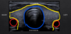
Which plane is shown in blue?

Coronal

Which plane is shown in red?

Saggital

Which plane is shown in green?

Axial aka transverse

Which structures are in red?

Carotid Arteries
Why do the structures circled in red appear black?
What is the term for this on an ultrasound?

They appear black because they are blood vessels filled with fluid
Structures that are black on an ultrasound are anechoic

Which structure is circled in blue?

The trachea

Which structure is outlined in yellow?

The thyroid

Which structure is labled by #1

Right Pulmonary Artery

Which structure is labled by #2

Pulmonary Trunk
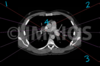
Which structure is labled by #3

Left Pulmonary Artery

Which structure is labled by #4

Ascending Aorta

Which number lables the right pulmonary artery?

1

Which number lables the pumonary trunk?

2

Which number lables the left pulmonary artery?

3

Which number lables the ascending aorta?

4

In which plane is this image taken?

Axial aka Transverse

