FDN2_SM_BodyWall+Cavity Flashcards
List the layers of the body wall from superficial to deep
- Skin
- Superficial Fascia
- Skeletal Muscle/Associated investing fascia
- Celomic lining
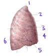
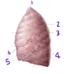
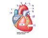
What is celomic lining in the thorax called?
Parietal pleura
What is parietal pleura?
The celomic lining in the thorax
What is visceral pleura?
Part of the celomic lining that directly adheres to each lung
What is investing fascia over muscle called?
Epimysium
What is investing fascia over bone called?
Periosteum
What is investing fascia in the thoracic cavity (deep to the ribs) called?
Endothoracic fascia
What are epaxial muscles?
Back muscles (part of the typcial body segment)
What are hypaxial muscles?
Muscles that extend aroudn the celom and form the body wall
Organized into 3 concentric layers
What are the 3 concentric layers of hypaxial muscles, as they present in the thorax?
Which direction do they go?
External intercostal muscles \//
Internal intercostal muscles //\
Transversus muscles (innermost intercostal + transversus thoracis) //\ (similar to intercostal muscles)
Note: Transversus thoracis is deep to the innermost intercostal muscles
What are the 3 concentric layers of hypaxial muscles, as they present in the abodomen?
Which direction do they go?
External abdominal oblique \//
Internal abdominal oblique //\
Transverse abdominis ==
How is the rectus layer of muscle different in the abdomen and thorax?
Abdomen: || (form the 6-pack)
Thorax: absent or vestigial
What is the function of the external intercostal muscles?
Elevate ribs in forced inspiration
Maintain rigidity of intercostal space
What is the function of the internal intercostal muscles?
Depress ribs in forced expiration
Maintain rigidity of the intercostal space
Where does innervation of the external intercostal muscles come from?
Ventral rami
What is the purpose of the rectus abdominus?
Flexes the trunk (like when you do a sit-up)
What is the function of the external abdominal oblique?
Both sides together flex the trunk
Lateral bend and flex
Rotation to the opposite side of the contracting muscle
What is the function of the internal abominal oblique?
Both sides together flex the trunk
Bend or rotate to the same side as the contracting muscle
What is the function of the transverse abdominis?
Compress abdominal viscera
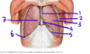
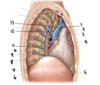
What is the neuromuscular bundle?
Where is it found?
The intercostal nerve, artery, and vein that belong to each intercostal space
The bundle sits in the costal groove, which is sheltered by the distal edge of the upper rib of the intercostal space
When you need to access the parietal pleura, why would you insert the needle just above the rib inferior to the intercostal space?
You want to avoid the neuromuscular bundle, which is associated with the rib superior to the intercostal space
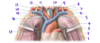
What are the relevant components of the intercostal space?
Boundaries: upper and lower rib
3 concentric layers of muscle (external, internal, innermost)
Each space is associate with a neuromuscular bundle (intercostal nerve, vein, artery)
Describe the path of an intercostal nerve
The intercostal nerve is a spinal nerve
Presynaptic: in CNS
Synapse: in sympathetic trunk
Postsynaptic: Leaves trunk, follows ventral ramus, innervates muscles in the thoracic body wall
Between which two layers of muscle would you find an intercostal nerve?
Between the internal and innermost intercostal muscles
What are the components of a typical body segment? (10 things)
Spinal cord
Vertabra
Dorsal Ramus
Ventral Ramus
Cutaneous nerves
Intercostal nerve
Epaxial (back) and Hypaxial (celomic) muscles
Celomic cavity (pleural or peritoneal)
Gut
Associated blood vessles
Which layers make up the superficial body wall?
Skin, fat, superficial fascia
Which nerves carry sensory information from the breast to the brain?
Anterior cutaneous nerves
What is the muscle immediately deep to the breast?
Pectoralis major
What is the function of the celom?
It allows for mobility of the organs inside of the body and the organism itself
How does the pericardial cavity form?
Pleurocardial folds unit behind the heart to form the fibrous pericardium.
This separates the pleural cavities from the pericardial cavity
What is inside of the pleural cavity?
Serous fluid
(The lungs are pressed against the pleural cavity, but not inside of it!)
Describe the layers of coelom covering the heart
From superficial to deep:
Fibrous pericardium (outside of the pericardial sac)
Parietal serous pericardium
Visceral serous pericardium aka epicardium
Where does the visceral layer of coelom come from?
Splanchnopleure, which is derived from the lateral plate
Where does the parietal layer of coelom come from?
Somatopleure, which comes from the lateral plate
Describe the innervation of the parietal coelom
Somatosensory neurons (sharp pain sensation!)
Describe the innervation of the visceral coelom
Visceral sensory neurons
We don’t feel localized or sharp pain in this layer
How does the coelom form?
The intraembryonic coelom forms as a cavitation in the mesoderm that separates the somatopleure and the splanchnopleure
After the trilaminar disc becomes a cylinder, organs grow and push against it, shaping it into its eventual adult form
What is the mediastinum?
The area between the two lungs that contains the heart, part of the trachea and esophagus, thymus gland, nerves, the great vessels, and lymph nodes
In a pneumothorax, where does air accumulate?
In the pleural cavity
If food “goes down the wrong tube,” which side does it go down? Why?
Right; the heart kind of pushes it that way
What structures are contained in the root of the lung?
Pulmonary arteries and veins, primary bronchi, bronchial arteries, pulmonary nerve plexus and lymphatics
What is the name for the place on the lung where there root enters?
Hilum
Describe the path of the phrenic nerve
The prhenic nerve is a somatic nerve
Cell body: in the brain
Path out of the CNS: Ventral Rami of C3-5
The right and left phrenic nerves travel anterior to the root of the lung, between the mediastinum and the mediastinal pleura to the diaphragm.
What are the arrows pointing to?

Right (lower arrow) and left (upper arrow) phrenic nerves

What does it mean when they say that the pleural cavity is a “potential space?”
There is normally nothing inside of it except for a thin film of serous fluid
For it to become an actual space, the surface tension between visceral and parietal pleura must be broken for the membranes to separate
What is the structure labeled by #1?

The apex of the lung

What is the structure labeled by #2?

Superior Lobe

What is the structure labeled by #3?

Horizontal fissure

What is the structure labeled by #4?

Middle lobe

What is the structure labeled by #5?

Oblique fissure

What is the structure labeled by #6?

Inferior lobe

What structure is this?

Right lung

What structure is this?

Left Lung

What structure is labeled by #1?

Apex

What structure is labeled by #2?

Superior Lobe

What structure is labeled by #3?

Oblique Fissure

What structure is labeled by #4?

Inferior Lobe

What structure is labeled by #5?

Lingula

What structure is labeled by #6?

Cardiac Notch

What structure is labeled by #1?

Mediastinal pleura

What structure is labeled by #2?

Costal Pleura

What type of pleura is labeled by #3?

Visceral plerua

What structure is labeled by #4?

Mediastinum (the pericardial sac is inside)

What structure is labeled by #5?

Diaphragmatic Pleura

What space is labeled by #6?

Costodiaphragmatic Recess

If #7 were pointing ot a space, what space would it be?

Costomediastinal Recess

What structure is labeled by #1?

Left Phrenic Nerve

What structure is labeled by #2?

Ligamentum Arteriosum

What structure is labeled by #3?

Left Recurrent Laryngeal Nerve

What structure is labeled by #4?

Left Vagus Nerve

What structure is labeled by #5?

Cervical Pleura

What structure is labeled by #6?

Left Brachiocephalic Vein

What structure is labeled by #7?

Left Subclavian vein

What structure is labeled by #8?

Left Phrenic Nerve

What structure is labeled by #9?

Left Internal Jugular Vein

What structure is labeled by #10?

Esophagus

What structure is labeled by #11?

Trachea

What structure is labeled by #12?

Right Internal Jugular Vein

What structure is labeled by #13?

Right Phrenic Nerve

What structure is labeled by #14?

Right Subclavian Vein

What structure is labeled by #15?

Right Vagus Nerve

What structure is labeled by #16?

Right Brachiocephalic Vein

What structure is labeled by #17?

Superior Vena Cava

What structure is labeled by #1?

Ascending Aorta

What structure is labeled by #2?

Brachiocephalic Trunk

What structure is labeled by #3?

Left Common Carotid Artery

What structure is labeled by #4?

Left Subclavian Artery

What structure is labeled by #5?

Aortic Arch

What structure is labeled by #6?

Ligamentum Arteriosum

What structure is labeled by #7?

Descending Aorta

What structure is labeled by #8?

Left Pulmonary Artery

What structure is labeled by #9?

Pulmonary Trunk

What structure is labeled by #10?

Left Atrium

What structure is labeled by #11?

Left Ventricle

What structure is labeled by #12?

Right Ventricle

What structure is labeled by #13?

Right Atrium

What structure is labeled by #14?

Superior Vena Cava

Which structure is the red arrow pointing to?

Pulmonary Trunk

Which structure is the blue arrow pointing to?

Aorta

Which structure is the green arrow pointing to?

Superior Vena Cava

What structure is labeled by #1?

Serous Parietal Pericardium

What structure is labeled by #2?

Mediastinal Pleura

What structure is labeled by #3?

Diaphragm

What structure is labeled by #4?

Serous Visceral Pericardium (Epicardium)

What structure is labeled by #5?

Fibrous Pericardium

What structure is labeled by #6?

Superior Vena Cava

What structure is labeled by #7?

Aortic Arch

What is the name of the colored structure as a whole?

Pericardial Sac

What structure is labeled by #1?

Trachea

What structure is labeled by #2?

Superior Vena Cava

What structure is labeled by #3?

Right Phrenic Nerve

What structure is labeled by #4?

Pericardial Sac

What structure is labeled by #5?

Diaphragm

What structure is labeled by #6?

Intercostal Muscles

What structure is labeled by #7?

Neurovascular Bundle
(contains intercostal vein, intercostal artery, intercostal nerve)

What structure is labeled by #8?

Costal Pleura

What structure is labeled by #9?

Greater Splanchnic Nerve

What structure is labeled by #10?

Esophagus

What structure is labeled by #11?

Splanchnic Nerves

What structure is labeled by #12?

Sympathetic Trunk aka Sympathetic Chain
(specifically a ganglion)

What structure is labeled by #13?

Right Vagus Nerve

What specific structure is labeled by #1?

Jugular Notch

What specific structure is labeled by #2?

Manubrium

What specific structure is labeled by #3?

Sternal Angle

What specific structure is labeled by #4?

Body

What specific structure is labeled by #5?

Xiphoid Process

As a whole, what is the main bone in this picture?

Sternum

What structures are labeled by #1?

Cooper’s (aka Suspensory) Ligaments

What kind of tissue is labeled by #2?

Glandular tissue
(epithelial)

What structure is labeled by #3?

Nipple

What structure is labeled by #4?

a Lactiferous Duct

What kind of tissue is labeled by #5?

Connective Tissue
(Intrerlobular connective tissue)

What layer is labeled by #6?

Superficial Fascia

What structue is labeled by #7?

Pectoralis Major

What structures are labeled by #8?

Intercostal Muscles

What structure is labeled by #9?

Pectoralis Minor

What is the structure labeled by #1?

Innermost Intercostal Muscle

What is the structure labeled by #3?

Parietal Pleura
(Costal Pleura)

What is the structure labeled by #4?

Internal Intercostal Muscle

What is the structure labeled by #5?

External Intercostal Muscle

What is the structure labeled by #6?

Serratus Anterior

What are the structures labeled by #7?

Internal thoracic veins

What is the structure labeled by #8?

Anterior Intercostal Vein

What is the structure labeled by #9?

Internal Thoracic Artery

What structure is labeled #1?
(View: imagine a camera where the heart is looking at the body wall)

Right Subclavian Artery

What structure is labeled #2?
(View: imagine a camera where the heart is looking at the body wall)

Brachiocephalic Vein

What structure is labeled #3?
(View: imagine a camera where the heart is looking at the body wall)

Internal Thoracic Vein

What structure is labeled #4?
(View: imagine a camera where the heart is looking at the body wall)

Internal Thoracic Artery

What structure is labeled #5?
(View: imagine a camera where the heart is looking at the body wall)

Transversus Thoracis

What structure is labeled #6?
(View: imagine a camera where the heart is looking at the body wall)

Xiphoid Process of the Sternum

What structure is labeled #7?
(View: imagine a camera where the heart is looking at the body wall)

Transversus Abdominus

What structure is labeled #8?
(View: imagine a camera where the heart is looking at the body wall)

Diaphragm

What structure is labeled #9?
(View: imagine a camera where the heart is looking at the body wall)

Body of the Sternum
