FDN2_SM_WK3_Nervous Flashcards

1
Q

Where are the cell bodies of all neurons in the CNS?

A

Grey Matter

Grey matter surrounds the cerebral cortex and makes up the inner core of the spinal cord

2
Q

What does grey matter contain?

A

Cell bodies of CNS neurons

3
Q

Where can grey matter be found?

A

The cerebral cortex of the brain

The inner core of the spinal cord

4
Q

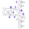
Which number(s) point(s) to the grey matter in the spinal cord?

A

2, 3, 4

5
Q

Which number(s) point(s) to the white matter in the spinal cord?

A

1

6
Q

What does white matter contain?

A

Myelinated axons (cell processes) in the CNS

7
Q

Where are the cell bodies of somatomotor neurons found?

A

Ventral horn of the spinal cord (in the grey matter)

8
Q

What is a ganglion?

A

Any cell body located outside of the CNS

9
Q

Where are the cell bodies of sensory neurons?

A

Dorsal Root Ganglia

Exception: Visceral scensory nerons in the vagus nerve have cell bodies in the brain (innervate the thorax and abdomen)

10
Q

Where do sympathetic nerves exit the spinal cord?

A

Lateral horn from T1-L2

11
Q

What cell bodies are found in the ventral horn?

A

Cell bodies of somatomotor neurons

12
Q

What cell bodies are found in the dorsal horn?

A

Interneuron cell bodies

13
Q

What cell bodies are found in the dorsal root ganglion?

A

Sensory cell bodies (general and visceral)

*Except visceral neuron cell bodies in the vagus nerve*

14
Q

Which sensory cell bodies are NOT found in the dorsal root ganglia?

A

Cell bodies of visceral sensory neurons in the vagus nerve

  • These signals will be coming from the abdomen and thorax
15
Q

What is the difference between the ventral and dorsal horns?

A

Besides location, the ventral horn contains mostly cell bodies of efferent neruons, while the dorsal horn contains mostly cell bodies of interneurons

16
Q

What is the difference between the ventral root and the ventral ramus?

A

The ventral root contains motor (efferent) neurons on their way out of the ventral horn to the rest of the body

The ventral ramus is a branch of a mixed spinal nerve that contains sympathetic, sensory, and somatomotor neurons that innervate the front and sides of the body

17
Q

What is the difference between the dorsal root and the dorsal ramus?

A

The dorsal root contains sensory (afferent) neurons on their way into the spinal cord to synapse with an interneuron in the dorsal horn

The dorsal ramus is a branch of a mixed spinal nerve that contains sympathetic, sensory, and somatomotor neurons that innervate the back of the body

18
Q

What makes up the Central Nervous System CNS?

A

The brain and spinal cord

(All cells with cell bodies in the brain and spinal cord)

19
Q

What makes up the Peripheral Nervous System (PNS)?

A

Cranial and spinal nerves, autonomic nerves in the gut

Any neuron with a cell body outside of the CNS

20
Q

What cell bodies are found in the sympathetic trunk?

A

Cell bodies of post-synaptic sympathetic neurons

21
Q

What cell bodies are found in the lateral horn?

A

Cell bodies of presynaptic sympathetic neurons, found in lateral horns from T1-L2

22
Q

Where would you find the cell body of a postsynaptic sympathetic neuron?

A

In the sympathetic trunk OR the collateral ganglion

Sympathetic trunk for anything except the thoracic and lumbar splanchnic nerves (Cardiopulmonary splanchnic neurons have cell bodies in the sympathetic trunk)

Collateral ganglion for neurons in the thoracic and lumbar splanchnic nerves

23
Q

Which division of the nervous system is considered “voluntary”?

A

Somatic

24
Q

What kind of nerves make up the somatic nervous system?

A

Spinal nerves and cranial nerves

25
Which division of the nervous system is "involuntary"?
Autonomic | (autonomic = automatic)
26
What makes up the autonomic nervous system?
Symathetic and parasympathetic nerves
27
Which division of the nervous system controlls our "fight or flight" reflex?
Sympathetic division of the autonomic nervous system
28
When activated, the sympathetic nervous system causes...
Activation of the sympathetic nervous system causes **increases in: blood pressure\*, heart rate, cardiac output, airway dilation, pupil dilation** (\*blood vessel constriction in skin, kidneys, digestive tract = more blood for the muscles you use to run away)
29
When activated, the parasympathetic nervous system causes...
Activation of the parasympathetic nervous system causes **D****ecreased blood pressure, heart rate** **Constriction of airways, pupils, GI smooth muscle (peristalsis)** **Increased glandular secretion**
30
Which components of the nervous system are derived from the neural crest?
All cell bodies outside of the CNS - Dorsal Root Ganglia - Sympathetic Trunk - Collateral Ganglia - Adrenal Medulla cells - Parasympathetic Ganglia - Melanocytes of skin
31
What does the **somatopleure** give rise to?
The body wall and all of the **spinal nerves** within it Therefore, **the somatopleurae gives rise to the somatic nervous system** and some **post-synaptic** **sympathetic neurons** that innervate the arteries of the body wall (remember that spinal nerves contain post-synaptic, sympathetic neurons)
32
What does the **splanchnopleure**
The **autonomic nervous system** - Nerves that innervate visceral organs
33
What are the components of a spinal nerve?
Sympathetic post-synaptic neurons Somatomotor neurons Sensory Neurons (general and most visceral)
34
What components does the cutaneous branch of a spinal nerve have?
Sympathetic post-synaptic neurons General sensory neurons (no somatomotor neurons because we can't move our skin, except in our face)
35
Describe the path of a nerve impulse that causes your toe to wiggle
Somatic Nervous System Cell body of the motor neuron is the **ventral horn** (gray matter) - \> Signal travels down 1 neuron to toe - \> Motor neuron releases ACh at motor endplate - \> ACh binds to its receptor to cause muscle contraction
36
Describe the path of a sympathetic nerve impulse to your back
Start: Presynaptic neuron with cell body in lateral horn of CNS - \> Leave lateral horn - \> Join ventral root - \> Join spinal nerve proper -\> Branch off with ventral, dorsal, or cutaneous rami (depending on target)
37
Describe the path of a "my feet are cold" signal
Sensory neuron in the foot w/cell body in dorsal root ganglion - \> Senses cold - \> Signal travels up axon, past cell body, into the dorsal horn - \> Synapse with interneuron in dorsal horn - \> interneuron carries the signal to the brain
38
What is a dermatome?
The section of skin that is innervated by 1 pair of spinal nerves
39
How many dorsal roots must be damaged in order to lose sensation in one dermatome?
3; the relevant dermatome, and the one above and below
40
Describe the consequence of losing innervation in one dermatome
Reduced sensation (not total loss)
41
What are the meninges?
The three membranes that enclose the spinal cord and brain
42
What are the three meninges?
Dura mater: tough outer layer Arachnoid: thin, spiderweb-like layer Pia mater: Clings tightly to the spinal cord
43
Where is the epidural space?
Superficial to the dura mater
44
Where is the CSF located with relation to the meninges?
In the subarachnoid space; between the arachnoid and the pia mater
45
What are the cauda equina?
Dorsal and ventral roots that extend down below the spinal cord The spinal cord ends at L1, but the roots travel down and exit through lower vertebrae (like a "horse's tail")
46
Why is a lumbar puncture done below L1?
The spinal cord ends at L1 Piercing the dura mater below L1 has a much lower risk of damaging the CNS
47
What is a lumbar puncture?
A needle pierces the dura mater and arachnoid and is inserted into the arachnoid space. CSF is sampled or drugs are delivered. Drug delivery is very fast but short-acting
48
What is the lowest point at which you can do a lumbar puncture? Why?
Anything above S1 (not inclusive of S1) The subarachnoid space ends at S1; If there is no space, you can't put a needle there
49
What is an epidural block?
A needle is insurted into the epidural space, superficial to the dura mater. This is usually used for drug delivery; you can't sample the CSF from here Drugs delivered to the epidural space are slower acting but longer lasting
50
Your attending physician tells you to administer drugs to a patient through the central nervous system. You can't remember whether they said to do a lumbar puncture or an epidural block, but you know for sure that it's a **sacral nerve block**. Which method do you use? Why?
You would use an epidural block You can't do a lumbar puncture in the sacrum because the subarachnoid space ends at S1.
51
Which type of nerve block would you use for a multi-hour hip surgery?
Epidural - it lasts longer than a lumbar block
52
Which division of the nervous system contains only efferent neurons?
Autonomic
53
If a neuron has its cell body in the central nervous system, which embryonic structure did it come from?
The neural tube
54
The neural tube gives rise to which cell bodies?
Cell bodies in the CNS
55
What structures are part of the "deep body wall"?
Bone, muscle, deep (investing) fascia, parietal pleura, peritoneum
56
A spinal nerve innervating the deep body wall will contain...
Sympathetic post-synaptic neurons General sensory neurons Somatomotor neurons
57
A spinal nerve innervating the superficial body wall will contain...
Sympathetic post-synaptic neurons General sensory neurons (no motor neurons!)
58
What structures are part of the "superficial body wall"?
Skin, superficial (Camper's) fascia, Scarpa's fascia
59
Where would you find a nerve that has short pre-synaptic and long post-synaptic neurons?
The Sympathetic Nervous System
60
Which division of the nervous system is most likely to innervate structures within body cavities?
The autonomic nervous system
61
In which part of the nervous system would you find the neurotransmitter ACh?
(almost) Everywhere! Somatic: All synapses (neuromuscular jcts) Autonomic: All Presynaptic -\> postsynaptic Parasympathetic postsynaptic -\> target
62
In which part of the nervous system would you find the neurotransmitter norepinephrine?
Autonomic, sympathetic nervous system postsynaptic -\> target
63
From where does _every spinal nerve_ receive postsynaptic sympathetic neurons?
The sympathetic trunk
64
What are the only two possible destinations for a presynaptic sympathetic neuron leaving the lateral horn?
- The sympathetic trunk - The collateral ganglion
65
Describe the path of a cardiopulmonary splanchnic nerve
Presynaptic **sympathetic** neuron w/cell body in lateral horn - \> Leaves lateral horn - \> Joins ventral root - \> Joins spinal nerve proper - \> Synapses with postsynaptic in the **sympathetic trunk** - \> postsynaptic travels to cardiopulmonary area
66
Describe the path of thoracic and lumbar splanchnic nerves
Presynaptic **sympathetic** neuron w/cell body in lateral horn - \> Leaves lateral horn - \> Joins ventral root - \> Joins spinal nerve proper - \> **Bypasses sympathetic trunk** - \> Travels to **collateral ganglion**, synapses with post-synatpic neuron - \> postsynaptic travels to target tissue
67
Which splanchnic nerves are sympathetic vs parasympathetic?
Sympathetic: cardiopulmonary, thoracic, lumbar Parasympathetic: pelvic
68
Which division of the autonomic nervous system has **thoracolumbar outflow**?
Sympathetic Nervous System **Sympathetic neurons only exit the CNS from T1-L2** Sympathetic presynaptic neurons can travel up or down from here to join spinal nerves at every vertebral level
69
Which division of the autonomic nervous system has **craniosacral outflow?**
Parasympathetic Nervous System **Parasympathetic presynaptic neurons only exit the CNS with cranial nerves and sacral spinal nerve roots**
70
Which nerves are associated with cranial outflow? What do they innervate?
Vagus nerve, cranial nerves 3, 7, 9 Destined for thoracic viscera, abdominal viscera or fore-mid gut (through ascending and transverse colon)
71
What is referred pain?
Pain that originates from a visceral nerve, but is sensed by the brain as pain in a dermatome in the body wall at the same level as the visceral organ
72
Which nerves are associated with sacral outflow? What do they innervate?
Pelvic splanchnic nerves Destined for viscera of pelvis and hindgut (descending colon)
73
What kind of neurons would you find in the ventral root of L3?
Somatomotor (sympathetic only exit T1-L2, parasympathetic are craniosacral)
74
What is a plexus?
A conglomoration fo sympathetic, parasympathetic, visceral scensory neurons. Nerves emanating from a plexus together usually coordinate their functions?
75
What allows for specificity of action of sympathetic neurons? i.e, Why do some smooth muscles contract while some relax in the "fight or flight" response?
Receptor type: Alpha or beta Smooth muscles with alpha receptors constrict in response to norepinephrine (Arteries of GI tract) Smooth muscles with beta receptors dilate in response to norepinephrine (increased airway to lungs, increased blood to the heart -\> increased cardiac output\*)
76
What happens to the smooth muscles in the arteries of skeletal muscle when the sympathetic nervous system is activated?
The beta-receptors on the smooth muscle cells of arteries cause the arteries to **dilate** in response to epinephrine This allows for increased blood flow to skeletal muscles so you can run away!
77
What is the ration of presynaptic : postsynaptic neurons in the sympathetic nervous system? Why is this significant?
Low: one presynaptic neuron can synapse with many post-synaptic neurons, leading to a rapid amplification of the original signal This is useful because if we have to run away from a bear, we watn to do it quickly!
78
What is the ratio of presynaptic : postsynaptic neurons in the parasympathetic nervous system? Why is this significant?
Around 1: this allows for the slower, more localized "rest and digest" response
79
Where do we find the cell bodies of postsynaptic parasympathetic neurons?
In the tissues they innervate
80
Give an example of a multipolar neuron
Somatomotor Autonomic (especially sympathetic) Interneurons
81
What elements would we find in the dorsal root?
Afferent (sensory) neurons - general and visceral
82
Where is the cell body of this neuron?
Dorsal root ganglion This means that it must be a sensory neuron
83
What elements would we find in the ventral root at T6?
Sympathetic presynpatic neurons Somatomotor neurons
84
Where are the cell bodies of neurons in the vagus nerve?
The brain
85
What neuron components would we find in the nerves in Scarpa's fascia?
Post-synaptic sympathetic neurons General sensory neurons
86
What neruon components would we find in the vagus nerve?
Presynaptic parasympathetic Visceral sensory (for general sensations that do not require localization)
87
What kind of neuron is pictured in red? Where did it originate?
Sensory Neuron Somewhere on the back of the body
88
What kind of neuron is pictured in red? Where did it originate?
Sensory Neuron Somewhere on the ventral or lateral sides of the body
89
The neuron in red is part of what kind of nerve?
Sympathetic nerve
90
What is structure #1
The sympathetic trunk, aka sympathetic chain
91
What is structure #2
Dorsal Root
92
What is structure #3
Ventral Root
93
What is structure #4
Dorsal Root Ganglion
94
What is structure #5
Spinal Nerve Proper
95
What is structure #6
Ventral horn
96
What is structure #7
Lateral horn
97
What is structure #8
Dorsal horn
98
Which cells have cell bodies in #4
Sensory neurons (general and visceral)
99
Which cells have cell bodies in #8
Interneurons
100
Which cells have cell bodies in #7
Sympathetic Presynaptic Neurons
101
Which cells have cell bodies in #6
Somatomotor neurons
102
Give examples of neurons that synapse in the collateral ganglion
Any sympathetic neuron destined for a thoracic or lumbar visceral cavity Examples: Stomach, digestive tract
103
Where is the cell body of a sympathetic neuron in the heart?
**Sympathetic trunk** This is a post-synaptic neuron Any sympathetic post-synaptic neuron destined for something in the cardiopulmonary cavity has its cell body in the sympathetic trunk
104
Where is the cell body of a presynaptic parasympathetic neuron destined for the descending colon?
Sacral spinal cord
105
Where is the cell body of a parasympathetic presynaptic neuron destined for the small intestine?
The brain The small intestine is innervated by the vagus nerve, whose cell body is in the brain