Exam 4 Nasal Cavity and PPF Flashcards
1
Q
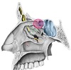
The Nose
- What forms the cartilaginous skeleton?
- What forms the bony skeleton?
- What makes up the bony portion of the nasal septum?
- What makes up the cartilaginous portion of the nasal septum?
A
- Cartilagionus skeleton made up by 2 lateral nasal cartilages and 2 greater alar cartilages
- Bony skeleton made up by two nasal bones, frontal processes of maxillae, and nasal part of frontal bones.
- Bony portion of nasal septum formed by: Perpindicular plate of ethmoid and vomer bones
- Cartilaginous portion formed by septal cartilage
2
Q
Nasal Cavity
- What is the nasal vestibule?
- What are the bony boundaries of the nasal cavity?
- Superior wall
- Inferior wall
- Medial wall
- Lateral wall
- What is the respiratory area of the nasal cavity?
- What is the olfactory area of the nasal cavity?
A
- Nasal vestibule: Initial space between nares and nasal septum (hairy)
- Bony boundaries:
- Superior wall: Frontal, nasal, ethmoid, sphenoid bones
- Perforated by cribiform plate of ethmoid bone
- Inferior wall: Palatine process of maxilla bone and horizontal plate of palatine bone (forms hard palate)
- Medial wall: Nasal septum
- Lateral wall: Maxilla, palatine, and sphenoid bones
- Superimposed by lacrimal, ethmoid, and inferior nasal conchae bones
- Superior wall: Frontal, nasal, ethmoid, sphenoid bones
- Respitory area: Lower 2/3 of nasal mucosa
- Olfactory area: Upper 1/3 of nasal mucosa
3
Q
Nasal conchae
- What bone does each conchae arise from?
- What spaces are between each conchae?
- What openings/features are found in each conchae?
A
- Superior and middle conchae: Part of ethmoid bone
- Inferior conchae: Separate bone
- Spaces (Meatuses)
- Inferior meatus: Inferior to inferior conchae
- Nasolacrimal gland opens here
- Middle meatus: Inferior to middle conchae
- Semilunar Hiatus: Crescent-shaped opening
- Ethmoidal Bulla: Bulge in middle meatus posterosuperior to semilunar hiatus
- Uncinate Process: Curved projection of ethmoid bone in middle meatus. Anteroinferior to semilunar hiatus
- Superior meatus: Inferior to superior conchae
- Sphenoethmoidal recess: Space superior to superior chonchae and inferior to superior wall.
- Inferior meatus: Inferior to inferior conchae

4
Q
Blood Supply of Nasal Cavity
- What are the arteries?
- Where do they branch off of?
- What else do they supply (if applicable)?
A
- Sphenopalatine artery: Terminal branch of maxillary artery
- Gives off posterior lateral nasal branches and posterior septal branches.
- Largest blood supply to nasal cavity
- Posterior and anterior ethmoidal arteries: Branches of ophthalmic artery
- Greater palatine artery: Branch of maxillary artery
- Supplies hard palate
- Lateral nasal artery: Branch of facial artery
- Superior labial artery: Branch of facial artery

5
Q
Nerves of Nasal Cavity
- What are they?
- What is their function?
A
- Olfactory nerves (CN I): Sense of smell
- Ophthalmic nerve (CN V1): Sensory information from anterosuperior portions of nasal cavity via anterior and posterior ethmoidal nerves
- Maxillary nerve (CN V3): Sensory information from septum via nasopalatine nerve and lateral wall via greater palatine nerve and posterior nasal nerve

6
Q
Sphenoid sinus
- Where is it located?
- What does it drain into?
- What is its innervation?
- What is its blood supply?
A
- Located in sphenoid bone
- Drains into sphenoethmoidal recess above superior nasal conchae
- Innervation: Posterior ethmoidal nerve (CN V1)
- Blood Supply: Posterior ethmoidal artery (off ophthalmic artery)
7
Q
Frontal Sinus
- Where is it located?
- Where does it drain?
- What is its innervation?
- What is its blood supply?
A
- Located between outer and inner layers of frontal bone
- Drains via frontonasal duct into ethmoidal infundibulum
- Then drains into anterosuperior portion of semilunar hiatus of middle nasal meatus.
- Innervation: Supraorbital nerve (CN V1)
- Blood supply: Supraorbital artery (off ophthalmic artery)

8
Q
Maxillary Sinus
- Where is it located? (Boundaries)
- Where does it drain into?
- What is its innervation?
- What is its blood supply?
A
- Boundaries:
- Roof: Floor of orbit
- Floor: Alveolar part of maxillae
- Medial walls: Border nasal cavity
- Drains into posterior end of semilunar hiatus of middle nasal meatus.
- Innervation: Anterior, middle, and posterior superior alveolar nerves (CN V2)
- Blood supply: Superior alveolar branches of maxillary artery
Largest of nasal sinuses.

9
Q
Ethmoidal Sinuses (Air cells)
- Where are they located?
- Where do they drain?
- What is their innervation?
A
- Several cavities located in ethmoid bone between nasal cavity and orbit
- Drainage:
- Anterior ethmoidal cell: Ethmoidal infundibulum into semilunar hiatus of middle nasal meatus.
- Middle ethmoidal cell: Drains directly into middle meatus via ethmoidal bulla
- Posterior ethmoidal cell: Drains directly into superior meatus.

10
Q
Boundaries and Communications of Pterygopalatine Fossa
- Anterior
- Posterior
- Lateral
- Medial
- Superior
- Inferior
A
- Anterior: Posterior border of maxilla
- Posterior: Pterygoid process of sphenoid bone
- Communications: Pterygoid canal, foramen rotundum, pharyngeal canal
- Lateral: Infratemporal fossa
- Communications: Pterygomaxillary fissure
- Medial: Perpindicular plate of palatine bone, nasal cavity
- Communications: Sphenopalatine foramen
- Superior: Greater wing of sphenoid bone
- Communication: Inferior orbital fissure and orbit
- Inferior: : Pyramidal process of palatine bone
- Communications: Palatine canal, greater &lesser palatine foramina

11
Q
Contents of Pterygopalatine Fossa
A
- 3rd part of maxillary artery
- Nerve of pterygoid canal
- Formed by greater petrosal and deep petrosal nerves
- Maxillary nerve (CN V2) and its branches
- Pterygopalatine ganglion (parasympathetic ganglion for CN VII)

12
Q
Branches of Maxillary Nerve (CN V2)
- What are they?
- What do they supply?
- What openings do they pass through?
A
- Greater palatine nerve: Innervates palatal mucosa and palatal (lingual) gingiva posterior to maxillary canines
- Passes through greater palatine foramen
- Lesser palatine nerve: Innervates mucous membranes and glands of soft palate, uvula, and palatine tonsil
- Passes through lesser palatine foramen
- Nasopalatine nerve (long sphenopalatine nerve): Innervates nasal mucosa of septum and palatal mucosa of anterior hard palate
- Exits PPF via sphenopalatine foramen
- Enters hard palate via incisive foramen
- Posterolateral nasal branches (short sphenopalatine nerve): Innervates mucous membrane of lateral wall of nasal cavity
- Exits PPF via sphenopalatine foramen
- Pharyngeal nerve: Innervates nasopharynx and sphenoidal sinus
- Exits PPF via pharyngeal canal
- Zygomatic nerve: Exits PPF via inferior orbital fissure
- Zygomaticofacial nerve: Innervates prominence of cheek
- Zygomaticotemporal nerve: Innervates skin of anterior temple region
- Posterior superior alveolar nerve: Innervates most of maxillary molars and maxillary sinus, and buccal gingiva (via gingival branches)
- Exits PPF pterygomaxillary fissure
- Enters maxillary sinus via posterior superior
- Infraorbital nerve: Exits PPF via infraorbital canal
- Middle superior alveolar nerve: Innervates maxillary premolars, maxillary sinus, buccal gingiva
- Anterior superior alveolar nerve: Innervates maxillary sinus, maxillary canines, and maxillary incisors
13
Q
Pathway of parasympathetics from CN VII to lacrimal gland
A
- Cell bodies arise in superior salivary nucleus
- Parasympathetics travel within CN VII
- Branch off CN VII by geniculate ganglion and travels within Greater Petrosal Nerve
- Passes through petrous temporal bone via hiatus of facial canal
- Enters pterygoid canal via foramen lacerum
- Joins deep petrosal nerve to become nerve of pterygoid canal
- Preganglionic parasympathetics synapse in pterygopalatine ganglion
- Postganglionic parasympathetics hitch a ride on zygomaticotemporal nerve of CN V2 (maxillary nerve)
- Zygomaticotemporal nerve gives connecting branch to lacrimal nerve of CN V1 (ophthalmic nerve) to supply parasympathetic fibers to lacrimal gland

14
Q
Pathway of Sympathetic Fibers of Maxillary Nerve (CN V2)
A
- Deep petrosal nerve comes off internal carotid plexus
- Joins greater petrosal nerve to form nerve of pterygoid plexus
- Passes through pterygopalatine ganglion without synapsing
- Joins branches of maxillary nerve (CN V2)
15
Q
Arteries in Pterygopalatine Fossa
- What are they?
- What do they supply?
- What openings do they pass through?
A
- Descending palatine artery: Travels through palatine canal. Branches into:
- Greater palatine artery: Supplies mucosa of hard palate
- Exits palatine canal via greater palatine foramen
- Lesser palatine artery: Supplies soft palate and palatine tonsil
- Exits palatine canal via lesser OR greater palatine foramen
- Greater palatine artery: Supplies mucosa of hard palate
- Sphenopalatine artery: Exits PPF via sphenopalatine foramen. Branches into:
- Posterolateral nasal branches: Supplies lateral wall of nasal cavity, portions of maxillary , ethmoidal, and sphenoidal sinuses
- Posterior septal branches: Supplies nasal septum and hard palate (via nasopalatine artery which exits via incisive canal)
- Artery of pterygoid canal: Supplies superior pharynx, auditory tube, and middle ear
- Exits PPF via pterygoid canal
- Posterior superior alveolar artery: Supplies molar and premolar teeth, maxillary sinus, and buccal gingiva
- Exits PPF via posterior superior alveolar foramina
- Infraorbital artery: Supplies lower eye structures. Gives off anterior superior alveolar artery.
- Exits PPF via infraorbital fissure -> infraorbital groove -> infraorbital canal -> infraorbital foramen
