EX3; Mandible and TMJ Practical Images Flashcards
What kind of ossification center is this

intramembranous
What kind of ossification center is this

endochondral
What is this image as a whole of (specifically the PDL)

growth site
What are these structures from Meckel’s cartilage

- incus
- malleus
- anterior mallelar ligament
- sphenomandibular ligament
- mylohyoid groove ligament
- enclosed in anterior lingual part of developing mandible
What is this an image of

ossification center of a mandible half

- tongue
- meckel’s cartilage
- bone
- lip

- Meckel’s cartilage
- bone

- epithelial buds
- Meckel’s cartilage

- bone
- Meckel’s cartilage
The developing mandibular bone is in this region as depicted by the arrows

area of mental foramen
What do the red and black arrows represent

red; developing primary second molar bud
black; mylohyoid muscle attached to medial sides of Meckel’s cartilages
What is the red arrow pointing to

the attachment of the mylohyoid on the medial Meckel’s cartilage

- malleus
- incus
- TMJ articular disc
- condylar cartilage
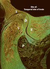
What kind of joint is this

freely movable (fetal elbow)
note; the single joint cavity, incomplete disc, and cartilage articular surfaces

A; soft connective tissue articulating surfaces
B; two joint cavities
C; complete articular disc

T; temporal bone
D; disc
C; condyle
P; lateral pterygoid
E; articular eminence
What do the arrows represent

red = semilunar ganglion
blue = blastama of TMJ
black = mandibular bone

- temporal bone
- blastema; TMJ
- mandible bone
- Meckel’s cartilage
- lateral pterygoid
- semilunar ganglion

articular disc

- temporal bone
- articular disc
- cavitation of joint
- articualr surface
- condylar cartilage
- lateral pterygoid

- articular disc
- lower joint caivty
- articular surface
- condylar cartilage
Which one of the condyles is of an adult older than 25 years, top or bottom?

bottom; note the bone tissue and absence of bone marrow
Identify the labels and the age of this TMJ

- articular dics
- articular surface
- compact bone
over 25, due to compact bone replacing cartilage
Which of these is from an individual 25 or younger, top or bottom

bottom
note; the hyaline cartilage and bone marrow
Which of these is the older/younger

left = young
right = old


