esophageal+gastric carcinoma 1/31 Flashcards
etiology of esophageal SCC
genetic factors appear less important. Dietery and environmental causes
US+Europe=EtOH and tobacco
other countries=carcinogens like fungus or nitrosamine containing food stuff
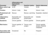
types of adenocarcinoma of esophagus and EG junction
of esophagus developing with BE
without BE
of gastric cardia
Risk factors:GERD,BE,Dysplasia
gastric polyps
hyperplastic/inflammatory polyps (non-neoplastic)
majority are associated with chronic gastritis,should resect if over 1.5cm
fundic gland polyps
can be sporadic or in people with FAP
associated with PPI
women>men
have cystically dilated irregular glands
gastric polyps picture
gastric adenoma (neoplastic polyp)
risks increase with age,men>women,FAP
associated with chronic gastritis with atrophy+intestinal metaplasia
see dysplastic intestinal-type epithelium+nuclear enlargement,elongation,hyperchromasia,irregular architecture (if high-grade)
risk of adenocarcinoma up if over 2cm in diameter
gastric adenoCA
more common in east and europe than US
due to dietery+environmental factors.Also with BE
seen in people with lower socioeconomic status and pts with mucosal atrophy and intestinal metaplasia
RF for gastric CA
nitrites,smoked salted and pickled food,lack of fruits+veggie
cigarettes
chronic gastritis
partial gastrectomy,menetrier’s disease,BE
blood groupA,family Hx,HNPCC
types of gastric CA
intestinal+diffuse
loss of E-cadherin fx is key for diffuse type
see increased risk of intestinal type in pts with FAP,also increased incidence in Japan
chronic inflammation promotes neoplastic progression in both types
histology of both types of gastric adenoCA
intestinal: gland formation.grossly,see large bulky tumor
diffuse: signet ring cells.grossly,see thickening of gastric wall(linitis plastica),can be difficult to see on slides
intestinal type gastric CA
predominates in high risk areas,develops from precursor flat dysplasia or adenoma
mean age is 55,men>women
linked to atrophic gastritis and intestinal metaplasia
decreased incidence drastically
diffuse type of gastric CA
no precursor lesions
men=women
uniform incidence across countries
clinical presentation and Tx of gastric CA
similar to chronic gastritis
later see weight loss,early satiety,anemia
may be tipped off by periumbilical subcutaneous nodule (sister mary joseph nodule)
Tx:if early detected–>resection, usually discovered late
gastric lymphoma
very rare
most are indolent extra-nodal marginal zone lymphomas of MALT
arise at sites of chronic inflammation,many HP+
low grade MALT lymphoma regresses if HP is treated
morphology of gastric lymphoma+presentation
see dense lymphocytic infiltrate in lamina propria, often infiltrates gastric glands
lymphoepithelial lesions
express B cell markers
can show monoclonality
can present with pain,later weight loss
carcinoid tumor
from endocrine cells,many seen in GI and next common site is lung
lesion is intramural or submucosal with overlying mucosa that can ulcerate
yellow or tan and firm tumor
see islands or strands of tumor cells that show ‘salt and pepper’ chromatin
stain for NE markers:chromogranin,synaptophysin,also see NE granules on EM
GI stromal tumor (GIST)
most common mesenchymal tumor of abd
peak age is 60
if seen in child,think Carney triad:young female,paraganglioma,pulmonary chndroma.OR NF type I
most have oncogenic gain of fx mutation of gene encoding tyrosine kinase c-KIT (receptor for stem-cell factor)–>use as marker in Dx
tumor can be very large,fleshy mass covered by mucosa,can have abd mets
2 types:spindle cell+epitheloid
source of GIST
interstitial cells of Cajal located in muscularis propria and serve as pacemaker for peristalsis
they express c-KIT,also known as CD117 and CD34
mutation of c-KIT is early event in sporadic GIST
Tx of GIST
complete resection if localized (gastric GIST less aggressive than intestinal GIST)
with imantinib which is a tyrosine kinase inhibitor of c-KIT
