Causes of Seizures 1/16 Flashcards

1
Q

Types of brain herniation

A
2
Q

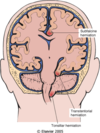
Subfalcine herniation

A

may compress ant. cerebral art.

3
Q

transtentorial herniation

A

may compress:

1) CNIII= see ipsilateral dilated pupil and impaired eye movement
2) post. cerebral art.= primary visual cortex
3) cerebral peduncle=kernohan’s notch, ipsilateral hemiparesis
4) pons and midbrain= duret hemorrhage due to vessel tearing–>life threatening

4
Q

tonsillar herniation

A

brain stem compression–>cardiac and respiratory centers

5
Q

NO LP if CT shows:

A

mid-line shift, loss of CSF cisterns, post. fossa mass–>high risk of herniation

6
Q

CT without contrast

A

hemorrhage/hematoma, skull fx/trauma, hydrocephalus, fluid accumulation/abscess

7
Q

CT with contrast

A

tumor(angiogenesis), vascular malformation, infection/inflammation

8
Q

intracranial hemorrhage

A

related to HTN and peak age is 60. most frequent sites: thalamus, basal ganglia, pons and cerebellum

9
Q

Charcot-bouchard micro aneurysms

A

associated with chronis HTN. Minute aneurysms in small vessels which can rupture

10
Q

subarachnoid hemorrhage

A

caused by bleeding from cerebral arterial aneurysms. bleeding is at arterial pressure. “The worst headache of my life” then loss of consciousness. re-bleeding is common among survivors.

Acute: vasospasm–>ischemia

chronic: meningeal fibrosis–>hydrocephalus

11
Q

berry aneurysm

A

bright red, shiny surface and a thin, translucent wall. often arise at junctions of vessels due to defect in vessel walls at branch points.

cerebral vessels have only one elastic lamina(internal elastic lamina)–>more prone to dilation

12
Q

picture of berry aneurysm development

A
13
Q

atherosclerotic aneurysm

A

arises as dilation of entire part of an artery weakend by atherosclerosis. less common than berry.

14
Q

mycotic aneurysm

A

arises in vessel walls weakend by microbial infx, usually bacteria, despite the name.

15
Q

epidural hematoma

A

due to rupture of middle meningeal art. due to traumatic skull fx–>seperation of dura from inner surface of skull–> compression of brain.

pts lucid for several hours then develop neurological signs. emergency drainage required

16
Q

picture of epi and subdural hematomas

A
17
Q

subdural hematoma

A

bridging veins from cortical convexities to sup. sagittal sinus travel through the space between dura and arachnoid layer (subdural).

elderly with cerebral atrophy are at risk due to stretching of veins. Infants are also at rick because veins are thin-walled and fragile

18
Q

signs and symptoms of chronic subdural hematoma

A

headache, confusion and focal signs

19
Q

CSF production

A

by choroid plexus in lateral and 4th ventricles–>enters subarachnoid space at cisterna magna through foramina of luschka and magendie.

CSF reabsorbed through arachnoid granulations of sup. sagittal sinus.

20
Q

hydrocephalus

A

obstruction, overproduction, loss of cerebral tissue.

if before closure of cranial sutures–>increased head circumference

after–>increased ICP

21
Q

non-communicating hydrocephalus

A

obstruction within ventricular system: mass lesions, aqueductal stenosis, obstruction of 4th ventricle

22
Q

communicating hydrocephalus

A

obstruction outside the ventricular system: subarachnoid hemorrhage or meningitis with secondary meningeal scarring

23
Q

hydrocephalus ex vacuo

A

if there is less brain tissue–>compensatory enlargement of ventricles (alzheimer’s)

24
Q

choroid plexus papilloma

A

results in csf overproduction

25
Q

Preference of location of brain abscesses

A

frontal lobe>parietal>cerebellum

26
Q

clinical findings of brain abscess

A

progressive focal deficits, increased csf, white cells and protein, normal glucose

27
Q

arterio-venous malformations AVM

A

largest intracerebral malformations, tangled network of wormlike vascular channels, prominent pulsatile arterio-venous shunt with high blood flow through the malformation.

tend to occur in cerebral hemispheres

28
Q

cavernous hemangioma

A

greatly distended, loosly organized vascular channels with thin, collagenized walls

29
Q

capillary hemangioma

A

rarely cause symptoms during life

30
Q

tuberculous meningoencephalitis

A

csf: moderate lymph or lymph+poly, protein elevated, glucose moderately reduced or normal

arteries in subarachnoid space may show obliterative endarteritis (heubner’s arteritis)

pus usually accumulates at base of brain

31
Q

Brain tumors in adults

A

supratentorial. Gliomas are most common primary tumors>meningioma

32
Q

brain tumors in chidren

A

secondary to hematopoietic tumors. Infratentorial. most are astrocytoma>medulloblastoma>ependymoma

33
Q

infratentorial tumors

A

adults: schwannomas>gliomas of brain stem> mets
children: astrocytoma>medulloblastoma>ependymomas

34
Q

extradural tumors of spine

A

usually within vertebral body. most common in adult: mets>primary tumors of bone

35
Q

intradural tumors of spine

A

between dura and spinal cord or within spinal cord=intramedullary

meningioma>ependymoma>astrocytoma

36
Q

astrocytoma high grade=glioblastoma multiforme=anaplastic astrocytoma

A

increased number of astrocytes that are anaplastic, necrosis with pseudopallisading of tumor cells. poor prognosis–>cerebral edema+herniation

37
Q

pilocytic astrocytoma

A

usually in cerebellum, but also optic nerve and 3rd ventricle. usually benign but 3rd ventricle pilocytic astrocytoma has poor prognosis

38
Q

oligodendroglioma

A

fried egg appearance, well-circmscribed, can be cystic with calcification

39
Q

ependymoma

A

tumor of 4th ventricle–>hydrocephalus, in young children

Can occur in spinal cord in middle life. See ependymoma rosettes

40
Q

medulloblastoma=pnet=peripheral neuroectodermal tumor

A

usually found in cerebellar vermis and can spread down the spinal cord=drop mets. See small cells with prominent nucleoli, scant cytoplasm and occasional rosettes

41
Q

meningioma

A

benign, from subarachnoid cell, see whorls with psammoma bodies, compresses the brain

42
Q

primary CNS lymphoma

A

from microglia, associated with AIDS and renal transplant, mostly B cell phenotype, diffuse infiltration of brain tissue, micro:see large cell lymphoma, poor prognosis

Lesions are often multiple in gray and white matter

43
Q

mets to brain

A

often multiple, well-circumscribed and at white-gray junction. Often come from lung, breast, kidney

44
Q

carcinomatous meningitis

A

tumor deposits throughout CNS in subarachnoid space–>widespread neurological dysfunction.

Seen most often with adenocarcinomas, including carcinomas from breast or stomach

45
Q

schwannoma

A

acoustic neuroma, CNVIII, Antoni A and B, verocay bodies (pallisading of nuclei), thickend vessels

46
Q

Neurofibroma

A

NF 1 mutation, fusiform noncircumscribed enlargement of nerve seen often at distal part of nerve. micro: see interlacing bands of spindle cells wih elongated nuclei. Hard to remove from nerve. Can infiltrate adjacent tissue.