Dermatology Flashcards
(123 cards)
State some basic functions of the skin
- Protective barrier
- Temperature regulation
- Sensation
- Vit D synthesis
- Immunosurveillance
State 3 layers of skin
- Epidermis
- Dermis
- Hypodermis/subcutaneous tissue
State the 5 layers of the epidermis
- Stratum corneum
- Statum lucidum
- Stratum granulosum
- Stratum spinosum
- Stratum basale
*Remember, Stratum Lucidum isn’t always present (not present in thin skin)

Which layer of epidermis are melanocytes most commonly found in
Stratum basale
Compare thick and thin skin

State the thinnest areas of skin on body
- Eyelids
- Scrotum
What is the dermis made of?
What 4 structures are found in the dermis?
- Collagen, elastin, fibrillin
- Contains nerve endings, glands, hair follicles and blood vessels
State 2 functions of subcutaneous tissue/hypodermis
- Insulation
- Protective padding
Remind yourself of the Fitzpatrick skin types
*Ignore bottom bit- referring to laser hari

What is the pH of normal skin?
5.5
State some key questions you should ask when taking a dermatology history
Presenting Complaint
- Site of onset
- Evolution
- Duration
- Distribution- asymmetrical/symmetrical, flexors/extensors, mucous membranes, sun exposed sites
- Describe appearance (if can’t see in practice)
- Symptoms e.g. itchy, sore
- Exacerbating & relieving factors
- Tried any treatments: where, how much used, how long for
PMH
- History of skin diseases or atopy
- History of systemic diseases
- History of sunburn, sunbathing, sunbed use
- FH of skin diseases including atopy
- FH of autoimmune diseases
- Drug history (including timeline)
Social History
- Social
- Occupation (sun exposure, contactants), does it improve when awary from work
- Travel history
- Sexual history
ICE
- Pyschosocial impact
Discuss some key components of a good dermatological examination
KEY IDEAS: inspect, palpate, describe, systemic check
- Ensure have adequete exposure & lighting (pt should remove makeup if wearing)
- Examination of affected areas and other areas such as hair/scalp, nails, mucous membranes
- Palpate
- Examine other systems if appropriate e.g. joints (e.g. is psorasis), lymph nodes (e.g.infection, malignancy)
Define the following terms:
- Papule
- Nodule
- Erythema
- Vesicle
- Bulla
- Pustule
- Telangiecta
- Alopecia
- Hirsutism
- Excoriations
- Striae
- Papule: small lump <5mm
- Nodule: larger lump 5-10mm
- Erythema: redness
- Vesicle: small water blister
- Bulla: large water blister
- Pustule: pus-filled vesicle
- Telangiecta: thread vein
- Alopecia: hair loss/thinning
- Hirsutism: hairness
- Excoriations: scratch marks
- Striae: stretch marks
Define the following terms:
- Pruritus
- Atrophy
- Macule
- Patch
- Plaque
- Erosion
- Ulcer
- Lichenification
- Pruritus: ithcing
- Atrophy: thinning
- Macule: non palpable area of discolouration
- Patch: macule >3cm
- Plaque: palpable flat topped area >1-2cm
- Erosion: loss of epidermis (superifcial)
- Ulcer: loss of epidermis & dermis (deep)
- Lichenification: thickening of skin with exagerated skin markings
Remind yourself of the SCAM mneumonic to help you describe skin lesions

Remind yourself of the framework for describing pigmented lesions
- Asymmetry
- Border (regular or irregular)
- Colour
- Diameter
Remember to see Sem 4: Integration for clinical application for further dermatology revision
Discuss the potential psychological, social and emotional impact of skin disorders
- Decreased confidence
- Self-conscious
- Decreased involvement in activities
- Impact on education
- Pressure to try and resolve condition
- Feeling like no-one understands
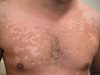
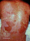
What is eczema?
At what ages is it prevelant?
Atopic eczema is a chronic, ithcy, inflammatory skin condition that can affect people of all ages although 70-90% present under 5 yrs with a high incidence in 1st year
What are signs & symptoms of eczema?
- Prurutic rash
- Erythematous rash
- Dry skin
- Excoriations
- History of atopy

Disucss how you diagnose eczema
What might make you suspect that someone’s eczema is infected?
Yellow crusting, signs of systemic infection e.g. pyrexia
State the7 different types of eczema and discuss how you can distinguish between them
- Atopic dermatitis:typical widespread itchy, erythematous, dry eczema
- Contact dermatitis: eczema in response to contact with iritant or allergen
- Neurodermatitis: usually confined to one or two patches of skin- continued scratching can irritate nerve endings in skin, intensifying both itching and scratching
- Dyshidrotic eczema: small, intensely itchy blisters on the palms of hands, soles of feet and edges of the fingers and toes
- Nummular eczema: scattered circular, often itchy and sometimes oozing patches
- Seborrheic dermatitis: appears on the body where there are a lot of oil-producing (sebaceous) glands like the upper back, nose and scalp
- Stasis dermatitis: gravitational dermatitis, venous eczema, and venous stasis dermatitis, happens when there is venous insufficiency, or poor circulation in the lower legs
It is possible to have more than one type of eczema on your body at the same time. Each form of eczema has its own set of triggers and treatment requirements, which is why it’s so important to consult with a healthcare provider who specializes in treating eczema. Dermatologists in particular can help identify which type or types of eczema you may have and how to treat and prevent flare-ups.
Discuss the management of eczema
- Education regarding triggers & management
- First line= emollients
- Second line= topcial corticosteroids
- Mild= Hydrocortisone 0.1-2.5%
- Moderate= Eumovate (active ingredient= clobetasone butyrate)
- Strong= Betnovate (betamethasone 17-valerate)
- Very strong= Dermovate (clobetasol propionate)
- Third line= non-sedating antihistamine
- Fourth line= oral corticosteroids (would be with specialist at this point)
- Antibiotics e.g. flucloxacillin if infected