Derm #2 Flashcards

1
Q

What is acanthosis, what can be seen with it, and what is it a common response to?

A

Thickening due to hyperplasia of the epidermis, especially in the stratum spinosum;

Down growth and enlargement of rete pegs is seen;

Common response to chronic irritation

2
Q

What is hyperkeratosis, is it acute or chronic, and what is its distribution?

A

Excessive thickness or hyperplasia of the stratum corneum;

Seen in mild, chronic irritation;

Can be focal (callus) or generalized

3
Q

What is focal hyperkeratosis called?

A

callus

4
Q

What is orthokeratosis?

A

Hyperkeratosis of normally appearing keratin layers

5
Q

What is parakeratosis?

A

Retention of cell nuclei within the keratin

6
Q

What change is seen here and is it acute or chronic?

A

Acanthosis, chronic

7
Q

What change is seen here?

A

Pustule - sub-cornual (sits beneath stratum corneum)

If only this is present then the change is very acute

8
Q

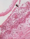
What is indicated by the arrow? The star? Is this change acute or chronic?

A

orthokeratotic hyperkeratosis (arrow);

There is so much keratin present that it has started to plug up the hair follicle (star);

Change is chronic

9
Q

What is acantholysis and what can it be caused by?

A

Loss of cohesion bt epidermal cells due to degeneration of desmosomes; caused by autoimmune disease (EX: Pemphigus)

10
Q

What is spongiosis and what does it lead to?

A

Intercellular edema in the epidermis; leads to vesicle formation

11
Q

What is ballooning degeneration and what can it lead to formation of?

A

Intracellular edema or hydropic degeneration of epidermal cells; areas so affected often coalesce to form vesicles

12
Q

When is ballooning degeneration commonly seen?

A

In pox diseases of skin

13
Q

What is seen here demarcated by the black arrow? Red arrows?

A

Ballooning degeneration (black arrow);

Skin pale and swollen, filled with water, starts to swell and eventually forms large vesicles (red arrows)

14
Q

What are the arrow heads pointing to?

A

acantholytic keratinocytes

15
Q

What is the arrow head pointing to?

A

acantholytic keratinocyte

16
Q

What occurs during Pemphigus foliaceus?

A

Acantholysis occurs in the subcorneal epidermis, resulting in release of freefloating keratinocytes in subcorneal vesicles and pustules

17
Q

What is change 1?

A

hyperkeratosis

18
Q

What is change 2?

A

Acanthosis

19
Q

What is happening in this horse and what is likely the cause?

A

Pustules; some have opened around the eyes and formed crusts; likely caused by Pemphigus foliaceus

20
Q

What is happening here?

A

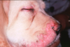
Juvenile pyoderma (face swells, animal gets febrile)

21
Q

What are characteristics of dermatitis?

A

Localized or generalized, primary or secondary, usually dermis and epidermis involved, location of inflammation - response to injury (patterns)

22
Q

Why should you look for patterns in dermatitis?

A

To group diseases that cause a particular pattern and to form differentials based on those patterns

23
Q

What are selected patterns of dermatitis?

A

Perivascular and vascular, follicular, epidermal hyperplasia, interface, pustular, and nodular

24
Q

How can you characterize perivascular inflammation?

A

Around blood vessels in the dermis, very common, can be caused by allergy

25
How can you characterize vascular inflammation and what is an example?
Inflammation is inside the vessel, vessel wall inflamed, can be caused by sepsis; EX: diamond skin disease in pigs
26
How can you characterize an interface pattern of inflammation and what can it be caused by?
Inflammatory cells located at the junction of dermis and epidermis; can be caused by autoimmune disease such as discoid lupus
27
How can you characterize a hyperplastic pattern of inflammation?
Acanthosis, very common, cannot tell much about the cause
28
What two patterns of inflammation go together?
luminal folliculitis and furunculosis
29
How can you characterize luminal folliculitis as a pattern of inflammation?
Damage inside a hair follicle, caused by demodex (mites), dermatophytes (ringworm), deep bacterial infections
30
How can you characterize furunculosis?
Luminal folliculitis that gets so bad that the follicle ruptures, anything that was causing it will go into the dermis and incites tremendous inflammation; it is painful, itches, and swells
31
How can you characterize an epidermal pustule?
Can be caused by superficial bacterial infections such as streps and staphs, can be caused by secondary bacterial infections, can be caused by Pemphigus (autoimmune)
32
How can you characterize a nodular to diffuse pattern of inflammation?
Usually has macrophages present, opportunistic pathogens come in through bite wounds or foreign bodies, can also be caused by systemic fungal infections (yeast, blastomycosis, pithiosis)
33
What type of dermatitis is seen here?
perivascular
34
What type of dermatitis is seen here and what disease might look like this?
interface; discoid lupus
35
What type of dermatitis is seen here?
Folliculitis that has progressed to furunculosis
36
What gross lesions can be seen with acute dermatitis?
Erythema, vesicles, pustules, wheals, exudation, crusts, ulcers, traumatic alopecia
37
What microscopic lesions can be seen with acute dermatitis?
Spongiosis, ballooning degeneration, vesicles, pustules, dermal edema, vascular dilation and congestion, necrosis, fibrin, thrombosis, acute inflammatory infiltrates
38
What can happen during resolution of acute dermatitis?
Complete restitution, scarring, or progression to chronic inflammation
39
What change is seen here?
Acute dermatitis, pyoderma, erythema, ruptured pustules
40
What change is seen here and what is the common name?
Acute, diffuse, serofibrinous and pustular dermatitis; AKA exudative epidermitis, AKA greasy pig disease
41
What causes greasy pig disease?
Severe superficial cutaneous infection with Staph. hyicus
42
What are gross lesions seen with chronic dermatitis?
scale, epidermal thickening - hyperplasia, lichenification, pigmentary changes, alopecia, fibrosis
43
What are micro lesions seen with chronic dermatitis?
Acanthosis, hyperkeratosis, fibrosis, chronic (mononuclear) cellular infiltrates – macrophages, plasma cells, lymphocytes
44
What can occur during resolution of chronic dermatitis?
Complete resolution (but more difficult than acute), persistent chronic inflammation, fibrosis and scarring, depigmentation
45
What can be seen here?
chronic dermatitis: alopecia, epidermal thickening, scale, crust
46
What is happening here?
Demodicosis: alopecia, thickened skin, crust
47
What is happening here in this bovine?
Thickened hyperplastic skin - coalescing nodules or plaque, hyperkeratinized (Stephanofilaria)