BONUS FIGURES Flashcards


Scalp plaque with scarring alopecia hyperpigmentation and depigmentation, discoid lupus erythematosus.

Macular depigmentation, vitiligo

Whitish grouped papules of lichen nitidus

Vesicles, bullae, and erosions; bullous pemphigoid.

Erythematous plaques studded with sheets of pustules, pustular psoriasis.

Ulcer of the lip, chancre of primary syphilis.

Annular, arcuate, and polycyclic configurations; granuloma annulare.

Acral small blue papule, blue nevus.

Scalp plaque with scarring alopecia hyperpigmentation and depigmentation, discoid lupus erythematosus.

eFig. 3.1 Lightning strike.

eFig. 3.2 Erythema ab igne from transcutaneous electrical nerve stimulation (TENS) unit, with device wire at lower right.
eFig. 3.3 Pernio (chilblain).

eFig. 3.4 Frostbite in a homeless person.

eFig. 3.5 Stellate pseudoscars.

eFig. 3.6 Colloid milium. (Courtesy Ken Greer, MD.)

eFig. 3.7 Berloque dermatitis.

eFig. 3.8 Phytodermatitis to lime in a bartender.
eFig. 3.9 Chronic actinic dermatitis.

eFig. 3.10 Chronic radiodermatitis after fluoroscopy.

eFig. 3.11 Chronic radiodermatitis.

eFig. 3.12 Angiosarcoma years after radiation therapy.

eFig. 3.13 (A and B) Calluses from sitting in yoga position. (Courtesy Dr. Shyam Verma.)


eFig. 3.14 Scars caused by “skin popping.”

eFig 3.15 Red tattoo reaction. (Courtesy Curt Samlaska, MD.)

eFig. 4.1 Eczema craquelé.

eFig. 4.2 Prurigo pigmentosa.

eFig. 4.3 Lichen simplex chronicus

eFig. 4.4 Prurigo nodularis. (Courtesy Lawrence Lieblich, MD.)

eFig. 4.5 Dermatitis caused by lip licking

eFig. 4.6 Samples brought in by patient with delusions of parasitosis.

eFig. 4.7 Factitial ulcer.

eFig. 4.8 Complex regional pain syndrome

eFig. 4.9 Diabetic foot ulcer.

eFig. 5.1 Dennie-Morgan folds (or Morgan folds).

eFig. 5.2 Nasal crease.

eFig. 5.3 Ear eczema secondary to allergic contact dermatitis.

eFig. 5 4 Pityriasis alba.

eFig. 5.5 Acute vesiculobullous hand eczema.

eFig. 5.6 Hyperkeratotic hand dermatitis.

eFig. 5.7 Napkin psoriasis.

eFig. 5.8 Nummular eczema.

eFig. 5.9 Eczematous eruption with purpura in Wiskott-Aldrich syndrome.

eFig. 6.1 Toxicodendron radicans subspecies radicans, a common poison ivy species found in the eastern United States. (Courtesy James WD: Textbook of Military Medicine. Office of the Surgeon General, United States Army, 1994 )

eFig. 6.2 Poison ivy dermatit s.

eFig. 6.3 Shoe dermatitis.

eFig. 6.4 Occupational dermatitis from rubber glove allergy.

eFig. 6.5 Fixed drug reactions. (Courtesy Dr. L. Lieblich )

eFig 6.6 Nonpigmenting fixed dry eruption caused by pseudoephedrine.

eFig. 6 8 Toxic erythema of chemotherapy.

eFig. 6.7 Lichenoid drug eruption caused by gold.

eFig. 6.9 Acneiform eruption caused by epidermal growth factor receptor (EGFR) inhibitor therapy.

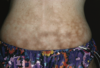
eFig. 6.10 Steroid-induced striae.

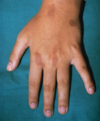
eFig. 6.11 Fat atrophy caused by superficial steroid injection.

