Anatomy - Heart Failure Flashcards
What are the origins of the first and second heart sounds?
First = AV valves closing = low pitched ‘lub’
Second = closing of aortic/pulmonary valve = high pitched ‘dup’
What are the properties of the jugular venous pulse?
- Pulsations in internal jugular vein reflect changes in right atrial pressure
- Visible 2cm above clavicle

What are the third and fourth heart sounds?
Third heart sound = vibration of ventricular wall when filling (hard to hear)
Fourth heart sound = ventricular filling during atrial systole (hard to hear)
When do the aortic and mitral valves open and close?
Aortic:
- Opens during systole
- Closes at start of diastole
Mitral:
- Opens during diastole
- Closes at start of systole
What are the types of abnormal heart sounds?
Murmurs –> excessive noise (high flow or flow in different directions)
Stenosis –> narrowing, creating steep pressure gradient so murmur when valve opens
Leaky valve –> flow can occur in different directions, so murmur when valves should be closed
What drives the intrinsic heart rate?
Pacemaker (SA node) and conduction (AV node)
What things influence heart rate?
Sympathetic nervous system - activation of b-adrenoceptors causes an increase in heart rate
Parasympathetic nervous system – activation of muscarinic receptors causes a decrease in heart rate
Hormones – e.g. adrenaline acting on b-adrenoceptors causes an increase in heart rate
Extra/intracellular ions – alterations in membrane potential (e.g. potassium)
What is the ESV and EDV?
ESV = end systolic volume = blood remaining in ventricle after ventricular contraction
EDV = end diastolic volume = blood in ventricle before contraction
How do you work out cardiac output?
What are the normal values at rest and during exercise?
Cardiace output (litres/min) = stroke volume (litres/beat) X heart rate (b/min)
Rest = 5 L/min
Exercise = 22 L/min
What 3 things determine stroke volume?
- Pre-load –> blood coming back into heart from veins
- Cardiac contractility –> determined by blood present in heart
- After-load –> pressure to push blood out of heart
What is stroke volume?
What influences cardiac output?
Blood ejected from each ventricle in a single heartbeat = difference between EDV and ESV
Influenced by venous return, alongside filling time and atrial contractility
What to remember about cardiac muscle and stretch?
What does the Frank-starling curve show and look like?
Force of contraction of cardiac muscle fibres is proportional to degree of stretch

What factors affect stroke volume and what increases and decreases stroke volume?
- Cardiac contractility
- Aortic pressure and after-load
Increase:
- SNS activity
- Calcium
- Positive inotropic drugs (digoxin)
Decrease:
- SNS activity
- Hypoxia
- Acidosis
- Heart failure
What does the graph of velocity against load look like?

What is happening at and between each letter?


What happens once baroreceptors detect fall in blood pressure?
- Sympathetic outflow from CNS for minute by minute response
- Kidney determines long-term bp restoration
What are all the functions of the kidney?
- Regulation of pH (H+ and HCO3-)
- Removing metabolic waste products
- Production of hormones (e.g. erythropoeitin)
- Activation of vitamin D
- Regulation of osmolarity (control of solute concentrations)
- Regulation of salt concentrations
- Regulation of extracellular fluid volume
What are the properties of the kidneys?
- Receive about 20% of the cardiac output at rest
- Shows good autoregulation i.e. the blood flow stays relatively constant over a wide range of arterial pressures.
- About 20% of the fluid arriving in the kidney is filtered into the renal tubule but then 99% of this is reclaimed.
- This process is intimately linked with sodium reabsorption and is under hormonal control.
What are the functions of the granular and lacula densa and what is the importance of them?

Granular = regulate bp and volume by detecting stretch
Macula densa = monitor salt concentrations
These factors influence first part of renin-angiotensin system
Low volume = renin release
What 3 things trigger renin release?
- Decreased renal perfusion pressure (detected by granular cells)
- Decrease NaCl concentration (detected at the macula densa)
- Increased sympathetic nerve activity (via activation of beta-1 adrenoceptors)
What is the cascade of reactions that happen once renin is released?

What are the actions of angiotensin ii?
- Potent vasoconstrictor – increases peripheral resistance and hence blood pressure
- Enhances sympathetic nerve function
- Increases the release of aldosterone (adrenal gland)
- Promotes thirst
- Release vasopressin (anti-diuretic hormone) from posterior pituitary
- Trophic effects in heart and blood vessels (? Sustain hypertension, cardiac hypertrophy)
- Increase in oxidative stress (? Endothelial cell damage)
What are the actions of vasopressin in CV system?
- Direct vasoconstriction
- Increases number of aquaporin-2 channels in the distal tubules/collecting duct of the kidney – increases fluid retention (produces a more concentrated urine)
- Hence vasopressin is also known as Anti-Diuretic Hormone (ADH)
What are the actions of aldosterone?
- Increase expression of sodium channels
- Activates the sodium potassium pump
- This results in retention of sodium (and water) in the body
What are pathological causes of decreased blood flow?
- Decrease in cardiac output – heart failure
- Renal stenosis or aortic stenosis (narrowing of the renal artery or aorta), produces renin-induced hypertension.
- Hypotensive shock –> a condition in which blood pressure is below the autoregulatory range for maintenance of cerebral and renal perfusion, such that consciousness is lost and vital organ perfusion critically impaired
What are general causes of decreased blood pressure?
- BP = Cardiac Output x Total Peripheral Resistance
- Therefore, low BP can be due to low CO and/or peripheral vasodilatation
- Haemorrhagic shock – blood loss, low CO
- Cardiogenic shock – myocardial infarction causing loss of myocardial power
- Endotoxic shock – bacterial toxins cause marked peripheral vasodilatation
- Anaphylactic shock – allergic reaction, histamine release causes vasodilatation and increased capillary permeability
What are the types of heart failure?
- Chronic heart failure
- Congestive heart failure
- Congestive cardiac failure
- May be valve or muscle failure
- Chronic or acute
What are the 3 main causes of heart failure?
- Ischaemic heart disease
- Hypertension
- Cardiomyopathies
What things precipitate heart failure?
- Pregnancy
- Anaemia
- Hyper and hypothyroidism
- Fluid retaining drugs (i.e. NSAIDs and glucocorticoids)
What happens during neurohormonal adaptation?
- Compensation for circulatory failure
- Sympathetic nervous system activation (worsen)
- Renin-angiotensin-aldosterone system activation (worsen)
- Antidiuretic hormone (worsen)
- Atrial natriuretic peptide –> + sodium excretion, + fluid excretion (better)
What does neurohormonal adaptation lead to?
- Increased afterload
- Increased circulating volume (increased preload & afterload)
- Increased resistance will lead to impaired renal function, more salt/water retention with further activation of RAAS
- A vicious cycle develops which further impairs the pump activity of the heart.
- Myocyte dysfunction
- Makes heart failure worse
What are the properties of left-sided heart failure?
- Secondary to hypertension usually
- Impairment of left ventricle = pulmonary oedema
- Dyspnoea
- Cough
- Orthopnoea
- Inspiratory crepitations
What are the properties of right-sided heart failure?
- Failure of right ventricle output
- Due to lung disease, pulmonary valvular stenosis
- venous pressure
- jugular venous pressure
- Enlarged liver
- Peripheral oedema (ankles)
What is biventricular heart failure?
- Both chambers affected by disease
- Left ventricular failure = pulmonary congestion = right ventricular failure
What are the symptoms of heart failure?
- Fatigue, listless
- Poor exercise tolerance (determines grade)
- Cold peripheries
- Low blood pressure
- Reduced urine flow
- Weight loss
How do you diagnose heart failure?
- Symptoms
- Examination
- Echocardiogram –> ejection fraction < 45%
- B-type natriuretic peptide levels (BNP elevated = heart failure indication)
- Chest X-ray –> enlarged heart, pulmonary oedema
How is atrial fibrillation relevant to heart failure?
- Complication of heart failure
- LV failure = + left atrial pressure = distention = AF
- Stasis of blood –> thrombus formation
- Need prophylaxis, warfarin or DOAC
What is the prognosis of heart failure?
- Dependent on stage (i – iV)
- Poor
- Median survival rate = 5 years
What are the goals of the treatment of heart failure?
- Identify / treat any cause (valvular disease; IHD)
- Reduce cardiac workload
- Increase cardiac output
- Counteract maladaptation
- Relieve symptoms
- Prolong quality life – reduce hospitalization
What is backward and forward heart failure?
Backward = fluid congestion
Forward = less blood flow to tissues
What are the 4 classes of heart failure?
Class I No limitation of physical activity
Class II Slight limitation of activity (breathlessness/fatigue with moderate exercise)
Class III Marked limitation of activity (breathlessness with minimal exercise)
Class IV Severe limitation of activity (symptoms at rest)
What are the 3 types of left ventricular heart failure?
- Insufficient pump power
- Problem with the heart muscle
- Obstruction of the blood flow outwards
- Valve, aorta, arteries, arterioles
- Obstruction to inflow
- Pericardial effusion, constrictive pericarditis
What are the causes of left ventricular heart failure?
- Acute ventricular dysrhythmias (VF)
- Myocardial infarction and ischaemic heart disease
- Longstanding hypertension
- Valve disease (aortic and mitral)
- Cardiomyopathies and drugs
- Congenital heart disease
What is high output cardiac failure and what are its consequences?
- Normal heart muscle, output can’t perfuse tissue adequately
- Arteriovenous fistula – blood bypasses tissue
- Septic shock – vasodilatation
- Anaemia- oxygen requirements not met
- Thyrotoxicosis – increased tissue demand
What do the dark and pale areas on liver represent?
Dark = centrilobular congestion
Pale = periportal fatty change
What does pulmonary oedema look like on histology slide?

What are the steps of left ventricular heart failure?

How do ACE inhibitors work?
- e.g. ramipril
- Reduce arterial and venous vasoconstriction (reduce after- and pre-load)
- Reduce salt/water retention, hence reduce circulating volume
- Inhibits RAAS, prevents cardiac remodelling
- Also used in hypertension
What to remember when prescribing ACE inhibitors>
- Low dose then titrate up
- Monitor creatinine / eGFR and K+ before and during treatment
- May cause severe hypotension
- Cause renal function deterioration with pre-existing renal disease
- Avoid in renovascular disease
- Dry cough in 10% of users à ACE inhibitors breakdown bradykinin, causing cough
What happens when there’s a blockage in a renal artery?
+ renin release
Maintenance of blood pressure
What are the properties of AT1 receptor antagonists?
- e.g. losartan, candesartan
- Angiotensin II acts at AT1 receptors
- AT1 receptor antagonists block the action of AII
- Far less likely give rise to a cough
- An alternative to ACE Inhibitors

What are the properties of beta blockers?
- Used to be contraindicated in CHF
- Now first line with ACEi’s
- Beta1 selective
- Metoprolol, bisoprolol, carvedilol (also an a-blocker/antioxidant)
- Now known to reduce disease progression, symptoms and mortality
- Start with very small dose, with gradual build-up
When and how are beta blockers used in heart failure?
- Of use in stable, moderate failure –> cautious with COPD
- Reduce sympathetic stimulation, heart rate and O2 consumption
- Antiarrhythmic: will control rate in atrial fibrillation
- Oppose the neurohormonal activation which leads to myocyte dysfunction
- Especially useful in failure associated with ischaemia
- Start with a low dose
- Symptoms may get worse at first
What are the properties of diuretics in heart failure?
- Loop diuretics (act on loop of Henley) such as furosemide (and to a lesser extent thiazides)
- Reduce circulating volume
- Reduces preload on the heart
- Relieve pulmonary and peripheral oedema
- Thiazides (esp) / loop diuretics may cause hypokalaemia (drop in potassium levels)
What are the proeprties of digoxin?
- Sometimes used in heart failure
- Favourable in patients with AF
- +ve inotrope (increase in contractility) by inhibiting Na+/K+ ATPase, Na+ accumulates in myocytes, exchanged with Ca2+ leading to increased contractility
How does digoxin work and when is it used?
- Impairs AV conduction and increases vagal activity (via CNS).
- The heart block and bradycardia are beneficial in heart failure with atrial fibrillation
- Slowing the heart rate improves cardiac filling
- Digoxin reserved for failure with AF or when ACEI + diuretic fails
- Titrate dose to ventricular rate>60 beats/min
What things must you monitor on patient taking digoxin?
- Renal function - elderly esp. may have eGRF impairment
- Potassium levels
- Hypokalaemia
What are the negatives of digoxin?
- Toxicity
- Narrow therapeutic window
- Can lead to anorexia, nausea, visual disturbances, diarrhoea
- Monitor pulse (> 60bpm) in AF
How do you know when to prescribe digoxin and when are diuretics prescribed?
Digoxin = prescribed as last resort
Diuretics = always prescribed in heart failure

What is arteriosclerosis, arteriolosclerosis, atheroma and atherosclerosis?
ARTERIOSCLEROSIS = thickening and hardening of the wall of an artery
ARTERIOLOSCLEROSIS = thickening and hardening of the wall of an arteriole
ATHEROMA = an important disease of large and medium arteries.
ATHEROSCLEROSIS = arteriosclerosis due to atheroma
What is hypertensive arteriosclerosis?
- Hypertrophy of media
- Fibroelastic thickening of intima
- Elastic lamina reduplication

Where does hypertension cause arteriolosclerosis to occur?
In arterioles and small arteries
What is hypertensive arteriolosclerosis?
Replacement of wall structures by amorphous hyaline material

What are the properties of atheroma?
- Disease of large and medium arteries
- Only occurs in high pressure systems ie systemic arterial system, NOT venous system
- Initially a disease of tunica intima, but later affects tunica media
- Is ubiquitous, but very mild in young people, worsening with age
What are the stages of atheroma formation?
- Fatty streak
- Lipid plaque
- Fibro-lipid plaque
- Complicated atheroma
What happens during the first stage of atheroma formation?
Blood lipids enter intima at site of endothelial damage

What happens during the second stage of atheroma formation?
Phagocytosis of lipids by macrophages in intima, forming raised fatty streaks

What happens during the third stage of atheroma formation?
- Lipid released by macrophages (lipid plaque)
- Macrophages secrete cytokines, stimulating myofibroblasts to secrete collagen
- Early elastic lamina and media damage

What happens during the fourth stage of atheroma formation?
- Collagen covers plaque surface (fibro-lipid plaque)
- Thinning of media, replacement of muscle fibres by collagen
- Calcification of lipid intima
- Ulceration of surface fibro-lipid plaque
- Weakness and inelasticity from media thinning (complicated atheroma)

What does fibro-lipid plaque look like under microscope?

What does fibro-fatty plaque look like under a microscope?

What are some of the complications of atheroma?
What are the potential complications of atheroma in coronary, leg, mesenteric and cerebral and vertebral arteries?
Coronary arteries = angina
Leg arteries = intermittent claudication
Mesenteric arteries = ischaemic colitis
Cerebral and vertebral arteries = cerebral ischaemic events
How does aneurysm form from media damage?
- Enlarging intimal atheroma plaque leads to atrophy of media
- Muscle and elastic fibres in media replaced by collagen
- Collagen strong but neither contractile nor capable of elastic recoil
- With each systolic pulse, wall of artery stretches and thins, PARTICULARLY WHEN BLOOD PRESSURE IS ELEVATED
- Most common in abdominal aorta
What is an aneurysm and the types?
- Aneurysm = abnormal permanent focal dilation of artery
- Most common = secondary to atherosclerosis in abdominal aorta
- Other types:
- Syphilitic
- developmental(“berry”) in cerebral vessels
- “dissecting aneurysms” of thoracic aorta
- mycotic aneurysms
What are the properties of mycotic aneurysms?
- Mostly caused by endocarditis (infection of heart valve)
- Bacterial septicaemia
- Infection of arterial wall
- Weakening and dilatation = aneurysm
- Risk of bleed
What do the 3 types of aneurysm look like?

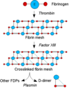
What is a thrombus?
Solid mass of blood constituents aggregated together in flowing blood in lumen of blood vessel
Main constituents = platelets and fibrin

What is the function of fibrinolysis in thrombosis?
What happens when fibrinolysis isn’t functioning?
Limits process of thrombosis
Thrombosis becomes a pathological process
What happens during the first stage of thrombosis?
- Vessel wall is breached
- Circulating platelets aggregate to plug gap
- Platelets release factors which trigger coagulation cascade
What happens during the second stage of thrombosis?
- Coagulation cascade converts fibrinogen to large molecules of insoluble fibrin
- Long fibrin molecules bind together platelets and entrapped red & white cells
What is fibrinolysis and what is the function of plasmin?
Dissolving of thrombus due to breakdown of fibrin
Is active enzyme which fragments fibrin, forming FDPs
What are the properties of plasmin?
- Plasma contains the inactive proenzyme plasminogen, which becomes plasmi
- Plasminogen is converted to plasmin by plasminogen activators, particularly tissue plasminogen activator (t-PA) secreted by endothelial cells
- When fibrin is formed, plasminogen and t-PA bind to it. The t-PA converts nearby plasminogen to plasmin, which begins to degrade the fibrin
- This controls the size of the thrombus
What is pathological thrombosis?
Pathological thrombosis occurs when the thrombus enlarges beyond vessel healing requirements, and continues to grow
What happens when fibrinolysis fails?
Thrombus grows by accretion of layer upon layer, forming mass in vessel lumen

What are D-Dimers?
Breakdown product of fibrin mesh, stabilised by factor XIII
+ blood levels in thrombosis
What is Virchow’s Triad?
- Damage to vessel wall
- (esp. endothelium)
- Stasis
- (slow or turbulent blood flow)
- Change in character of blood
- (esp. increased platelets, increased red cell numbers, increased viscosity)
Where does thrombosis occur and properties?
- ARTERIES
- main predisposing factors are VESSEL WALL DAMAGE
- VEINS
- STASIS most important
- HEART
- Ventricles - chamber wall damage most important
- Atrium - stasis most important
- Heart valves - valve surface damage most important
What are the potential outcomes of thrombosis?
- It may be lysed by intrinsic fibrinolysis - RARE
- It may completely block the lumen (occlusion)
- It may undergo organisation & recanalisation
- It may extend locally (propagation)
- It may fragment or detach completely and travel elsewhere in the circulation. This is called THROMBO-EMBOLISM
What is infarction and congestion?
Infarction = blood supply cut off
Congestion = venous blockage by thrombus, preventing drainage so blood pools (+ haemorrhaging infarction)
What happens in an organised thrombus?
- New vessels grow into the thrombus
- Vascular granulation tissue develops
- Fibroblasts invade & deposit collagen
- Fibrovascular granulation tissue develops
- Recanalisation occurs if vessels link up

What is an embolism and what are the most important matters to embolise?
Transfer of abnormal material by bloodstream with eventual impaction of material in vessel distal to site of origin
Cancer (metastasis) and thrombus cells
What is a thromboembolism?
Where thrombus breaks off and occludes a distal vessel
Where does thrombus in artery of left side of heart embolise to and what are the potential consequences?
Systemic arterial system
- of brain arteries -> STROKE
- of lower limb arteries -> GANGRENE OF LEGS
- of mesenteric arteries -> BOWEL NECROSIS
- of renal arteries -> KIDNEY INFARCT
- of splenic artery -> SPLENIC INFARCT
What is the outcome of a thrombus in a systemic vein?
Pulmonary embolus
Small embolus = small peripheral lung infarct
Large embolus = sudden death
How does venous thromboembolism risk assessment work?
- Active cancer or cancer treatment
- Age > 60
- Dehydration
- Known thrombophilias
- Obesity (BMI >30 kg/m2)
- One or more significant medical comorbidities
- heart disease
- metabolic, endocrine or respiratory pathologies
- acute infectious diseases
- inflammatory conditions
What are other factors included in VTE risk assessment?
- Personal history or first-degree relative with a history of VTE
- Use of hormone replacement therapy
- Use of oestrogen-containing contraceptive therapy
- Varicose veins with phlebitis
- Pregnancy or < 6 weeks post partum (see NICE guidance for specific risk factors)
What are other important materials to embolise?
- Fat and marrow
- Air
- Nitrogen
- Amniotic fluid
