Actual images Flashcards

1
Q

What does this picture of esophagus show?

A

kissing ulcers = sign that pill caused esophagitis

2
Q

What is this finding on endoscopy?

A

candida esophagitis

3
Q

What bug is causing this finding in the esophagus?

A

CMV esophagitis

4
Q

What bug is causing this finding in the esophagus?

A

THis is herpest esophagitis

  1. Cell-cell detachment
  2. Multinucleation
  3. “Ground glass” nuclei
5
Q

What do these findings on endoscopy suggest?

A

shows transverse rings = trachealization

possible sign of eosinophilic esophagitis

6
Q

What is this lesion finding in the esophagus?

A

barrett’s esophagus = have intestinal like columnar epithelium with goblet cells

7
Q

What normal part of the body is this?

A

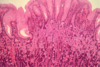
this is stomach = shows gastric fundic glands

8
Q

What gastric cell type is this?

A

parietal cells

9
Q

What disease of the stomach is this?

A

acute erosive gastritis

10
Q

What is this finding in the stomach?

A

chemical gastropathy

see corkscrew gastric pits, dilated capillaries, loss of epithelial mucin

11
Q

What kind of chronic gastritis is this?

A

This is autoimmune gastritis

  • have intestinal and antral metaplasia
  • chronic inflammation
  • reduced glands
  • loss of parietal cells
12
Q

What type of malignancy is characterized by these cells?

A

these are signet ring cells

suggest diffuse adenocarcinoma

13
Q

What type of gastric malignancy is this?

A

intestinal type gastric carcinoma

have gland formation

14
Q

What disease of malabsorption is this?

A

abetalipoproteinemia = b/c theres fat filling the cells

15
Q

What is this image show in the liver

A

this is alpha 1 antitrypsin deficiency

tiny pink dots are a1a that is abnormally folded and retained in the hepatocyte

16
Q

What is this finding in the liver?

A

hepatocellular carcinoma

17
Q

What is this finding in the liver?

A

hepatocellular carcinoma producing bile

18
Q

What is this finding in the liver?

A

angiosarcoma = you can see the RBCs

19
Q

What does this picture of the small intestine show?

A

cobblestoning of the intestinal wall = sign of Crohns

20
Q

What does this finding in the intestine suggest?

A

Noncaseating granuloma –> suggests crohns

21
Q

What is this disease?

A

ulcerative colitis = can easily see the line where it starts/stops

22
Q

What type of IBD could this be?

A

ulcerative colitis = don’t see intramural inflammation like you would in crohns

23
Q

What type of intestinal disease is this?

A

This is IBD = see crypt atrophy

24
Q

What is this finding in the intestine?

A

This is cryptitis = see inflammation of the crypts with neutrophils; suggests IBD

25
What is this finding? Disease?
This is creeping fat; seen in crohns
26
What is this complication? What disease?
This is toxic megacolon complication of UC
27
What is this finding in the colon?
This is dysplasia
28
What causes this finding?
This is C Diff colitis
29
What is this finding?
This is pseudomembranous colitis
30
What is this finding?
colonic ischmia = you can tell by the dusky red color
31
What is this finding in the colon?
colonic ischemia
32
What is this finding in the colon?
diverticula
33
What type of colitis is this?
collagenous colitis
34
What type of colitis is this?
lymphocytic colitis = see the lymphocytes in the epithelium
35
What does this show in the colon?
lots of polyps = FAP
36
What type of adenoma?
tubular
37
What type of adenoma?
villous
38
What type of adenoma?
villous sessile adenoma
39
What type of polyp?
40
What type of polyp?
hyperplastic
41
What type of polyp?
sessile serrated polyp
42
What type of polyp on left vs right?
43
What does this image show?
see adenomatous tissue on left with cancer on the right
44
What is happening to these colon glands?
desmoplasia with dysplastic cancerous glands
45
What is differnet between these two cancers?
right = poorly differentiated left = moderately differentiated
46
What type of colonic neoplasm is this?
47
What type of colorectal cancer is this?
microsatellite unstable stained for CD3 lymphocytes that are classic for MSI carcinoma
48
What is this finding in the appendix?
49
What is this finding in the appendix?
mucinous cystadenoma
50
What is this bug?
giardia
51
What is this bug?
entamoeba histolytica
52
What is this finding in an infant?
meconium ileus
53
What is this finding in the esophagus?
squamous cell carcinoma = found in upper 2/3 of esophagus
54
What is this finding in the esophagus?
squamous cell carcinoma
55
What is this finding in the esophagus?
squamous cell dysplasia = precursor to carcinoma
56
What is this finding in the esophagus?
Candida esophagitis
57
What is this finding in the esophagus?
herpes esophagitis = most common cause of esophagitis in immune competent
58
What is this finding in the esophagus?
59
What is this finding in the stomach?
ECL cell hyperplasia; possible carcinoid tumor stained by chromogranin
60
What is this finding in the smalll intestine?
celiac disease = villous atrophy
61
What is this disease of the colon that presents with normal endoscopy?
62
What is this disease of the colon that presents with normal endoscopy?
lymphocytic esophagitis
63
What is on the left and right in these images of liver?
left = normal; right = hepatocellular carcinoma