Abdominal Organs and the Celiac Trunk Flashcards
What are the two layers of the abdominal cavity
What is there innervation
Parital- lines the body wall and covers the retroperitoneal organs
Visceral- encloses the surface of the intraperitoneal
Innervated by the lower intercostal nerves and the ilioinguinal and the iliohypogastric nerves of the lumbar plexus
Describe the mesenterary and its function
DOuble layered peritoneal membrance that suspends part of the GI tract from the body walll and allows for movement of the GI as needed. Also allows vessels, nerves and lymphatics to reach the GI tract
What are the four important remnants of the mesentery in the abdomen
The less omentum–attach the liver to the lesser curvature of the stomach and the duodenum.
The greater omentum–attach to the greater curvature of the stomach.
The Mesocolon—attach the body wall to the transverse and sigmoid colon.
Ligaments—reflections of mesenteries between organs or the body wall that are named according to their attachments.
What are the layers of fascia of the peritoneum and their function and what defines them?
The fascia create space, cover organs and allow limited movement.
- Transversalis fascia– deep to the muscles of the abdominal
wall. - Extraperitoneal fascia- deep to the transversalis fascia (lines the abdominal
cavity. ) - Peritoneal membrane-defines the peritoneal cavity. has two layers
A. parietal peritoneum (paries = wall) lines the wall of the abdomen. Parietal peritoneum is
very sensitive to somatic pain* and is innervated by the lower intercostal nerves and
nerves of the lumbar plexus. e.g. Inflammation of the parietal peritoneum (peritonitis)
results in sharp pain that is localized over the area. —- reason for severe pain during peritonisis
B. visceral peritoneum-(viscus = organ) encloses the surfaces of the intraperitoneal and is not senstitive to somatic pain
intraabodomen fascia identify


what does the mesentery form from
Remember that mesenteries form from the ventral and dorsal mesogastrium
Major organs- identify


The ________plus__________ equals Lesser omentum
The epiploic foramen of Winslow connects the_______
to the _______
.
The lesser omentum connects the __________ to the ___________.
The _Heptagastric ligament____plus_heptaduodenal ligament____ equals Lesser omentum
The epiploic foramen of Winslow connects the_lesser sac__ to the greater sac_______
.
The lesser omentum connects the liver__________ to the _lesser curvature of the stomach and the first part of the duodenu,__________.
Explain the different regions in the abdominal cavity
- The abdominal viscera are line by the peritoneum
- Some organs (e.g. ascending and descending colon) never grow into the cavity and are pressed against the body wall in their retroperitonealposition.
- Other organs like the pancreas become
secondarily retroperitoneal
.


How is the peritoneal sac cavity divided during developement?
What borders the lesser sac?
How do they communicate?
Peritoneal cavity
- During development the peritoneal cavity is subdivided into the greater and lesser sacs.
• - The lesser sac is bordered by the posterior abdominal wall (posteriorly) and the gastrocolic ligament, stomach and lesser omentum (anteriorly).
• - The two sacs communicate through the epiploic foramen of Winslow
IMportant relationships within the foramen of winslow
Anterior– Hepatoduodenal ligament and the hepatic portal vein
•
Posterior—Inferior vena cava
•
Superior—Caudate lobe of the liver
•
Inferior—First part of the duodenum
What organs are intraperitoneal
Stomach
Tail of the pancreas
Liver
Gallbladder
Spleen
1st part of the duodenum
Jejunum
Ileum
Transverse colon
Appendix
Sigmoid colon
What organs are retroperitoneal?
What does that mean?
They never had a mesentary– even during development
Head, neck and body of the pancreas
Ascending and descening colon
2nd and 3rd part of the duodenum
Upper rectum
What organs are secondarily retroperitoneal?
What does that mean?
They had a mesentary but lost it during development (A.I.L. plus kidney stuff)
Kidneys
Adrenal gland
Ureters
Aorta
Inferior vena cava
Lower rectum
The stomach has a ___________curvature, which is connected to the_________
of the liver by
the ___________ (_____________), and a left
______________ from which the ____________ is suspended.
The stomach has a right lesser curvature, which is connected to the porta hepatis of the liver by the lesser omentum (hepatogastric ligament), and a left greater curvature from which the greater
omentum is suspended.
Explain what main thing happens at each part of the stomach
What two organs does the stomach attach to by the lesser omentum
Fundus- dome- shaped upper portion of the stomach, which is normally filled with air. Rest on left dome of diaphragm
Body- The main central part of thestomach .
The pyloric portion- (under the body) of the stomach has a thick muscular wall and narrow lumen that empties into the duodenum approximately in the transpyloric plane (L1 vertebra)
Liver and gallbladder by lesser omentum
When does a sliding hiatal occur?
What can be damaged?
Sliding hiatus occurs when the cardia of the stomach herniates through the esophageal hiatus of the diaphragm. This can damage the vagal trunks as they pass through the hiatus
Parts of the stomach


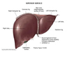
What are the livers two surfaces?
What surronds it?
Where is it place and what protects it?
Superior diagphgramtic surface and inferior visceral surface
Surronded by visceral peritoneum
Protected by the rib cage and in the right abdominal cavity
What are the reflections of visceral of the liver?
falciform ligament– between the diaphragmatic surface of the liver and the diaphragm. attaches to the anterior abdominal wall
•coronary ligament– the reflection point from the liver and has right and left
triangular ligaments.
• The lesser omentum–extends between the visceral surface of the liver (hepatogastric ligament) and the first part of the duodenum (hepatoduodenal ligament.
Explain the two lobes in terms of size and relation to the other two lobes
Right larger than left
Quadrate and cuadate lobes are anatomically with right lobe but functionally with the smaller left lobe
Identify Superior Surface of LIver