AA2 Point Locations Flashcards
SI9
- Location
- Direction
- Depth
- Position
SI9
Location
1 cun above the posterior axillary fold when the arm is adducted, in between the teres muscles
Direction
Posterior to anterior. Do not direct the needle medially.
Depth
0.5 - 1.5 cun
Position
Sitting

SI13
- Location
- Direction
- Depth
- Position
SI13
Location
Medial corner of the supraspinatus fossa
Direction
Perpendicular or oblique, direction toward supraspinous fossa. Too medial or too deep may cause pneumothorax. Stay on scapula.
Depth
0.3-0.5 cun
Position
Sitting

SI14
- Location
- Direction
- Depth
- Position
SI14
Location
3 cun lateral to the midline at the level of the lower border of the spinous process of T1
Direction
oblique; inferior to superior and medially. Pick up tissue similar to GB 21. Avoiddeepperpendicularinsertiontoavoidpneumothorax.
Depth
0.3 - 0.5 cun
Position
Sitting

LI14
- Location
- Direction
- Depth
- Position
LI14
Location
On the line connecting LI 15 and LI 11; 3 cun below LI 15. The point is located anterior and superior to the deltoid tubercle on the anterior margin of the deltoid.
Direction
perpendicular or oblique inferior to superior
Depth
0.8 - 1.5 cun
Position
Sitting

LI16
- Location
- Direction
- Depth
- Position
LI16
Location
In the depression medial to the acromion and posterior to the acromioclavicular joint.
Direction
superior to inferior. For shoulder joint problems, direct the needle obliquely from medial to lateral and superior to inferior
Depth
1-1.5 cun
Position
Sitting

LU2
- Location
- Direction
- Depth
- Position
LU2
Location
Medial to the coracoid process and just inferior to the clavicle in the infraclavicular fossa, 6 cun lateral to the midline.
Direction
anterior to posterior, slightly medial to lateral and superiorly
Depth
0.5-1 cun
Caution: Risk of pneumothorax if needled in wrong direction or depth.
Position
Sitting

LU3
- Location
- Direction
- Depth
- Position
LU3
Location
On the anterolateral aspect of the arm, 3 cun below the axillary fold, in the groove between the biceps and the humerus, on the radial side of biceps brachii. Anterior and inferior to LI 14.
Direction
lateral to medial on a horizontal plane
Depth
0.5-1 cun
Position
Sitting

SI8
- Location
- Direction
- Depth
- Position
SI8
Location
Midpoint between the medial epicondyle and the olecranon in the ulnar groove. Locate the point with the elbow flexed.
Direction
perpendicular, or proximal to distal or distal to proximal along the ulnar nerve. Needle direction should be toward the pain.
Depth
0.3 - 0.8 cun
Position
Supine

HT3
- Location
- Direction
- Depth
- Position
HT3
Location
Midpoint between the medial epicondyle and the bicipital aponeurosis on the transverse cubital crease, in the depression.
Direction
Perpendicular
Depth
0.5-1 cun
Position
Supine

LU5
- Location
- Direction
- Depth
- Position
LU5
Location
On the transverse cubital crease on the lateral side of the bicipital aponeurosis.
Direction
intra-articular, perpendicular (use Betadine® or Stanhexidine® for deep insertion)
Depth
0.5 - 1 cun
Position
Supine

LI12
- Location
- Direction
- Depth
- Position
LI12
Location
1 cun superolateral to LI 11 on the supracondylar ridge of the humerus, at the insertion of brachioradialis, and extensor carpi radialis longus.
Direction
perpendicular
Depth
1 -1.5 cun
Position
Supine

LI5
- Location
- Direction
- Depth
- Position
LI5
Location
At the base of the anatomical snuff box, just distal to the radial styloid process, between the tendons of extensor pollicis longus and brevis.
Direction
perpendicular
Depth
0.3 - 0.5 cun
Position
Supine

LU7
- Location
- Direction
- Depth
- Position
LU7
Location
1.5 cun proximal to the radiocarpal joint (LI 5), on the radial side of the radial styloid process, just proximal to the insertion of the brachioradialis muscle, in the cleft between the tendons of brachioradialis and abductor pollicis longus.
Direction
oblique, proximal to distal
Depth
0.3 - 0.5 cun
Position
Supine

SI3
- Location
- Direction
- Depth
- Position
SI3
Location
When a loose fist is made, the point is found on the ulnar aspect of the hand, at the end of the skin fold just proximal to the head of the 5th metacarpal bone, where the skin changes color and texture.
Direction
perpendicular
Depth
0.5 - 0.7 cun
Position
Supine

SI6
- Location
- Direction
- Depth
- Position
SI6
Location
On the dorsal aspect of the forearm, with the palm of the hand facing the chest, in the groove between the ulnar head and the radius, just proximal to the radio-ulnar ligament.
Direction
angle of 45 degrees obliquely between the radius and the ulna
Depth
1.0 – 1.5 cun
Position
Supine

Baxie
- Location
- Direction
- Depth
- Position
Baxie
Location
With the hand in a loose fist, points are located on the web between the heads of the metacarpals at the junction of the red and white skin. Between the first and second fingers, the point is closer to the thumb. There are eight points in all.
Direction
towards the interspaces of the metacarpal bones
Depth
0.5 - 1.5 cun
Position
Supine

GV15
- Location
- Direction
- Depth
- Position
GV15
Location
On the midline of the neck, directly above the spinous process of C2, approximately 1 cun below the lower margin of the external occipital protuberance.
Direction
Aim towards the tip of the nose so that the needle remains on a horizontal plane
Depth
0.5 – 1 cun
Position
Sitting or prone

BL10
- Location
- Direction
- Depth
- Position
BL10
Location
On the level above the C2 spinous process (same as GV 15) 1.3 cun lateral to the midline, in the depression on the lateral aspect of the trapezius.
Direction
perpendicular posterior to anterior
Depth
0.5 – 1 cun
Position
Sitting or prone

GV16
- Location
- Direction
- Depth
- Position
GV16
Location
On the midline of the neck between the occiput and C1. Identify the lower margin of the occiput and insert just below. The point is also approximately 1 cun above the upper margin of C2 spinous process.
Direction
Aim towards the tip of the nose so that the needle remains on a horizontal plane
Depth
0.5 – 1 cun
Position
Sitting

C4 & C5
- Location
- Direction
- Depth
- Position
C4 & C5
Location
The landmarks used to identify these points are the mastoid process and the posterior margin of the sternocleidomastoid muscle. All the points are located along the posterior margin of the sternocleidomastoid muscle at the following levels:
C4 Upper margin of the thyroid cartilage
C5 Lower margin of the thyroid cartilage
Direction
Lateral to medial
Depth
1 cun
Position
supine with neck rotated to contralateral side is ideal; side lying or sitting are other options

GB21
- Location
- Direction
- Depth
- Position
GB21
Location
On the midpoint of the line connecting the lower margin of C7 spinous process and the lateral aspect of the acromion.
Direction
Tap the needle through the skin, then pick up the skin and underlying muscle and insert the needle posterior to anterior and inferior to superior into the muscle.
Depth
0.5 - 0.8 cun
Position
Sitting

BL31
- Location
- Direction
- Depth
- Position
BL31
Location
Approximately 1 cun lateral to the midline in the first sacral foramen.
Direction
perpendicular to the skin, into the first sacral foramen
Depth
1 - 1.5 cun
Position
Prone

BL32
- Location
- Direction
- Depth
- Position
BL32
Location
Approximately 1 cun lateral to the midline in the second sacral foramen at the level of the lower border of the SI joint.
Direction
perpendicular to the skin, into the second sacral foramen
Depth
1 - 1.5 cun
Position
Prone

BL53
- Location
- Direction
- Depth
- Position
BL53
Location
3 cun lateral to the midline at the level of the S2 foramen or the lower edge of the SI joint, just lateral to the sacrum.
Direction
perpendicular to the skin
Depth
1 - 1.5 cun
Position
Prone

BL54
- Location
- Direction
- Depth
- Position
BL54
Location
3 cun lateral to the midline at the level of the S4 foramen, about 1 cun above the sacral hiatus
Direction
perpendicular to the skin
Depth
1.5 – 2 cun
Position
Prone

CV3
- Location
- Direction
- Depth
- Position
CV3
Location
On the anterior midline, 4 cun below the centre of the umbilicus, or 1 cun above the superior borderof the symphysis pubis.
Direction
obliquely, generally in the direction of the flow of the meridian, inferior to superior
Depth
0.5 - 0.8 cun. Do not go past the resistance felt at the linea alba.
Position
Supine

ST21
- Location
- Direction
- Depth
- Position
ST21
Location
2 cun lateral to the midline, at the level of CV 12 (4 cun above the centre of the umbilicus)
Direction
oblique, 45° angle to the skin or less, towards the midline
Depth
0.7 – 1 cun
Position
Supine

ST25
- Location
- Direction
- Depth
- Position
ST25
Location
2 cun lateral to the midline, level with the centre of the umbilicus
Direction
oblique, 45° angle to the skin or less, towards the midline
Depth
0.7 - 1.2 cun
Position
Supine

ST2
- Location
- Direction
- Depth
- Position
ST2
Location
In the infraorbital foramen (palpate for depression) in line with the pupil when the eye is looking straight ahead, approximately 1 cun below the pupil.
Direction
perpendicular, slightly oblique inferior to superior
Depth
0.2-0.3 cun
Position
Supine

ST6
- Location
- Direction
- Depth
- Position
ST6
Location
1 cun anterior and superior to the angle of the mandible. For easier localization, ask the patient to clench the teeth to bulge the masseter muscle. The point is right in the prominence of the masseter muscle.
Direction
Perpendicularly
Depth
0.3 – 0.5 cun
Position
Supine

SI19
- Location
- Direction
- Depth
- Position
SI19
Location
On the face, anterior to the tragus of the ear, posterior to the condyle of the mandible, in the middle of the groove / depression appearing when the mouth is slightly open.
Direction
Perpendicularly, lateral to medial.
Depth
0.5 – 1.5 cun
Position
Supine

BL2
- Location
- Direction
- Depth
- Position
BL2
Location
On the medial end of the eyebrow at the medial superior corner of the orbital fossa near the supratrochlear notch.
Direction
oblique either medial to lateral along the eyebrow or superior to inferior.
Depth
0.3-0.5 cun
Position
Supine

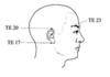
TE23
- Location
- Direction
- Depth
- Position
TE23
Location
In the depression on the lateral end of the eyebrow at the lateral superior corner of the orbital fossa.
Direction
obliquely, superior to inferior or lateral to medial along eyebrow
Depth
0.3 - 0.5 cun
Position
Supine
GB14
- Location
- Direction
- Depth
- Position
GB14
Location
On the forehead, in line with the pupil when looking straight ahead or the midpoint of the eyebrow or the superior orbital margin and 1 cun above the superior orbital margin (2 cun below the hairline), right on the supraorbital foramen.
Direction
oblique superior to inferior, towards the middle of the eyebrow or toward the pain.
Depth
0.5-1 cun
Position
Supine

GV20
- Location
- Direction
- Depth
- Position
GV20
Location
On the midline of the head, midway between the external occipital protuberance (EOP) and the anterior hairline. This distance is measured as 10 cun; therefore, the point is 5 cun anterior to the superior border of the EOP and 5 cun posterior to the anterior hairline.
Direction
oblique ~15-20° posterior to anterior aiming into the loose connective tissue of the scalp
Depth
0.5-1 cun
Position
Prone

GB29
- Location
- Direction
- Depth
- Position
GB29
Location
On the midpoint of the line between the ASIS and the most prominent aspect of the posterior portion of the greater trochanter. Locate this point in side lying.
Direction
intra-articular, lateral to medial and slightly inferior
Depth
1 – 3 cun
Position
Sidelying

BL36
- Location
- Direction
- Depth
- Position
BL36
Location
Midpoint of the gluteal fold
Direction
posterior to anterior
Depth
1.5 – 2 cun
Position
Prone

LR10
- Location
- Direction
- Depth
- Position
LR10
Location
2 cun distal to the pubic ramus or 3 cun distal to the superior border of the symphysis pubis, in the lateral aspect of adductor longus, either on the muscle or lateral to it
Direction
perpendicular
Depth
1 - 1.5 cun
Position
Supine

SP12
- Location
- Direction
- Depth
- Position
SP12
Location
On the midpoint of the line between the ASIS and the pubic symphysis (CV 2), just inferior to the inguinal ligament (in the femoral triangle) and lateral to the femoral artery (needle may pulsate).
CAUTION: Insertion must be inferior to the inguinal ligament, as above the inguinal ligament is into the abdominal cavity.
Direction
perpendicular to the skin, palpate for the femoral artery pulsation and insert just lateral
Depth
1 - 1.5 cun
Position
Supine

Medial knee eye
- Location
- Direction
- Depth
- Position
Medial knee eye
Location
With the knee flexed to 90°, medial to the patellar tendon in the depression above the tibia and below the femur at the knee joint.
Direction
Intra-articular,anteriortoposterior,aimingsomewhatlaterally
Depth
1 - 1.5 cun
Position
Supine

ST35
- Location
- Direction
- Depth
- Position
ST35
Location
With the knee flexed to 90°, lateral to the patellar tendon in the depression above the tibia and below the femur at the knee joint.
Direction
intra-articular, anterior to posterior, aiming somewhat medially. Use special caution with increased fluid in joint
Depth
1 - 1.5 cun
Position
Supine

SP10
- Location
- Direction
- Depth
- Position
SP10
Location
When the knee is flexed, the point is 2 cun proximal to patella, in line with the medial border of the patella, just above the medial condyle of the femur on the bulge of vastus medialis muscle.
Direction
perpendicular
Depth
0.7 - 1.5 cun
Position
Supine with knee flexed

BL37
- Location
- Direction
- Depth
- Position
BL37
Location
6 cun below BL 36 on the midline of the posterior aspect of the thigh, or 8 cun above the middle of the popliteal crease.
Direction
perpendicular
Depth
1 - 1.5 cun
Position
Prone

KI5
- Location
- Direction
- Depth
- Position
KI5
Location
1 cun inferior to K3 in the depression anterior and superior to the medial side of the calcaneal tuberosity.
Direction
medial to lateral
Depth
0.3 - 0.5 cun
Position
Supine

KI6
- Location
- Direction
- Depth
- Position
KI6
Location
In the groove, 1-1.5 cun below the most prominent point of the medial malleolus, where the skin changes colour and texture.
Direction
perpendicular
Depth
0.3 - 0.5 cun
Position
Prone

Bafeng
- Location
- Direction
- Depth
- Position
Bafeng
Location
Midpoint between the toes (interdigital space) where the skin changes color and texture
Direction
oblique, aim between the metatarsal bones.
Depth
0.5 – 1 cun
Position
Supine

ST41
- Location
- Direction
- Depth
- Position
ST41
Location
On the anterior ankle joint line (identify by dorsiflexing the foot) in the depression between the tendons of extensor digitorum longus on the lateral side and extensor hallucis longus on medial side.
Direction
intra-articular, anterior to posterior
Depth
0.5 - 0.7 cun
Position
Supine

ST44
- Location
- Direction
- Depth
- Position
ST44
Location
Between the second and third toes, 0.5 cun proximal to the margin of the web, in the depression distal and lateral to the second MTP joint.
Direction
perpendicular
Depth
0.5 - 0.7 cun
Position
Supine

