75: Tarsal Coalition - Smith Flashcards
what is a tarsal coalition?
Tarsal coalition exists when a union causes restricted motion or absence of motion between two or more tarsal bones.
bar vs. bridge
-
Bar: Extraarticular coalition that occurs outside a normal joint
- Calcaneonavicular Bar - these bones do not normally articulate
-
Bridge: Intraarticular coalition that occurs at a normal joint site
- Talocalcaneal Bridge
most common cause of tarsal coalition in pediatric pt
most common cause of tarsal coalition in adult pt
congential
acquired (ex: fx calcaneus and healing forms union b/w talus and calcaneus)
congenital etiologies
- Pfitzner – Incorporation of accessory ossicles into the normal tarsal bones on either side of a joint or in close approximation with one another.
- issue: coalitions seen in fetuses
- Leboucq – Failure of differentiation and segmentation of primitive mesenchyme
- Heritable defect (autosomal dominant)
- Insult in first trimester
syndesmosis vs. synchondrosis vs. synostosis
-
Syndesmosis: Fibrous union
- Incomplete union with motion
-
Synchondrosis: Cartilaginous union
- Incomplete union with motion
-
Synostosis: Osseous union
- Complete union without motion
Downey Classification
_***********_
- Juvenile (osseous immaturity)
- Type I: extrarticular
- A: no secondary arthritis
- B: secondary arthritis
- Type II: intraarticular
- A: no secondary arthritis
- B: secondary arthritis
- Type I: extrarticular
- Adult (osseous maturity)
- Type I: extrarticular
- A: no secondary arthritis
- B: secondary arthritis
- Type II: intraarticular
- A: no secondary arthritis
- B: secondary arthritis
- Type I: extrarticular
highest incidence joint
talocalcaneal 48.1%
calcaneonavicular 43.6%
clinical s/s tarsal coalition
- Pain
- deep aching in area of coaliton, often near sinus tarsi or entire rearfoot, may also have secondary arthritis
- relieved with rest
- esp w/ walking over rough uneven terrain
- Limitation of motion and rigid flatfoot
- Most likely cause of unilateral pediatric flatfoot
- Muscle spasm
- No symptoms in 22% of patients
- Patients often mistreated for ankle sprains and sinus tarsi syndrome
when do the following coalitions ossify?****
talonavicular
calcaneonavicular
talocalcaneal
- Talonavicular Coalition: 3-5 years
- Calcaneonavicular Coalition: 8-12 years
- Talocalcaneal Coalition: 12-16 years
most obvious clinical finding
limitation of motion STJ and MTJ
what will happen in hubscher maneuver with rigid flatfoot?
no chang ein arch height with dorsiflexion of hallux w/ pt standing
why do the peroneal tendons spasm with rigid flatfoot?
- firing peroneal tendons to keep foot in everted position (it hurts to invert)
- spasm due to rapid firing
- may need to give common peroneal n block to examine the pt
- peroneal spastic flatfoot does not always develop in tarsal coalition and there are other etiologies to peroneal spastic flatfoot

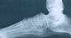
- Calcaneonavicular Bar
- oblique radiograph
- comma sign or anteater sign
- osseous easier to diagnose
- soft tissue coalitions
- close proximity of calcaneus and navicular
- irregualr lateral navicualr cotical surface
- flattening of navicualr laterally
- hypoplasia of head of talus

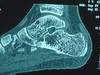
- Talocalcaneal Coalition
- Lateral Radiograph
- diminished posterior or middle facet
- halo sign: sclerotic enhancement of sustentaculum tali
- narrowing of posterior facet
- talonavicular beaking
- flattening of lateral talar process
- calcaneal axial
-
facet obliquity: parallel or less than 25 degrees normally
- abnormal pictured below
-
facet obliquity: parallel or less than 25 degrees normally


- Talonavicualar Coalition
- looks like a long talar neck
- seen on AP and lateral radiograph

talocalcaneal coalition of middle facet
MRI
incomplete calcaneonavicular coalition
CT scan
ball and socket ankle joint
secondary arthritic change assoc with tarsal coalition
ankle starts to remodel to gain motion lost from coalesced jt

talonavicular beaking
secondary arthritic change assoc with tarsal coalition
what type of orthotics would you want to use as conservative tx?
orthotics that restrict STJ and MTJ motion to reduce pain and muscle spasm (keep foot everted)
surgical treatment recommendations based on Downey classification
_****_
- Juvenile
- IA: resection with interposition of EDB m (Badgley 1927)
- IB: triple arthrodesis
- IIA: resection with interposition of arthroereisis, isiolated/single arthrodesis, triple arthrodesis
- IIB: triple arthrodesis
- Adult
- IA: resection with interposition of EDB m, triple arthrodesis
- IB: resection with isolated/single arthrodesis, triple arthrodesis
- IIA: isolated or single arthrodesis, triple arthrodesis
- IIB: triple arthrodesis
arthrodesis indicated in older pts with coalition
describe resection/interpositon with EDB
- Badgley 1927
- Ollier Incision
- EDB dissected from Calcaneus and reflected distally
- Resection of coalition (until you get enough STJ ROM)
- EDB placed into defect
- Silicone or fat may also be used
- Kieth needle passed out through bottom
- Bone Wax
- on cut ends to create hemostasis and inhibit bone healing
