21. Joint Disease Flashcards

1
Q

How does joint space thickness vary over time in the presence of joint disease?

A
  • Typically:
    EARLY: Expansion (effused)

LATE: Collapse (due to cartilage degradation)

=> Later phase rarely dx due to lack of weight bearing rx and variable projection angles

2
Q

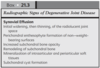
Radiographic features of JOINT DISEASE

A

Image with examples…

3
Q

List three examples of large accumulations of articular or periarticular mineralisation?

A

1) Intraarticular OSTEOCHONDROMA
2) Meniscal mineralisation (cats)
3) “Pseudogout” - calcium pyrophosphate deposition disease (dogs)

4
Q

List three distinct categories of articular calcified bodies?

A

1) Avulsed fragments -> articular or periarticular bone
2) Osteochondral components of disintegrating joint surface
3) Small synovial osteochondromas

5
Q

Pathophys of osteophyte formation?

A
  • Degradation of articular cartilage
  • Degradation products -> synovial hyperplasia
  • Synovial hyperplasia -> Osteophyte (initially cartilage, later ossified)

=> Bony outgrowths at periphery of articular cartilage

6
Q

What are entheses?

A
  • INSERTION of tendon, ligament, joint capsule, or fascia TO BONE

Enthesophyte = Bony spondylopathy at enthesis

7
Q

Common entheses of carpus x 5

A
8
Q

Main enthesis of shoulder x 3

A
9
Q

Main entheses of stifle X 5

A
10
Q

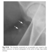
What is Vacuum phenomenon? What are the posible causes of intrarticular gas (4 broad categories)?

A
  • Vacuum = Intrarticular diffusion of nitrogen from ECF following negative pressure
11
Q

In what % of shoulder with humeral head OCD radiographs is vacuum phenomenon seen?

A

20%

NOT a feature in normal contralateral radiographs!

12
Q

List 4 clinical conditions associated with intraarticular gas

A
  • Osteochondrosis
  • Degenerative IVDD
  • Vertebral instability
  • DJD
13
Q

Sites of sesamoids in the dog TABLE

A
14
Q

What % of feline menisci have mineralisation in stifle rads? Which site most commonly affected?

A

46%

Cranial horn, medial meniscus

=> % mineralisation significanly associated with degree of cartilage damage in medial femoral and tibial condyles. THEREFORE associated with medial compartment DJD

15
Q

MCP sesamoid fragmentation is associated with which bones and which breeds?

A

2nd and 7th MCP sesamoids

ROTTIS (several large breeds)

=> up to 44% in one group of rottis; 73% in one group up to 12mo with CS associated in 65%

Another group: cause of FL lameness in 50% young rottis

16
Q

Transverse fragmentation of the digital sesamoids reported in which breed?

A

Racing greyhounds -> possibly fracture is cause

17
Q

What is the most common example of sesamoid displacement? Which breeds are predisposed?

A

Patella luxation!

Toy breeds and Devon Rex
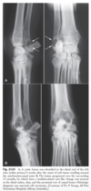
18
Q

What are these

A

Iliopubic sesamoids!

19
Q

What are the three most frequent locations of canine DJD?

A
  • Hips (dysplasia
  • Shoulder
  • Stifles
20
Q

What is the incidence of shoulder and stifle DJD reported at rx / necropsy?

A

Shoulder: 33-50%

Stifle: 20%

21
Q

What are the two sesomoids of the tarsus!! Odd….

A

Lateralplantar tarsometatarsal sesamoid

Intra-articular tarsometatarsal sesamoid

22
Q

What is the most reliable feature to assess for grading of stifle DJD?

A
  • Number and size of osteophytes

=> other features less reliable e.g. effusion, sclerosis, mineralisation

23
Q

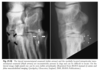
What is the timeline of onset of osteophytosis after cranial cruciate transection?

A
  • Commence as early as 3 days -> margins of femoral trochlea seen radiographically at 2 weeks

Prox / dist fem trochlea -> femoral and tibial condyles and patella

Enthesophytes: CrCr and collateral

24
Q

List 3 specific views that can be used to identify early trochlear osteophytes?

A

1) Flexed mediolateral
2) Craniomedial - caudolateral
3) Caudomedial - craniolateral

25
What feature is the strongest indicator of coxofemoral DJD? What measure is used to evaluate this feature? Normal values?
- Joint subluxation - DISTRACTION INDEX: DJD varies with breed and DI Unlikely to develop DJD if \<0.3 in GSD \<0.4 in lab / Rotti
26
Radiographic signs of DJD
BOX
27
Causes of OA in cats 3x primary 7 x secondary
BOX
28
How do radiographic signs of DJD differ in cats compared to dogs?
- More intraarticular soft tissue calcification - Less joint effusion - Less periarticular soft tissue thickening
29
What is the estimated heritability of canine hip dysplasia?
0.2-0.6 =\> GSDs updated from 0.46% to 61%!
30
What are the earliest recognisable signs of hip dysplasia? And how are they manifested radiographically?
- Synovial effusion, synovitis, round ligament hypertrophy and perifoveal cartilage erosion =\> LAXITY (precipitated by effusion)
31
What are the main features of DJD with HD?
1) Perichondral osteophytes 2) Remodelling of femoral head and neck 3) Acetabular remodelling 4) Sclerosis of acetabulum / femoral head 5) Morgan line (curvilinear caudocentral osteophyte) 6) Collar of perichondral osteophytes (circumferential femoral head osteophyte)
32
Incidence of feline HD?
6.6% in DSH, 12.3% in purebreds =\> Maine coon, 18-21% When coxofemoral laxity evaluated with stress radiography, may be as high as 32%! BUT LOWER THAN DOGS
33
Rx criteria for feline HD?
- Subluxation - **Acetabular enthesophyte formation** **=\> MOST degen changes at craniodorsal margin!** - Degen changes of neck and head
34
What is the distraction index?
- Calculation of coxofemoral laxity - INTRABREED value - Good predictor of DJD - Can be performed with good predictive value at early age
35
Describe Orthopaedic Foundation for Animals (OFA) HD scheme rads
Straight, entire femur and pelvis, patellae over distal femurs. , centred over coxofemoral joints
36
Describe PennHIP
3 radiographs 1) Hips in NEUTRAL position to simulate standing - Avoid spiral tension on synovium which reduces subluxation in extension - Compress joints -\> most congruent position 2) Neutral + distraction -\> device used =\> Compare these 2 images to generate DI 3) VD extended -\> Look for OA
37
3 advantages of PennHIP
- QUANTIFIES joint laxity - Can be performed on young dogs, with constant predictive value after 6 mo - Predicts a DI below which DJD unlikley to occur: 0.3 in GSD, 0.4 in labs /Rottis
38
List which salter harris fractures are articular. Also ONE SPECIFIC fracture which is intrarticular
- Type 3 - Type 4 NOTE: ALL FEMORAL CAPITAL PHYSEAL FRACTURES ARE INTRAARTICULAR -\> as physis is intracapsular
39
List rx features of sprains affecting joints
1) Periarticular ST swelling 2) Avulsion 3) Joint instability / sublux 4) spatial derangement of osseous components
40
GIve 5 examples of stress radiography for sprained joints
1) Carpal hyperextension -\> compressive stress 2) Cr drawer -\> Shear stress 3) Capital physis -\> Traction stress 4) Scapulohumeral instability -\> Traction stress 5) Collateral ligs -\> wedge stress
41
PennHIP vs OFA
42
Name 2 tendons which travel through joints
- Bicipital tendon - Long digital extensor tendon
43
Which 2 features of bicipital tenosynovitis can be seen radiographically?
- Chondromalacia of bicipital groove and osteophytes at margin - Mineralisation of tendon
44
Where is the origin of the biceps brachii?
-SUPRAGLENOID TUBERCLE
45
List the radiographic lesions associated with pathology of following tendons: Abductor pollicus longus Extensor carpi radialis Flexor carpi ulnaris
1) Pollicus: Osteophytes along medial sulcus of radius -\> may be stenosing 2) Extensor: Insertion on prox / dors aspect MC 2 and 3 -\> enthteophytes on lateral rads 3) Flexor: Insertion on accessory carpal, check ligaments at distal surface -\> Enthesophytes
46
Which 5 tendons make up to common calcaneal tendon?
- SDF - Gastroc - Biceps fem - Semitendon - Gracilis
47
Features of hypervitaminosis A
- Dietary oversupplementation (e.g. bovine liver), more often cats - Can happen in 10 weeks. - Connective tissue disorder - CS: Obtundation, apprehension, reluctance to jump, hypersensitive to neck palp, lame - Rx: Ankylosing spondylopathy of cervical and cranial thoracic vertebral column, and periarticular entehsopathy / DJD of shoulder and elbow - NON-REVERSIBLE!
48
Features of mucopolysaccaridosis
- Tissue accumulation of GAGs - MPS VI best studied - Normal to short legged dwarfs with facial dysmorphism TWO PHENOTYPES: Less severe -\> Shoulder and stifle DJD Classical -\> Dwarfism and dysmorphia Rx: Epiphyseal dysplasia,osteopaenia, distortion of subchondral bone, ST mineralisation, vertebral malformation (Small and square bodies, elongated pedicles, malformed processes), new bone around process and anklyosising spnodylopathy;
49
Features of scottish fold chondro-osseous dysplasia (SFCOD)
- Auto dom -\> association with folded ears - Defective cartilage formation - Rx: Joints and entheses, and some long bones (MC, MT and phalanges) affected =\> Large enthesophytes around joints, ankylosis arthropathy =\> Malformed bones in manus / pes (short and fat) =\> Vertebral malformations in tail -\> short and wide caudal vert. spondylopathy
50
Features of haemarthrosis
- ST swelling of joint, may be extensive - Cartilage eroded and thin -\> Irreg subchondral bone
51
List 6 examples of triggers for septic POLYarthritis
Septic focus + bacteraemia: - Disco - Endocarditis - Omphalophlebitis Systemic infection: - Mycoplasma arthritis - Leish - Caliciviral lameness
52
Which joints are typically affected by septic arthritis vs Immune mediated?
Septic: Proximal appendicular large joints IM: Distal appendicular joints
53
List 4 causes of MONOARTICULAR joint sepsis
- Extension of osteomyelitis - FB - Trauma - Iatrogenic (sx / therapy)
54
What is the typical progression of joint sepsis radiographically?
- Initially: Effusion and periarticular swelling - Later: Subchondral and perichondral bone destruction
55
TABLE OF POLYARTHROPATHIES x 10 GROUPS!!!
56
List 5 infectious causes of feline polyarthropathies
- Mycoplasma (felis, gateae) - Pasteurella (bacterial L-form infection) - Calici - Coronavirus (FIP) - Fungal (Crypto, histoplasm)
57
Features of rheumatoid arthritis
- Dogs and cats - Severe, progressive, EROSIVE polyarth Rx include: 1) Subchondral bone destruction and cyst 2) Joint space narrowing 3) Progressive decreased opacoty of epiphyses 4) Destruction of subchonral and perichondral bone 5) Mushrooming of ends of metacarp/tars = SC bone collapse 6) Joint sublux / lux +- Other features of OA
58
Features of SLE, including most commonly affected systems (%)
MULTISYSTEMIC DISEASE - Dogs and cats - typically effusive nonerosive polyarthopathy, less common monoarthopathy - JOINTS MOST COMMONLY AFFECTED SYSTEM (69%) Haem (53%)\>Renal (50%)\>Cutaneous (33%)\> Intrathoracic (17%) =\> RADIOGRAPHIC SIGNS OFTEN ABSENT OR MINIMAL
59
Features of feline noninfectious polyarthritis
- Bit of a random paragraph... - Male cats, 1-5yrs - Nonerosive or Erosive =\> Subcats of erosive: Periosteal proliferation form OR erosive / "feline rhematoid arthritis" form =\> Subcats of non-erosive: Feline SLE; Idiopathic (x4 -\> see seperate question) - Dx of erosive form: + rhematoid factor, or confirmatory synovial biopsy
60
What are the 4 types of idiopathic nonerosive feline polyarthritis?
1) Uncomplicated 2) Reactive -\> Secondary to disease elsewhere 3) Enteropathic 4) Malignant-related -\> associated with myeloproliferative disease
61
Features of hypertrophic osteopathy
- Generalised osteoproductive disorder of periosteum -\> LONG AND SHORT TUBULAR BONES - Usually caused by intrathoracic mass or cardiopulmonary disease. Abdo masses (particularly urinary) have been described - Pathophys not understood - TYpically distal, progressing proximally - Periosteal surface nodular or spiculated
62
Differentiate HYPERTROPHIC OSTEOPATHY HYPERTROPHIC OSTEODYSTROPHY (=Metaphyseal osteopathy) LUNG-DIGIT SYNDROME
- Hypertrophic osteopathy: Thoracic mass + periosteal reactions - Hypertrophic osteodystrophy: Metaphyseal disease of young dogs (double physis line etc.) - Lung digit: Pulmonary neoplasia and distant skeletal mets
63
Ddx for periochondral erosive lesions x 4
- Villonodular synovitis - Synovial osteochondromatosis - Synovial neoplasia - Rhematoid arthritis
64
Features of villonodular synovitis
- Intracapsular joint disorder - Nodular synovial hyperplasia -\> may reflect response to trauma - HORSES AND DOGS - Carpus, hip, stifle - Rx: ST swelling, erosion of bone at chondrosynovial junction +- cyst-like with opaque border. =\> In people, femoral lesions like "Apple core"
65
Features of synovial osteochondromas
- Islands of cartilage -\> pedunculated / seperated - Dogs and cats, BURMESE over-represented - Rx: usually well-deinfed, rounded, often multiple mineralised intrarticular nodules =\> Can cause severe lameness, reported in dogs DDx in cats: Mild MPS 6 and hypervit A
66
Features of synovial sarcoma
- Uncommon in dog, rare in cat -\> Tends to be medium to large breeds, middle age - Stife and elbow MOST COMMON - Rx: Either side of joint, ST mass / partially calcified, spiculated periosteal response, erosion cortical none. MAY APPEAR NON-AGGRESSIVE INITIALLY - Locally invasive and potential to metastasise -\> lungs in approx 50%
67
List 8 neoplasms that are radiographically similar to synovial cell sarcoma
- Fibrosarc - Rhabdomyosarc - Fibromyxosacr - Lymphoma - Malig fibrous histiocytoma - Liposarcoma - Undiff sarcoma - Primary bone tumours (when in close prox to joint)
68
Table of intraarticular calcified bodies