Wrist and hand Flashcards
Label the image
What movements occur at radioulnar joints?
Which muscles are involved?
Which nerves are involved?

- Proximal radioulnar joint articulation between head of radius and radial notch of the ulna
- Distal radioulnar joint located near wrist articulation between ulnar notch of radius and ulnar head.
- Both synovial pivot joints that allow pronation and supination
- Pronation –> pronator teres and pronator quadratus -> innervation median nerve
- Supination –> supinator and biceps brachii -> innervation radial and musculocutaneous respectively.

Label the image


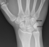
Label carpal bones


What are the structures shown?
What structure attach to these points?

The flexor retinaculum attaches to the hook of hamate and pisiform on the ulnar side. It attaches to trapezium and scaphoid on the radial side.
What is the structure shown and what is its relevance?

- Dorsal tubercle of radius (also known as Lister’s tubercle).
- It separates extensor compartments 2 and 3, separating the tendons of extensor carpi radialis longus and brevis and extensor pollicis longus.
- Dorsal tubercle of radius acts as a pulley for EPL tendon.

What forms the wrist joint?
What type of joint is the wrist joint?
- The radiocarpal (or wrist joint) is the articulation of the distal radius with the scaphoid, lunate and triquetrum
- Wrist joint = condyloid synovial joint
- Conyloid synovial joint is where an ovoid articular surface is received into an elliptical cavity.
- it permits flexion, extension, abduction and adduction.
What separates the distal ulna from the proximal carpal bones?
What supports the wrist joint?
- Distal ulna is separated from the proximal carpal bones by a fibrocartilaginous disc.
-
The wrist joint is supported by a
- Medial collateral ligament –> from ulnar styloid to triquetrum
- lateral collateral ligament –> from the radial styloid to the scaphoid bone

What would pain in the anatomical snuffbox indicate?
What would you be particularly worried about?
- Pain in the anatomical snuffbox indicates scaphoid bone fracture
- Scaphoid is the most commonly fractured bone in the hand is often fractures by a FOOSH.
- characteristic feature is pain and tenderness in anatomical snuffbox
- Scaphoid is at risk of avascular necrosis due to retrograde blood supply which enters distally and travels proximally
- fracture in the middle of scaphoid puts the proximal part of the scaphoid at risk of necrosis, increasing risk of development of osteoarthritis in patients with missed scaphoid fractures.

What is a bennet’s fracture?
A bennets fracture is a fracture thorugh the first metacarpal base (base of the thumb) resulting from forced abduction of the first metacarpal.
Defined as intra-articular two part fracture of base of 1st metacarpal bone.

What is a colles fracture?
What is a smiths fracture?
- Colles and smith fractures are fractures of the distal radius. Commonly caused by FOOSH.
- Colles fracture = extraarticular fracture of the distal radius whereby radial fragment displaced dorsally/ posteriorly.
- Colles fracture is most common type of wrist fracture (90%) and common with osteoporotic bones when wrist is dorsiflexed.
- Smiths fracture = fracture of distal radius where bone is displaced anteriorly.

Describe the borders of the anatomical snuffbox
what structures pass over/ through?
what would tenderness indicate?
- Medial border formed by tendon of extensor pollicis longus
- lateral border formed by two tendons 1) abductor pollicis longus 2) extensor pollicis brevis
- Floor = scaphoif and trapezium
- proximal border = styloid process of radius
Contents:
- Radial artery passes under the two lateral tendons and travels via the snuff box to enter the dorsum of the hand
- Radial artery pulsation can be felt in the snuffbox
- Cephalic vein (houseman’s vein) passes superficially in the snuffbox.
- Cephalic vein is commonly cannulated and runs with superficial (cutaneous) branch of radial nerve and superficial to radial artery.
- Tenderness in the snuffbox can indicate a scaphoid fracture

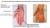
What tough fibrous structure covers the palm of the hand?
What is its function?
Palmar fascia covers the centre of the hand forming a palmar aponeurosis which is continuous with the palmaris longus tnedon and flexor retinaculum
Palmar aponeurosis protects underlying muscle compartments and fans out distally into 4 fibrous digital sheaths.
Fibrous digital sheaths cover the synovial sheaths which contain the flexor tendons in the digits, preventing bowstringing. This ensures movement is performed efficiently.
Palmar aponeurosis also binds and secures superficial skin allowing grip.

What is dupytren’s contracture?
What should it not be confused with?
- Dupytrens contracture is passive flexion of one or more of the medial digits and and inability to fully extend the affected digits
- due to thickening and fibrosis of the palmar aponeurosis
- often treated surgically to alleviate the tension
- Do not confuse dupytrens contracture with stenosing tenosynovitis or ulnar claw hand.

What is stenosing tenosynovitis?
What is ulnar claw hand, how does it present and why?
- Stenosing tenosynovitis also known as trigger finger is painful inflammation and progressive restriction of the tendon sheaths of the fingers.
- Results in thickening of the tendon sheath distal to the pulley leading to painful digital base, limitation of finger movements
- Ulnar claw hand = Due to damage to the ulnar nerve at the wrist.
- Ulnar nerve innervates the intrinsic muscle of the hand and the medial two lumbricals (digits 4/5). Lumbricals flex MCP and extend the PIP and DIP.
- loss of ulnar innervation leads to “ulnar claw” appearance with unopposed extension of MCP and flexion of PIP and DIP of digits 4/5. (remember lumbricals 2/3 are innervated by median nerve).
- Do not confuse ulnar claw with hand of benediction. Hand of benediction due to damage to the median nerve at the elbow, loss of innervation of the flexors to the wrist and digits and thenar muscles. Except ulnar innervates flexor carpi ulnaris and digits 4/5 of digitorum profundus. Therefore when patient attempts to make a fist only digits 4 and 5 can flex, appearance similar to ulnar claw.

What is the golden rule of the innervation of the hand?
- Everything is Ulnar (C8 and T1 supplied) except:
- Thenar muscles
- Lumbricals to digits 2 and 3 (lateral lumbricals).
What do the hypothenar and thenar eminences both contain?
- Hypothenar and thenar eminences both contain:
- a flexor
- abductor
- opposer
What muscles form the thenar eminence?
What are their actions?
Innervation?
-
Opponens pollicis –> largest and lies underneath the other two. Originates of the trapezium and flexor retinaculum, inserts onto base of 1st metacarpal.
- Opposes the thumb by medially rotating and flexing
- Innervation median nerve
-
Abductor pollicis brevis –> Originates from scaphoid and trapezium tubercles and flexor retinaculum, inserts lateral side proximal phalanx
- abducts thumb
- median nerve
-
Flexor pollicis brevis –> originates from tubercle of trapezium and flexor retinaculum, attaches to base of proximal phalanx of thumb
- flexes MCP of thumb
- median nerve

What muscles form the hypothenar eminence?
Action and innervations?
- Opponens digiti minimi lies deep to other hypothenar muscles
- originates from hook of hamate and flexor retinaculum, inserts onto medial margin metacarpal v
- rotates little finger towards palm producing opposition
- ulnar nerve innervated
- Abductor digiti minimi
- most superficial of hypothenar muscles
- originates pisiform and tendon of FCU attaches to base of proximal phalanx of little finger
- abducts little finger
- ulnar innervated
- Flexor digiti minimi brevis
- lies laterally to abductor digiti minimi
- originates hook of hamate/ flexor retinaculum inserts into base of proximal phalanx of little finger
- flexes MCP of little finger
- ulnar nerve

What is the action and innervation of the lumbricals?
- The lumbricals flex the MCP joints and extend the PIP and DIP joints
- They originate from the tendons of flexor digitorum profundus and pass dorsally and laterally to insert into the extensor hood
- The lateral two lumbricals / radial lumbricals are innervated by median nerve
- the medial two lumbricals/ ulnar lumbricals are innervated by the ulnar nerve

What are interossei?
What groups are there?
What are their actions?
what is the innervation?
- located between the metacarpals, dorsal and palmar interossei.
- Palmar interossei adduct (PAD) and dorsal interossei abduct (DAB).
- interossei also assist in flexion of MCP and extension of PIP/DIP joints
- 3 palmar interossei and 4 dorsal interossei
- Both palmar and dorsal interossei innervated by ulnar nerve
- Abduction and adduction of the fingers tests the ulnar nerve

What is froment sign?
- Froment sign indicates weakness in adduction of the thumb, loss of adductor pollicis
- Adductor pollicis innervated by the ulnar nerve
- Froments sign is when there is excess thumb flexion whilst pinching between thumb and index finger.
- patient asked to hold paper between thumb and index finger, should be able to hold it with no difficulty
- positive test is when patient unable to adduct thumb and instead flex the thumb at the IP joint to try and hold the paper.

Why test thumb abduction?
- Thumb abduction done mainly via abductor pollicis brevis which is median nerve innervated.
- Loss of ability to abduct the thumb may indicate median nerve damage or carpal tunnel syndrome.
What covers the tendons of the hand?
What is the clinical relevance?
- Tendons of the hand are covered by both a synovial and fibrous sheath.
- tendons of the flexor digitorum superficialis and profundus cross the palm and enter the fibrous sheaths on palmar aspect of digits
- Fibrous sheaths prevent bowing of the tendons when digits are flexed.
- synovial sheath of thumb and little finger are continuous with the sheaths associated with the tendons in the carpal tunnel.
- Common flexor sheath (ulnar bursa) extends distally along 5th digit and through the carpal tunnel into the proximal forearm.
-
Clinical relevance:
- Sheaths can be involved in infection spread
- ganglion cyst formation
- tenosynovitis (inflammation of tendon and sheath)

What spaces are there within the hand?
Clinical relevance of these spaces?
- The thenar and midpalmar spaces are between the deep flexor tendons and metacarpals/ interossei in the palm
- Clinical relevance: Deep space infections
- Thenar space located between deep flexors and adductor pollicis
- Midpalmar space between flexor tendons and 3/4/5th metacarpal.
- often by penetrating trauma
- Will have pain and swelling