Bone structure/formation/growth/ repair Flashcards
What type of tissue is bone?
what are the general roles of this tissue type?
- Bone is a specialised form of connective tissue
- Connective tissue has 5 main roles:
- Structural support –> underlies epithelia, encapsulates organs, forms bone/cartilage/ligaments/tendons
- Metabolic support –> forms blood vessels
- Cell adhesion
- Exchange –> signalling molecules, nutrients, waste
- Defense, protection and repair –> physical barrier to microorganisms, site of immune cell action, repair by fibroblasts

Describe the composition of connective tissue
Describe the properties of some of these constituents and what this allows
- Connective tissue made up of cells and ECM
- Cells =
- fibroblasts –> secrete ECM
- Immune cells
- cartilage cells –> chondrocytes/blasts
- bone cells –> osteoclasts/ blasts
- ECM made up of Fibrous proteins (collagen and elastin) and Ground substance
- Ground substance made up of:
- 90% water
- proteoglycan –> GAG bound to protein, highly hydrophobic and hydrated, resist compressive forces
- glycoprotein –> allows cells to adhere to ECM

Describe the two key features of bone
- Rigidity –> ability to resist forces and shape change, provided by the crystalised ECM formed of hydroxyapatite (Ca10 (PO4) 6 (OH)2)
- Resilience –> ability to absorb forces w/out breaking and return to original shape, provided by type 1 collagen fibres
IMPORTANT –> need a balance between rigidity and resilience

What are the 5 key functions of bone?
- Movement, support, protection
- mineral homestasis
- site of haematopoesis

What is the periosteum?
What layers are it formed from?
- Periosteum = non calcified dense irregular connective tissue layer covering bone where tendons and ligaments insert
- Formed of outer layer and inner cellular layer (osteoprogenitors and osteoblasts).
- absent on cartilage covered surfaces and sesamoid bones

What are the functions of periosteum?
- Forms attachment point for ligaments and tendons
- helps bones grow in thickness as contains osteoprogenitors/ blasts
- helps noursih and protect bone
Label the parts of the bone


What are the two types of bone?
How does their microscopic appearance differ?
- Woven bone = immature/ primary bone
- 1st bone formed at any site
- collagen fibres arranged randomly
- occur at sites of fracture healing
- Lamellar bone = mature bone
- collagen fibres remodelled into orderly arrangement –> STRENGTH

Describe bone structure –> the two types of tissue arrangement of lamellar bone
- Outer cortical structure –> compact, dense, strong and heavy
- Inner trabecular structure (Also known as cancellous/ porous bone) with beams and struts orientated in line with stress
- Spaces inbetween filled with bone marrow

Describe the microstructure of cortical bone
- Cortical bone formed of lamellae
- Lamellae = bony plates made up of collagen fibres arranged in parallel
- outer circumferential lamellae –> surrounds inner Haversian systems = osteons
- Haversian systems/ osteons formed of concentric lamellae around a central canal/ haversian canal:
- haversian canal contains blood/ lymph/ nerves
- connected to each other via Volkmann canals
- Haversian systems/ osteons joined via interstitial lamellae
- Inner layer of cortical bone lined with inner circumferential lamellae.

What is a volkmann canal?
- Volkmann canals run transversely or obliquely inbetween haversian canals, allowing communication between haversian canals, the periosteum and marrow cavity.

What is shown in these images?
Describe the arrangement

- Haversian canals/ osteons
- Collagen fibres in each concentric lamallae layer are in parallel with each other but at right angles to the fibres in the next layer
- Black dots = bone cells within the lamallae
Describe the microstructure of lamellar bone- Trabecular
- beams and struts orientated in line with stress forces
- large areas intercommunicating spaces (marrow spaces) for haematopoesis

Describe the neurovascular supply to bone
- Central main supply via nutrient artery
- Ends supplied by epiphyseal arteries
- supported by periosteal and metaphyseal arteries

Describe where bone cells come from
Their pathway from one cell type to another
what is secreted and forms bone matrix
- Bone cells come from mesenchymal cells
- Mesenchymal cells give rise to osteoprogenitor cells –> osteoblasts –> osteocytes
- Osteoblast cells line the inner surface of bone and secrete organic bone matrix (osteoid) –> containing type 1 collagen, proteoglycans, glycoproteins.
- Also contains enzyme alkaline phosphatase that secretes hydroxyapatite, osteocalcin (binds Ca2+) and osteopontin –> mineralisation of ECM.
- As they become surrounded by bone matrix osteoblasts become osteocytes

What are periosteal layers of bone made up of?
Why is this important for function?
Periosteal layer of bone is made up of resting osteoblasts which can divide in the event of fracture, important for fracture healing.

What are osteocytes formed from?
What is their role?
Where are they found?
How do they communicate?
- Osteocytes are formed from osteoblasts after they secrete bone matrix
- Osteocytes are mature, non dividing cells that occupy lacunae surrounded by bone matrix
- Communicate with each other via dendritic processes that pass through canaliculi (Radiating from central lacunae) and anastomose with other dendritic processes
- Dendritic processes communicate via gap junctions which allows the sharing of nutrients and ions
- Main role in mechanotransduction, matrix maintenance and calcium homeostasis.
- Mechanotransduction = detection of forces in bone allow laying down of bone in response to stress

What are osteoclasts? What do they look like
Where are they derived from?
What is their main role?
What do they form?
What other cell do they need to be in balance with?
- Osteoclasts are bone resorbing cells, large multinucleated cells with ruffled border
- They are derived via macrophage/ monocyte pathway (haematopoetic pathway)
- Main role –> resorption of bone matrix, secrete enzymes and acid
- involved in bone remodelling, growth and repair
- From howship’s lacunae = resorption craters
- Osteoclasts must be in balance with osteoblasts in bone remodelling

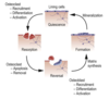
What is bone remodelling? Talk about the 4 Phases
What is coupled during this process?
- Bone remodelling = continual process throughout life that occurs in response to changing mechanical stress or microfractures of bone
Phases:
1) Quiescence (beginning of OC: Recuritment, Differentiation & Acitvity)
2) Resorption (beginning of OC: Apoptosis & Removal)
3) Reversal (beginning of OB: Recuritment, Differentiation & Activity –> Matrix synthesis)
4) Formation- Matrix mineralisation
* Coupling of bone resorption and formation
Describe how cortical bone is remodelled
- Note only cortical bone has lamellae arrange in haversian systems, trabecular bone has lamellae in struts and rungs.
- Cortical bone can be remodelled by osteoclasts that form cone shaped tunnels through the bone
- These tunnels become occupied by blood vessels, osteoprogenitors and osteoblasts that secrete bone matrix in concentric lamellae around the blood vessels
- Forming new haversian systems

Describe the signalling molecules involved in the regulation of bone resorption / osteoclast differentiation and how hormones are linked to this.
- Osteoclasts differentiate and activate under the control of various signalling molecules secreted by osteoblasts:
- RANKL (receptor activator nuclear kappa B ligand)
- RANKL binds to receptor RANK on osteoclast surface –> activation –> bone resorption
- Osteoprotegerin –> acts as a decoy receptor for RANKL
- Balance of osteoprotegerin and RANKL determines resoprtion/ formation
- Under control of other hormones e.g. Oestrogen favours osteoprotegerin and bone formation (declines in menopause -> osteoporosis) whereas PTH and calcitriol favours RANKL and resorption.

What are the two routes of bone formation?
- Intramembranous ossification –> where mesenchyme differentiates directly into bone cells
- Endochondral ossification –> where mesenchyme becomes cartilage, cartilage –> bone
Note: mesenchyme = undifferentiated embryonic connective tissue

Where does intramembranous ossification occur?
- Occurs in flat bones of the skull, clavicle, mandible
- Direct replacement of mesenchyme with bone tissue, no cartialge precursor

Describe the steps in intramembranous ossification
- Region of mesenchyme differentiates directly into osteoprogenitor cells –> osteoblasts –> secretes osteoid –> primary ossification centre
- invasion of blood vessels
- Establishment of sponge like trabecular bone and vascular CT transformed –> bone marrow
- Mesenchyme on the outside differentiates into fibrous periosteum and bone cells form inner cellular layer
- Compact bone deep to periosteum forms and trabecular bone forms in between
- Occipital bone = large bone with several ossification centres that fuse


