Gluteal region, hip and thigh Flashcards
What are the 4 regions of the lower limb?
- Gluteal region
- Thigh region -> from hip joint to knee joint
- Leg region -> leg joint to the ankle joint
- Foot region -> distal to ankle joint

What does the lower limb require? How does this compare to the upper limb?
What are the 4 main functions of the lower limb?
- The lower limb requires strength and stability in preference to range of movement
- Lower limb –> low mobility, high stability
- The upper limb –> High mobility, less stability
4 main functions of lower limb:
- Support body weight
- Maintain upright posture
- Locomotion (gait)
- Accomodate shock loading

What 4 phases are there of walking?
What is the last thing to leave the floor before you enter swing phase of walking?
- Heel strike
- Support
- Toe off
- Carry through / swing phase
- Last thing to leave the ground before the swing phase is your Hallux (big toe), able to propel whole body forward.

What shape is the pelvic girdle?
What forms it?
What does it allow?
What can cause fracture?
- Pelvic girdle is a basin shaped rigid ring of bone that is partly formed by the sacrum (axial skeleton) and two pelvic bones (appendicular skeleton).
- Enables distribution of weight of the axial body to the lower limbs
- Enables locomotion and standing
- Fracture of the pelvic girdle can be caused by HIGH FORCE TRAUMA
- Force transmission mostly via compression

What type of joint forms the hip joint?
How does this differ to the shoulder joint?
What is required to stabilise it?
What type of joint exists at the knee and ankle?
- Ball and socket joint forms the hip joint, formed by the head of the femur and the acetabulum
- Acetabulum forms a deep depression, hence higher stability than upper limb.
- However we require soft tissues, ligaments and muscles to support the deep ball and socket joint, without these we would have no ability to walk or shock absorb.
- Knee joint = modified hinge joint, allows some rotation to allow locking of the knee during standing = stability
- Ankle joint = synovial hinge joint formed between tibia, fibula and talus

In which direction is the hip dislocated in each of the following pictures?
What signs would a patient show with each type of dislocation?

- Left hand image = posterior dislocation as the femur head has gone backwards
- Posterior dislocation displays with:
- limb shortening on the side of the dislocation
- hip flexion
- internal rotation (hip flexors pull femur into flexed and internal rotated position).
- Right hand image = anterior dislocation as femur head has protruded forwards
- Anterior dislocation displays with:
- Abudction
- external rotation (lateral rotators pull femur into outwards position).

What joints exist between the sacrum and the hip bones?
What happens on injury to sacroiliac joints?
What joint joins the two pelvic bones anteriorly?
What type of joint is this?
- Sacroiliac joints exist between hip bones and sacrum
- Sacroiliac joints are synovial joints = bones joined together by fibrous joint capsule continuous with periosteum of bones
- Fibrous joint with a lot of ligament reinforcement
- injury to sacroiliac joints –> equisite pain, radiates up back, into gluteal and perineal region due to innervation of sacroiliac joints via same nerve
- Anteriorly joined by pubic symphysis, fibrocartilaginous joint and a secondary cartilaginous joint
- Secondary cartilaginous joint = fibrocartilaginous and hyaline joints usually occurring in the midline (eg. intervertebral discs).

What is the pelvic inlet?
What is the pelvic outlet?
- Pelvic inlet marks boundary between greater and lesser pelvis
- Markerd posteriorly by the sacral wings, laterally by the arcuate line of the ilium, anteiorly by pubic symphysis
- Pelvic inlet determines size and shape of the birth canal
- Pelvic outlet located at end of lesser pelvis bordered posteriroly by tip of coccyx, laterally by ischial tuberosities, anteriorly by the pubic arch

What two main ligaments support the pelvic girdle?
What do these two ligaments form?
- Sacrospinal ligament –> from the sacrum to the spine of the ischium (more superficial)
- Sacrotuberous ligament –> from the sacrum to the ischial tuberosity
The sacrospinal and sacrotuberous ligaments form the greater and lesser sciatic foramen.
What splits the greate sciatic foramen into two parts?
What does the greater sciatic foramen communicate with?
What does the lesser sciatic foramen communicate with?
- Piriformis muscle (powerful lateral rotator of hip) splits the greater sciatic foramen into an upper and lower portion
- Greater sciatic foramen connects the pelvic cavity to the gluteal region
- Lesser sciatic foramen connects the perineal region to the gluteal region

What 3 bones form the hip bone?
Where do they unite? When does full fusion occur?
- Ilium, ischium and the pubic bones form the hip bones
- They unite at the acetabulum, prior to puberty they are separated by the triradiate cartilage
- Fusion begins age 15-17, only complete by young adulthood
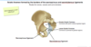
Label the image
How is the pelvic girdle angled?

- Pelvic girdle angled forwards such that the ASIS and pubic tubercles should be in alignment in a vertical plane.

Label the image and highlight the importance/ functions of each component part of the femur

Left = anterior view of the L femur:
- Head –> articulates with the acetabulum forming ball and socket joint
- Neck –> clinically relevant, most likely region of fracture in osteoporitic/ penia patients when they fall, inherently weaker, region of most stress
- Greater trochanter –> attachment point for gluteus medius and gluteus minimus, stabilises the pelvis when walking
- Adductor tubercle –> attachment point of adductor muscles, palpable
Right = posterior view of L femur
- Lesser trochanter –> Attachment point for iliopsoas muscle, highly important hip flexor, allows us to walk and get up from sitting. Formed by iliacus and psoas muscles, extends from lumbar vertebrae, ilium and inserts onto femur.
- Linea aspera --> rige of bone formed by attachment of many powerful thigh muscles, and 3 intermuscular septa.
- Supracondylar region and condyles –> J shaped regions for the knee, articulates with tibia

What is wolff’s law?
Wolff’s law = Bone is deposited and reabsorbed in accordance with the stressed placed upon it (remodels with mechanical stress).
Less stress = less bone
Remember bone = living tissue relatively flexible under stress

What helps stabilise the hip joint?
- Hip joint is designed so that the acetabulum directly presses down onto the femoral head, which creates a more stable joint
- Hip joint also supported by muscles, ligaments, bone shape and the acetabular labrum

What is the acetabular labrum?
What can produce pain in this region?
- Acetabular labrum is a horseshoe shaped ring of fibrocartilage that surrounds the acetabulum.
- It increases the depth of the joint and the surface and strength of the hip joint
- The acetabular labrum is closely apposed between the acetabulum and head of the femur and is both innervated and has its own blood supply
- Acetabular labrum can become impinged, trapped between the two surfaces of femoral head and acetabulum

Describe the membranes that cover the hip joint
- Synovial membrane of the hip attaches to the margins of the articular surfaces of the femur and the acetabulum
- This synovial membrane is covered by a fibrous membrane
- Synovial membrane and fibrous membrane keep the ball and socket joint a unit

What three ligaments further support the hip joint?
- Outside of the synovial and fibrous membrane 3 ligaments reinforce the external surface of the fibrous membrane and stabilise the joint
- Iliofemoral –> from ilium the femoral head
- Pubofemoral –> from pubic ramus to femoral head
- Ischiofemoral –> from the ischium to the femoral head
- All of these ligaments are twisted, amd are tightest upon standing and moving
- Loosest when sitting, the ligaments become lax and untwisted –> highest risk of posterior hip dislocation

Describe the blood supply to the femoral head:
What type of fracture increases the risk of avascular necrosis of the femoral head?
- The common iliac artery splits into an internal iliac (supplies pelvic region) and external iliac which becomes the femoral artery after it passes the inguinal ligament
- The femoral artery gives off deep femoral branch laterally
- This deep femoral branch gives off two circumflex arteries, the medial and lateral femoral circumflex arteries
- These circumflex arteries give off retinacular arteries which go on to supply the joint capsule and femoral head
- Intracapsular fractures are fractures occuring within the joint capsule, cuts off supply of the circumflex arteries and therefore increases your risk of avascular necrosis (medical emergency! Risk of loss femur head).
- Extracapsular fractures are fractures occuring outside the joint capsule, the circumflex arteries branch off the deep femoral artery proximal to the fracture and are still able to supply the femoral head.

What ligament exists between the femoral head and the acetabulum?
- Ligamentum teres/ ligament of head of femur attaches femur head to acetabulum
- offers joint support and has a tiny artery within it that supplies small region of the articular surface
- It is not big enough to provide blood supply to the femoral head if the circumflex branches are lost. (intracapusular fracture).

What is shenton’s line? Why is it clinically useful?
Is this X-ray from a female or male? How can you tell?

- Shenton’s line is a smooth curved, continous arched line that can be drawn from the femoral head to the obturator foramen
- Clinically useful as it can help detect subtle hip fractures without displacement
- X-ray is from a female, wider pelvic inlet, wider angle of the ilium, wider subpubic angle, soft tissue shadow shows mons pubis (lacking male genitalia).

Is this X-ray from a male or female patient?
Is shenton’s line normal?

- X-ray is from a male patient, narrow pelvic inlet, higher arch and more angled ilium, acute subpubic angle, soft tissue shadow of male external genitalia
- Shentons line is normal

Which fracture is extracapsular and which is intracapsular?
Which is at greatest risk of avascular necrosis of femoral head?

- Left hand image shows extracapsular fracture, running between greater and lesser trochanter (intratrochanteric fracture). Femoral circumflex arteries are more proximal to the fracture therefore still able to supply femoral head
- Right hand image shows intracapsular fracture of the R femoral neck, will cut off circumflex arteries, greater risk of avascular necrosis. –> medical emergency, attend quickly to prevent loss of femoral head

What are the two main terms used to describe deformity in joints?
Which direction are these deformities?
If this were to occur in the hip, what would the angle be and where would the knee point?
- Valgus = distal part of the limb is directed AWAY from the midline
- Varus = distal part of the limb is directed TOWARDS the midline
- In the hip a valgus deformity would mean the angle of the femoral neck is larger than 130 degrees, the knees are moved away from the midline
- A varus deformity of the hip would mean the angle of the femoral neck is less than 130 degrees and the knees would be turned inwards (knock knees).


