Week 2 Flashcards

1
Q

What are anaesthetics?

A

Drugs that cause loss of consciousness, usually with loss of reflexes and some alagesia

2
Q

What are analgesics?

A

Drugs that relieve pain

3
Q

What are psychotropics?

A

Drugs that affect mood and/or behaviour

4
Q

What are anxiolytics/ hypnotics/ sedatives/ minor tranquilisers?

A

Drugs that relieve anxiety and reduce excitability. In higher doses may induce sleep.

5
Q

What are major tranquilisers/ antipsychotics/ neuroleptics?

A

Drugs used in humans for the treatment of schizophrenia, in animals used to produce profound sedation

6
Q

What are antidepressants?

A

Drugs that alleviate the symptoms of depressive illness in humans and are used in veterinary medicine to treat compulsive behavioural disorders

7
Q

Where do virtually all drugs act in the CNS?

A

Virtually all drugs that act in the CNS produce their effects by modifying some step in chemical synaptic transmission

  1. action potential in presynaptic fibre
  2. Synthesis of transmitter
  3. Storage of transmitter
  4. Metabolism (breakdown) of transmitter
  5. Release of transmitter
  6. Reuptake of transmitter
  7. Degradation of transmitter
  8. Binding of transmitter to receptor
  9. Receptor induced increase or decrease in ionic conductance
8
Q

What are the chemical mediators in the CNS? What kind of effects can they produce?

A
  1. Neurotransmitters- released by presynaptic terminals and produce rapid excitatory response in post synaptic neurons
  2. Neuromodulators- released by neurons, and produce slower pre- or post- synaptic responses, mediated mainly by G-protein coupled receptors
  3. Neurotrophic factors- released mainly by non-neuronal cells, act on tyrosine-kinase-linked receptors that regulate gene expression

** chemical mediators in the brain have long slow lasting effects, can act diffusely at considerable distances from the site of release and can have diverse effect on transmitter synthesis, on expression of neurotransmitter receptors and on ionic conductance

9
Q

What are some fast neurotransmitters? What do they operate through?

A

Glutamate, glycine, GABA, ACh– that operate through ligand gated ion channels

* Speed depends on the receptor that it binds to. E.g. the same agent may act through both ligand gated and GPCR like glutamate or ACh

10
Q

What are the slow neurotransmitters and neuromodulators? What do they operate through? What ultimately causes a transmitter to be fast or slow?

A

Dopamine, 5HT, ACh, neuropeptides– operate through G-protein-coupled receptors.

* Speed depends on the receptor that it binds to. E.g. the same agent may act through both ligand gated and GPCR like glutamate or ACh

11
Q

What is an example of an amino acid neurotransmitters that are excitatory?

A

Glutamate, the principal fast “classical” excitatory transmitter and is widespread through the CNS. Several different glutamate receptors have been characterized according to particular agonists that bind to them e.g. NMDA, AMPA, kainate

12
Q

What are three examples of an amino acid neurotransmitters that are inhibitory?

A

Gamma-amino butyric acid (GABA) is a major inhibitory neurotrasmitter in the CNS. GABA receptors are of two main types GABA (A) receptor is ligand gated to Cl- ion channels and is the site of action of many neuroactive drugs e.g. barbituates, benzodiazepines, steroid anaesthetics GABA (B) receptor is G-protein-coupled receptor, coupled to biochemical processes and regulation of ion channels

* Glycine is an inhibitory transmitter acting mainly in the spinal cord. Strychnine is a competitive glycine antagonist.

* Acetyl choline- ACh is widely distributed in the brain. Both nicotinic and muscarinic receptors occur in the CNS. Muscarinic receptors mediate the main behavioural effects associated with ACh- arousal level, learning and short term memory

13
Q
A
14
Q

What are three examples of monoamines?

A

* Dopamine- neurotransmitter as well as being a precursor of NA. But distribution of dopamine in the brain is very different from NA. There are two main families of dopamine receptors- D1 and D2. D2 receptors are implicated in the pathophysiology of Parkinson’s disease and schizophrenia.

* Noradrenaline- Pathways for NA neurotransmission in the CNS are essentially the same as in the PNS. Adrenergic receptors in the CNS are alpha1, alpha2, beta. NA is important in the “arousal” system, controlling wakefulness, blood pressure regulation, control of mood. Drugs acting on noradrenergic transmission in the CNS include alpha 2 agonists, antidepressants, cocaine, and amphetamine.

* 5-hydroxytryptamine (serotonin)- an important CNS transmitter with complex and varied effects. 5-HT can exert excitatory or inhibitory effects, acting presynaptically or post synaptically. 5-HT pathways are involved in physiological and behavioural functions, namely hallucinations and behaviour changes, sleep, wakefulness, and mood, control of sensory transmission. A variety of new drugs influence 5-HT pathways in selective ways: for example selective serotonin reuptake inhibitors (SSRIs), ondansetron (an anti-emetic) 5-HT3 receptor antagonist, buspirone (anxiolytic) 5-HT1a receptor agonist.

15
Q

What are four examples of non adrenergic non cholinergic (NANC) transmitters?

A

* Histamine- fulfills the criteria of CNS neurotransmitter. H1, H2, H3 receptors are widespread in the brain. Many of the functions are unclear, but H1 receptor antagonists are strongly sedative and anti-emetic.

* Purines- (ATP and adenosine)

*nitric oxide

*arachidonic acid metabolites

^all have established roles as CNS transmitters and modulators.

16
Q

What are neuropeptides? Three examples?

A

* Many neuropeptides that act on specific CNS receptors.

EX: Opioid peptides that modulate pain pathways, substance P, neuropeptide Y

17
Q

Why is it difficult to predict the therapeutic effect of a particular pharmacological agent?

A

Because of the complexity of neuronal interconnections in the CNS and because of secondary adaptive responses. For example, an increase in transmitter release may lead to a decrease in transmitter synthesis, an increase in transporter expression, or in decreased receptor expression.

18
Q

What are the classes of analgesics used in veterinary medicine?

A

* local anaesthetics

* NSAIDs

* Opioid analgesics

* Centrally acting non-opioid analgesics

* alpha 2 adrenergic agonists

19
Q

In nociceptive afferent neurons in a peripheral sensory nerve, what are the two possible fibre types?

A

* C-fibres that are non-myelinated, have a low conduction velocity (< 1 m/sec) and sense dull pain.

* A fibres that are fine myelinated and have faster conduction velocities (6-30 m/sec). Sense sharp, localized pain. Terminate in the dorsal horn of the spinal cord.

20
Q

What is the gate control mechanism in the dorsal horn?

A

This centre modulates pain transmission. Inhibitory interneurons in teh substantia gleatinosa of the dorsal horn act to inhibit the transmission pathway.

* Inhibitory interneurons are activated by descending pathways from the mid-brain and brainstem, as well as non-nociceptive afferent input.

* Inhibitory interneurons are inhibited by persistent C-fibre activity, hence “wind up” phenomenon- increasing duration of stimulation leads to increased transmission of pain signals.

21
Q

What is the “wind up” phenomenon in regards to pain signals?

A

* Inhibitory interneurons are inhibited by persistent C-fibre activity, hence “wind up” phenomenon- increasing duration of stimulation leads to increased transmission of pain signals.

22
Q

What are the two parts of the brain involved in pain modulation?

A

*Mid-brain/ pons “peri aqueductal grey area”

* Lower pons/ medulla “nucleus raphe magnus”

* initiate descending pathways that exert a strong inhibitory effect on the dorsal horn. This descending inhibition is mediated by enkephalins, 5-HT, noradrenaline, and adenosine.

23
Q

What are the major inhibitory neurotransmitters in nociceptive pathways?

A

* Endogenous opioids

There are more than 20 endogenous opioids, the most important of which are beta- endorphin, enkerphalins, and dynorphin

24
Q

What are the three types of opioid receptors?

A

1) mu- receptors: two types mu-1 mediate most analgesic effects, euphoria supraspinal; enkephalins are enodgenous mu 1 ligands. mu-2 mediate most of the undesirable side effects such as respiratory depression and constipation
2) delta receptors: are important in the periphery where they contribute to analgesia
3) kappa receptors: mediated analgesia primarily at the spinal cord level and have fewer side effects- less respiratory depression, miosis, etc. They tend to cause sedation and dysphoria rather than euphoria.

25
Q
A
26
Q

What is an opioid? What is an endogenous opioid? What is the first synthetic opioid?

A

* An opioid is any substance whether endogenous or synthetic that produces morphine like effects that are blocked by antagonists such as naloxone. They vary in their receptor specificity and their efficacy at different types of receptors.

* endogenous (aka opioid peptides): endorphins (bind to opioid receptors)

* first exogenous opioid derived from the resin of the opium poppy- Opiates

27
Q

What do pure agonists have affinity for?

A

Mu receptors

This group includes most of the morphine like drugs e.g. morphine, pethidine, fentanyl.

Pure agonists may be strong (morphine, heroin, fentanyl, etorphine) or mild/ moderate (codeine, pethidine)

28
Q

What receptor affinity do mixed agonist- antagonists and partial agonists have?

A

Combine agonist effect on one receptor subtype with antagonist activity on another receptor type.

For example, butorphanol- is an antagonist at the mu-receptor, and an agonist at the kappa-receptor. Pentazocine is a weak mu antagonist and kappa agonist buprenorphine a partial mu receptor agonist (it has high affinity for the receptor, but only partial activity)

29
Q

What effect do opioid antagonists produce?

A

Produce very little effect when given on their own but block the effects of opioids. They are used to treat overdoses to opioids.

30
Q

What are the cellular actions of opioids?

A

Opioid receptors are linked to G-proteins. On neurons opioids have two different actions:

* Close voltage gated Ca++ channels on presynaptic terminals, and thereby reduce transmitter release

* Open K+ channels on post synaptic neurons, thereby causing hyperpolarization and inhibition of action potentials in post synaptic neuron.

31
Q

What are the sites of action of opioid drugs?

A

* Dorsal horn of spinal cord, so powerful analgesic effect direct on spinal cord

* supraspinal sites- pain modulating descending pathways opioids directly inhibit neurons

* opioids activate neurons that inhibit pain transmission

* Exogenous opioids stimulate release of endogenous opioid peptides that have actions at and receptors

* Peripheral opioid action- at sites outside the CNS, particular in pain associated with inflammation

32
Q

What are the pharmacological actions of opioids on the CNS?

A

* analgesia (mu mediated) reduces nociception at periaqueductal grey level at spinal level; reduces stress at limbic system

* euphoria (mu mediated) contentment and well being; balanced by kappa mediated dysphoria

* excitement, constant motor response, and convulsions. Seen more often in domestic animals particularly horses and cats, than in humans. Presumed mu mediated as seen with morphine and pethidine but not agonist-antagonists such as butorphanol.

* respiratory depression (mu-mediated) decreased sensitivity of respiratory centre to pCO2 that occurs at therapeutic doses

* depression of coughing reflex, mechanism uncertain. Codeine gives cough suppression in subanalgesic doses

* Nausea and vomiting (mu mediated) through activation at chemoreceptor trigger zone

* Pupillary constriction- centrally mediated via oculomotor nucleus. Important emergency diagnostic in humans

* Marked species variability

33
Q

What are the pharmacological action of opioids on the GI tract? And any other actions aside from CNS?

A

* Increased tone and reduced motility (mu, kappa, and delta mediated) leading to constipation, delay in gastric emptying, mediated mainly intramurally and partly centrally

* morphine stimulates release of histamine from mast cells, which can cause urticaria, bronchoconstriction (risk for asthmatics), hypotension

* Bradycardia and hypotension with high doses through a direct effect on medulla

34
Q

What is Methadone? Why is it necessary at times? What are opioid analgesics classified as?

A

* Methadone is a weak, long acting mu receptor agonist used to relieve withdrawal symptoms

* Tolerance develops rapidly along with physical withdrawal syndrome. Some opioid analgesics are much less likely to cause dependence- codeine, buprenorphine, butorphanol.

* Because of their dependence characteristics all opioid analgesics are Schedule 8 poisons

35
Q

What are the pharmacokinetics of opioids?

A

* Generally administered by injection in domestic animals

* They are metabolized by the liver, by conjugation with glucuronide- these glucuronides may be pharmacologically active

* The low conjugating capacity of neonates means opioids have much longer duration of action in neonates

36
Q

What are eyes protected by?

A

Adipose tissue, lacrimal glands, and eyelid

37
Q

What is binocular vision?

A

* Occurs where visual field of both eyes overlap

* allows for focus on near objects and for depth perception

* greater in carnivores than in their prey (i.e. herbivores)

monocular vision does occur in some species- this is the field of view does not overlap between the two eyes (most herbivores)

38
Q

What are the three tunics that comprise the eyeball? What else is part of the eyeball?

A
  1. outer fibrous tunic
  2. middle vascular tunic
  3. internval nervous tunic

* lens for focussing light on receptors, partly liquid, parly gelatinous centre

39
Q

What is the fibrous tunic?

A

* Dense collagenous tissue

* Resists internal pressure

* Consists of sclera (posteriorly) and cornea (anteriorly) which meet at limbus

40
Q

What is sclera?

A

* Opaque, penetrated by fibres of optic nerve, continuous with dura mater of optic nerve, provides attachment for tendons of extrinsic muscles of eye

41
Q

What is the cornea?

A

* Specialized transparent, dense connective tissue

* covered by anterior and posterior epithelial layers

* transparency depedent on: highly organized layers of collagen fibres, continuous pumping out of interstitial fluid by posterior epithelium

* avascular- nutrition by diffusion from lacrimal fluid (tears) and aqueous fluid of anterior chamber

* Very sensitive due to nerve endings (branches of ophthalmic division of CN V)

42
Q

What is the limbus?

A

The point at where the sclera and cornea meet

43
Q
A
44
Q

What is the vascular tunic?

A

* Also known as uvea- consists of choroid, ciliary body, and iris (from posterior to anterior)

45
Q

What is the choroid?

A

* Lines sclera from optic nerve almost to limbus

* contains blood vessels in pigmented connective tissue

* provides nutrients for outer layers of retina

* contains avascular tapetum lucidum: area of dorsal choroid, reflective, iridescent, not present in pigs (or humans), probably assists in nocturnal vision

46
Q

What is the ciliary body?

A

* Thickening of choroid anteriorly in radial ridges (ciliary processes)

* Provides anchoring point for zonular fibres, which suspend lens

* Produces aqueous humour

47
Q

What is the iris?

A

* ring of tissue suspended between cornea and lens

* Opening in centre is pupil

* consists of pigmented connective tissue covered with epithelium (pigmented posteriorly) - connective tissue layer contains smooth muscle sphincter (circular- PS) and dilator (radial-symp.), which regulate pupillary size

* Separates anterior from posterior chamber

48
Q

What is the internal tunic?

A

Retina

* contains light sensitive receptor cells (posterior two thirds of retina)

* lines vascular tunic from pupillary margin posteriorly to optic nerve

* thin outer layer (pigmented except over tapetum) and thick inner layer (neuroepithelial)

* neuroepithelial layer (from outer to inner):

  • photoreceptor layer
  • horizontal cells
  • bipolar cells
  • amacrine cells
  • ganglion cells
49
Q
A
50
Q

What are photoreceptor cells?

A

* Contain light sensitive receptors in extended membrane of outer segment

* Two types: rods (highly sensitive for night vision) and cones (colour perception); dogs and horses have fewer rods and limited colour perception compared to humans

* synapse with bipolar cells, which synapse with ganglion cells

51
Q

What are ganglion cells?

A

* nonmyelinated axons extend across inner surface of retina to optic disc where they meet to form the optic nerve i.e. ganglion cells are cell bodies of CN II

52
Q

What are horizontal cells?

A

Modify transmission between photoreceptors and bipolar cells

53
Q

What are amacrine cells?

A

Modify transmission between bipolar cells and ganglion cells

54
Q

Where do blood vessels enter the eyeball?

A

At the optic disc with optic nerve

55
Q

What is the anterior chamber of the eye? What is the posterior chamber?

A

anterior chamber: fluid filled space inside the eye between the iris and the cornea

posterior chamber: narrow space behind the peripheral part of the iris and in front of the suspensory ligament of the lens and the ciliary processes

56
Q

What is the lens?

A

* Soft, transparent, biconvex structure

* Composed of “lens fibres” - epithelial cells running in layers from anterior to posterior pole of lens

* Avascular- nourished by aqueous and vitreous humour

* focuses images on retina

* surrounded by an elastic capsule (basement membrane)

57
Q

What is aqueous humour?

A

* fills space between cornea and lens (anterior and posterior chambers)

* Produced by cells of ciliary body

* Passes from posterior chamber to anterior chamber

* drains into venous sinuses in sclera at iridocorneal angle

58
Q

What is the vitreous body?

A

* Occupies space between lens and retina

* gel like mass consisting of stroma of fine transparent fibres filled with glycosaminoglycans and water

59
Q

What is the adnexa?

A

* Structures that protect and move the eye within the orbit

60
Q

What is the periorbita?

A

* Fibrous fascial sheath, which blends with periosteum medially and dorsally

* inserts in eyelids and surrounds eyeball and extrinsic muscles of the eye

* attached to skull near optic foramen

* contains smooth muscle which keeps eyeball slightly protruded with normal tone (under symp. control)

* contains and surrounded by orbital fat (cushions contents of orbit)

61
Q

What do oblique muscles of the eye do?

A

* rotate eye about visual axis

* contraction of the dorsolateral aspect of eye medially- ventral oblique contraction pulls ventrolateral part of the eye medially

62
Q

What is the retractor bulbi?

A

* Several slips of muscle which insert on posterior aspect of eye

* surrounded optic nerve

* retracts globe in socket

* Absent in humans

63
Q

What muscle raises eyelid?

A

* Levator palpebrae superioris

64
Q
A
65
Q
A
66
Q

What is another name for the upper and lower eyelids?

A

Palpebrae

* meet at medial and lateral angles (canthi)

* surround palpebral fissure

67
Q

What layers make up the eyelids?

A

* formed from:

  • skin
  • musculofibrous layer: contains orbicularis oculi, fibrous periorbita, and smooth muscle and tarsal glands-open at free edge of lid
  • conjunctiva: thin mucous membrane lining posterior surface of eyelids (palpebral conjunctiva) and reflected onto sclera (bulbar conjunctiva)
  • puncta lacrimalia: minute slits on upper and lower lids adjacent to lacrimal caruncle, openings to canaliculi leading to nasolacrimal duct (in lateral wall of nasal cavity)
68
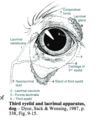
Q

What is the third eyelid?

A

* aka nictitating membrane

* between lower lid and eyeball

* covered with conjunctiva on both surfaces

* supported by T-shaped piece of cartilage

* gland of third eyelid surrounds stem of T: secretes towards eyeball from posterior surface

* while eyes open, normally held retracted by smooth muscle (symp. control)

* provides additional protection and moisture for eyeball

69
Q

What is lacrimal fluid for? What stimulate secretion?

A

* Moisten and nourish cornea and flush away foreign objects

* secretion stimulated by conjunctival, corneal, or nasal irritation

70
Q

What three layers does lacrimal secretion consist of?

A

* outermost lipid layer (from tarsal glands) spread tear film evenly and prevents evaporation

* aqeous layer from: lacrimal gland and gland of third eyelid

* inner mucoid layer (from goblet cells in conjunctiva) -binds tears to cornea

71
Q

How does lacrimal fluid drain?

A

Lacrimal puncta and canaliculi to nasolacrimal duct

72
Q

What is the vascular supply of the eye? Venous drainage?

A

* external ophthalmic artery- prinicipal supply- branches from maxillary artery then penetrates apex of periorbita

* internal ophthalmic artery- supplies CNII and spreads over retina from optic disc

* venous drainage via vorticose veins that emerge through sclera

73
Q

Nerve supply to the eye

A

CN II- perception of light

CN III- somatic efferent: dorsal medial and ventral recti, ventral oblique, levator palpebrae superioris

-visceral efferent: PS innervation to smooth muscle of iris and ciliary body

CN IV - dorsal oblique

CN V- (opthalmic and maxillary divisions)- sensory to eyeball (especially cornea), eyelids and conjunctiva

CN VI- lateral recturs, retractor bulbi

CN VII- motor to orbicularis oculi

Sympathetic innervation- exits cranial thoracic spinal cord and ascends in vagosympathetic trunk to cranial cervical ganglion near tympanic bulla

  • postsynaptic neurons travel with CNIII to supply smooth muscle of eye- periorbita (protrudes eyeball), pupillary dilator, eyelids (keeps palpebral fissure open), third eyelid (retracts)
74
Q

Where does the retina develop from?

A

Outgrowth of prospective diencephalon (optic cup); connective to forebrain remains as optic nerve

* Vascular and fibrous tunics- from neural crest derived mesenchyme

* Lens- formed by invaginations of ectoderm overlying optic cup

* Eyelids- grow over cornea from adjacent ectoderm and fuse; lids separate again either just before birth (ungulates) or some days after birth (carnivores)

75
Q

What is pain really?

A

It’s the signal that actually reaches the cortex

76
Q

How do you know an animal is in pain?

A
77
Q

What can inhibit nociceptor afferents?

A

* Inhibitory neurons in the dorsal horn of the spinal cord

* Inhibited by descending inhibitory pathways from medulla and midbrain (efferents!)

* inhibited by noradrenergic pathways (pons)

(can also be wound up by persistent nociceptor stimulation)

78
Q

In terms of sensitizers, what do you need for hyperalgesia?

A

Pain is the sum of a range of different stimuli

79
Q

Speak through the ascending nociceptive pathway

A
80
Q

Speak through the total potential nociceptive pathways (ascending and descending)

A
81
Q

How is pain the sum of different signals?

A
82
Q

Where are the descending inhibitory pathways of nociception initiated?

A

* midbrain/pons

* lower pons/ medullay

83
Q

Why is it difficult to develop the golden bullet of analgesia?

A

There are so many neurotransmitters and so many receptors.

But all lead to either:

* Decreased Ca influx pre-synaptically therefore reduced transmitter release

* Increased Cl or K influx post-synaptically therefore hyperpolarisation

** DOES THE AP get generated or not?

84
Q

How do opioid inhibit pain?

A

1) on ascending pathways- inhibit release of transmitter on presynaptic– decreased intracellular Ca OR by post synaptic membrane hyper polarization (increased intracellular Cl)– therefore making it less likely to generate an AP
2) on descending pathways- in the CNS, opioids stimulate increased excitation of inhibitory neurons

85
Q

Where are opioid receptors?

A

(have no managed to create a selective mu-1 antagonist)

86
Q

What is a major side effect of opioids?

A

* respiratory depression

* reduced GI motility

* dysphoria - state of unease (altered reality)

* dependence (physiological, not just psychological- can demonstrate at the tissue level)

(recall from table- mu is all over the CNS)

* species differences- humans on morphine- pinpoint non-responsive pupils (= overdose). Cats- dilated pupils.

87
Q

What are actions of opioids?

A

* excitement- species dependent (cats and horses)

88
Q

What is the receptor subtype for morphine, buprenorphine, and butorphanol, fentanyl?

A
89
Q

What are alpha2 adrenergic agonists?

A

Produce a dose dependent sedation, analgesia, and muscle relaxation. Alpha adrenergic agonists bind to and stimulate alpha2 adrenoceptors. These receptors are found in CNS and periphery. They may be presynaptic, where their activation inhibits release of NA, or postsynaptic, for example on vascular smooth muscle, where binding of the drug and activation of alpha2 receptors cause contraction.

** Sedation is dose dependent and varies from anxiety relief to profound sedation. Analgesia is of much shorter duration than sedation (approx 15-20 mins of 60-120 mins and Muscle relaxation due to inhibition of inter neuronal transmission at the CNS level– more pronounced in ruminants and horses)…

90
Q

In the CNS alpha2 adrenergic agonists have differing impacts depending on where it is acting… What does presynaptic inhibition of NA lead to? What about post synaptic? What about central NA and dopamine?

A

* presynaptic inhibition of NA leads to decreased nociception and therefore analgesia

* At post synaptic receptors in the spinal cord, binding of alpha 2 agonist leads to activation of pathways inhibitory to nociception

* Centrally decreased central NA and dopamine release leads to decreased arousal and sedation.

91
Q

What do alpha2 adrenergic agonists cause in cats? And some other species.

A

Cause emesis due to direct stimulation (that cannot be blocked by adrenergic or dopaminergic blockers) of Chemoreceptor Trigger Zone.

92
Q

What are the cardiovascular effects of alpha 2 agonists?

A

Activity at peripheral alpha1 and alpha2 receptors as well as central alpha2 receptors.

BP: initially increase (hypertension) due to binding of drug to the more accessible peripheral post synaptic alpha receptors and consequent peripheral vasoconstriction. This is followed by a period of hypotension due to central alpha 2 activity (or possibly to decreased cardiac output)

HR: Alpha 2 agonists cause a profound bradycardia as a result of central (medullary) inhibition of cardiac rate, (and possibly also due to increased reflex vagal activity as a result of initial pressor effect on blood vessels). The bradycardia may result in reduced cardiac output, is often accompanied by AV block, which can increase the risk of arrythmias.

93
Q

What effect do alpha 2 agonists have on the kidneys? And metabolic effects?

A

Cause diuresis, due to inhibition of renin release by Juxtaglomerular cells.

* Metabolic effects:

  • hyperglycaemia due to inhibition of insulin release and inhibition of lipolysis as a result of stimulation of alpha receptors on fat cells
  • body temperature: temperature regulation mechanisms may be impaired (hyper or hypothermia) for up to 24 hours post administration
94
Q

Where are alpha 2 agonists used?

When should they not be used?

A

Administered either alone or in combination with other agents e.g. opioids, to provide sedation in a range of setting- for performing minor, painful procedures, or to provide pre-op sedation. They are also used in combination with other drugs (such as with ketamine in cats) to provide surgical anaesthesia.

* Should not be used in heart disease, arterial hypotension, and shock, renal or hepatic impairment, final trimester pregnancy (oxytocic effect), epilepsy, in fact… any debilitated animal.

95
Q

What are some important things to keep in mind with alpha 2 agonists?

A

* a false sense of security- an aggressive animal may appear sleepy but can still react

* They are additive with other CNS drugs, particularly anaesthetics: barbiturate dose must be reduced by ONE HALF or more following alpha 2 agonists

* Cattle are ten times more sensitive to these agents than horses, dogs, and cats

96
Q

How do Phenothiazines act?

A

Sedatives- widely used to calm fractious animals to facilitate handling and as a preanaesthetic.

“dirty drugs” because they act on a wide variety of receptors therefore causing a wide variety of effects

* Blockade of Dopamine receptors- (deficiency of dopamine produces a parkinsonian syndrome in humans and catalepsy in animals (a medical condition characterized by a trance or seizure with a loss of sensation and consciousness accompanied by rigidity of the body.). Phenothiazines cause decreased motor activity in animals and at higher dosese catalepsy, rigidity, tremor, akinesia

* vasodilation and hypotension- due to blockage of adrenaline at peripheral sites

*inhibition of adenosine uptake into neurons- imp. in the anxiolytic properties of phenothiazines

* blockade of emesis at the CTZ in the medulla through the blockade of dopamine receptors

* Weak anti-muscarinic actions and antihistaminic (H1) sedative actions

* NO analgesic properties!!

97
Q

What are the principal effects of benzodiazepines on CNS?

A

Hypnotic (promoting sleep), sedative (reducing arousal), anxiolytic, anticonvulsant, skeletal muscle relaxant, amnesic

(Diazepam aka valium)

98
Q

What is the mechanism of action of benzodiazepines? Pharmacological effects?

A

Involves an allosteric effect: binds to postsynaptic membrane- changes the shape of the GABA receptor site, enhancing binding of GABA to its receptor, opening chloride channels and increasing inhibitory discharge

* pharm effects: promote centrally induced skeletal muscle relaxation. Minimal cardiorespiratory effects

** Used clinically in behaviour modification due to anxiolytic properties, as premediants and anticonvulsants… also Diazepam is an appetite stimulant in cats (presumed because of GABA inhibition of 5HT activity)

99
Q

When are butyrophenone derivatives used?

A

Azoperone- used in pigs to reduce population stress and fighting when litters are mixed to prevent sows from hurting their young at farrowing, reducing aggressionin boars.

** produced reduced motor activity and catalepsy due to a bloackade of the central effects of dopamine and noradrenaline.

100
Q

Why do vets use sedatives?

A

* restraint

* safety of handler and patient

* minimise stress to patient

101
Q

Why do anathesiologists use a number of agents?

A

To reduce risk of overdose, but to achieve proper sedation, analgesia, and anaesthetic state (renders patient unconscious)

102
Q

What are Xylazine (rompun, xylase), Medetomidine (Domitor), and Romifidine (sedivet) classified as?

A

alpha2 adrenergic agonists

103
Q

Where are alpha 2 receptors?

A

In CNS: brainstem locus coeruleus (modulator of wakefulness) sedation, in brainstem (cardiovascular regulating centre), in dorsal horn of spinal cord (analgesia), thalamus (analgesia), sympathetic neurones (analgesia)

In periphery: in autonomic ganglia, in blood vessels *(pre and post synaptic alpha 2)*

104
Q

What does activation of alpha 2 receptors lead to?

A

* Presynaptically- inhibition of NA release

* Postsynaptically- contraction of vascular smooth muscle

105
Q

What do alpha2 adrenergic agonists cause?

A

Sedation, analgesia, muscle relaxation (emesis in cats! muscle relaxation greater in cows than horses)

106
Q

What is the limiting factor for alpha2 adrenergic agonists?

A

Initial increase in BP due to initial peripheral binding on blood vessels… then decrease in BP to normal or hypotensive… due to feedback to CV control center which increases PS and decreases HR (reflex bradycardia)

*decrease in peripheral circulation (“muddy” mucous membranes)

* Animals can look unwell after giving these drugs

* You have lost the baroreflex- so the HR cannot increase if there is greater demand.

* Metabolic issues: hyperglycaemia, impairment of temperature regulation (problem in 40 degree heat), kidneys diuresis (inhibition of renin release)

107
Q

Why is it so important to consider pre-medicating agents with alpha 2 adrenergic agonists?

A

Additive effect- could kill patient

Good news is there are reversing agents

108
Q

Phenothiazines- mechanism of action

A

Sedatives- widely used to calm fractious animals to facilitate handling and as a preanaesthetic.

“dirty drugs” because they act on a wide variety of receptors therefore causing a wide variety of effects

* block peripheral alpha adrenoceptor therefore causing hypotension

* decreases PCV (because RBCs are sequestered in the spleen)

* No analgesia

* Do not use in an animal with shock (increased sympathetic drive–>therefore NA binds to beta receptors therefore leading to vasodilation exacerbating hypotension), colic, seizure threshold, potentiation of anaesthetic effect

109
Q

Mechanism of action of benzodiazepines. What is a common use due to lack of CV effects?

A

Allosteric effect on the GABA receptor so that GABA binds more effectively the site (working on the inhibitory pathway in the CNS)

* minimal CV effects therefore good for high risk patients (opioids are also a good thing to use as well)

* skeletal muscle relaxation

* appetite stimulant in cats (small IV dose of diazepam)

110
Q

Where do emobli/ infarctions often occur if in the brain?

A

At the junction of the grey and white matter due to the loop of vessels

* Venous infarction is uncommon

111
Q

Why are domestic animals less predisposed vs. humans to cerebral injury?

A

* Birth injury in humans whereas animals are much bigger shoulder or hip wise (different with brachiocephalic dogs)

*domestic animals have large frontal sinuses compared to humans- protective

* We have a bigger brain mass relative to our skull mass

112
Q
A
113
Q

Anisocoria

A

Variation in pupil size. Can indicate increased cranial pressure.

114
Q

Clinical signs of ICP

A

* anisocoria, mydriasis (pupillary dilation), non-responsive pupils, dull mentation, or altered state of consciousness, rigid paresis, abnormal respiratory pattern, bradycardia, coma

115
Q

What is happening in concussion?

A

* Unlikely to have any lesions

* temporary “jangling” of CNS components (temporarily upsetting axonal transport mechanisms)

* rapid acceleration and deceleration of brain in the skull

116
Q

What happens with repeat concussions?

A

* chronic traumatic encephalopathy (Dementia pugilista)- with repeated events you can lose neurons from your cerebral cortex

117
Q

Which trauma to the brain could be worse with stable head or moving head?

A

a still head in general less damage then a head that is moving when it receives a blow because the still head, the force can be absorbed by the skull.

118
Q

What is the most common cerebral contusion?

A

Traumatic haemorrhage into the leptomeninges (arachnoid and pia- subarachnoid space). Epidural or subdural traumatic haemorrage is less common because dura mater is tightly adherent to the calvarium.

119
Q

What are coup contusions vs. contrecoup contusion?

A

Coup contusion- contusion located at the site of impact

* contrecoup contusion- a contusion located on the opposite side from the site of impact (occurs because you are tearing of the meningeal and cortical blood vessels opposite the impact site)

(you can tell based on bruising & often contrecoup contusions are more severe)

120
Q

What determines how severity of trauma to the head?

A

* physical rigidity of the bone (influence by age, nutrition, presence of metabolic bone disease)

* mass

* velocity

* direction of applied force

* ability of the impacted tissues to move in response to applied force

121
Q
A

Glial scar, astroglial scar-filled with reactive astrocytes are the main cellular components. After injury, they extend their processes and increase sythesis of GFAP– they form a dense web of their plasma membrane extensions. SO they fill the space of the dead or dying neuronal cells (astrogliosis)

122
Q
A

Basisphenoid fracture- some skull fractures can be difficult to find

* visual deficits are common - an area with the pituitary, optic nerves

123
Q

What is Acute Brain Swelling?

A

* Swelling of the brain due to blood within blood vessels supplying the brain due to disregulation of normal arterial flow into the capillary beds of the meninges into the head

* can happen quickly after injury to the brain

= ICP

* can turn into true cerebral oedema

124
Q
A

Cerebral oedema

* acute or chronic

* in grey matter impossible to pick with the naked eye- grey matter is more dense than white matter with the cell processes

* White matter will show it better

* chronic oedema- smaller, yellow, flattened gyri, narrow sulci

125
Q
A

Coning of the cerebellum due to cerebral oedema– due to contact with the occipital bones

When the vermis is actually herniating- it is known as lipping of the vermis

126
Q

What higher centres are involved in the ANS? What is the primary integrating center of the ANS? Where does it originate?

A

* Consists of a higher centres within the thalamus, midbrain, pons and medulla

* Hypothalamus is the main integrating centre of the ANS (nuclei in the rostral potion of the hypothalamus control the PS division, nuclei in the caudal portion control the symp division)– the hypothalamus receives afferents from the cerebral cortex via the thalamic nuclei, and from ascending GVA pathways. In turn the hypothalamus then influences acitity of the metabolic centres in the reticular formation of the midbrain, pons and medulla

* Beginning of many of the pathways originate in the tectum of the midbrain

127
Q

Sympathetic system

A

* Referred to as the thoracolumbar system, since this is the region of the spinal cord where most of the preganglionic neurons start

* Has the ganglion housing the postganglionic neuron cell body located (relatively) close to the CNS and thus has very long postganglionic axons

* has noradrenaline as the neurotransmitter communicating between the pre- and postganglionic neurons

* is designed to aid the fight or flight response in animals in situations of perceived threat or danger

128
Q

Parasympathetic system

A

* aka craniosacral system, since the preganglionic neuron cell bodies live either in the brain or sacral spinal cord grey matter

* has the ganglion between neuron 1 and neuron 2 located fairly close to the effector (target) organ

* has acetylcholine (Ach) as the neurotransmitter communicating between the pre- and postganglionic neurons

* is designed to aid the body’s homeostatic mechanisms in day to day situations

** 2 major functions of the ANS for clinical purposes– neuro exam– control over pupil size and control over bladder function

129
Q

PS pathway for control of pupil size

A

* starts with the pathway for vision (CN II-optic)- approx 20% of these fibres bypass the LGN (lateral geniculte nucleus) and synapse in the pretectal nucleus or tectum.

* axons decussate to terminate in the PS nucleus of CN III (some stay ipsilateral)- they decussate twice at the optic chiasm and after the pretectal nucleus but the ipsilateral are the strongest stimulus)

* From PS nucleus of CN III, neuron 1 (preganglionic) tavels to the ciliary ganglion, located just caudal to the eye, where it synapses with the postganglionic neuron (NB PS- ganglion located near the effector organ). The postganglionic neurn supplies the sphincter of the pupil

130
Q

What is the sympathetic pathway of pupil size?

A

Originates from the tectum of the midbrain, neurons here synapse on the second neuron in the pathway, whose cell body lives in the tegmentum of the midbrain. The combo of these two neurons then forms the lateral tectotegmentospinal tract which runs down the cervical spinal cord to the T1, T2, and T3 spinal cord segments, where it synapses on neuron 1 in the sympathetic system.

* Neuron 1 (preganglionic) has its cell body within the spinal cord GM and sends axons down through the ventral nerve root, along the ramus communicans and then up the vagosympathetic trunk in the neck (bypassing the stellate and caudal cervical ganglia) to synpase at neuron 2 whose cell body lives in the cranial cervical ganglion (located near the tympanic bulla). Neuron 2 (postganglionic) axons then project to the smooth muscle of the orbit and eyelids, the third eyelid, the smooth ciliaris muscle and the smooth dilator muscle of the pupil

131
Q

What is pupil size a function of?

A

Balance between PS and symp nervous systems

* Also a balance between the amount of light in the environment and the emotional status of the animal

* PS pathway regulates the response of teh eye to environmenta light

* the symp pathway regulates the response of the pupil to environmental factors that elicit stress (e.g. excitement, fear, anger)

132
Q

Signs of dysfunction with lesions affecting the PS branch of CN III only are very rare–more common to see lesions that pick off the whole of CN III– lesions would be?

A

* Anisocoria (unequal size pupils between the two eyes)

* Absent direct papillary light reflex in the affected eye

* Strabismus (deviation of the eyeball within the orbit) and an abnormal oculocephalic reflex

133
Q

Lesions in the cervical spinal cord, thorax, neck, or tympanic bullae can affect sympathetic supply… Horner’s syndrome:

A

* Meiosis (small pupil on affected side)

* Ptosis (drooping upper eyelid on the affected side)

* Enophthalmos (sunken eyeball, with third eyelid protrusion as a result)

134
Q

What is the sympathetics role in bladder function? PS role? What are other components of the system?

A

* (efferent supply) sympathetic NS predominates while the bladder is filling up

* (efferent supply) PS NS predominates when the bladder is being emptied

** Higher control centres play a part as well, LMNs (efferent supply) that control striated muscle, afferent supply (GVA)– to detect how full the bladder is

135
Q

What is the role off the GVA neurons in bladder control?

A

Dendritic zones of the GVA neurons are located in the wall of the bladder

* Mechanoreceptors respond to stretch and distension- their axons travel through the pelvic nerve and enter the sacral spinal cord segments (S1-3). Cell bodies of these neurons are located in the dorsal root ganglion, and the axons send collaterals that terminate in 3 places: Interneurons in the dorsal horn of GM of S1-S3– communicate with PS (GVE) neurons in the S1-S3 segments, interneurons in the dorsal horn GM of L1-5 (commo with symp (GVE) neurons in these segments), some collaterals that travel into the dorsal column white matter and ascend to synapse in the thalamus which then projects axons to the cerebral cortex for conscious perception of bladder filling

136
Q

What is the role of the efferent supply in bladder control- sacral segment LMNs (GSE neurons)? SOMATIC NS

A

(SOMATIC NS) The sacral segment LMNs are providing voluntary control

over the striated muscle of the external sphincter of the bladder neck and pelvic urethra. Cell bodies of these neurons live in the ventral horn GM of the S1-S3 spinal cord segments. Axons travel through the ventral spinal nerve roots and via the sacral plexus into the pudendal nerve to synapse ultimately on the urethral sphincter to provide voluntary contraction- this allows for continence and storage of urine.

137
Q

What is the efferent supply- autonomic neurons (GVE neurons) of the PS supply (sacral segments) of bladder control?

A

* the PS supply to the bladder has the preganglionic cell bodies located in the GM of the sacral spinal cord segments (in the lateral horn). The axons travel out and form the pelvic nerve that lies on either side of the lateral surface of the rectum. The preganglionic PS axons are then joined by postganglionic sympathetic axons within the hypogastric nerve- together the hypogastric and pelvic nerves form the pelvic plexus. The PS preganglionic axons synapse on the neuronal cell bodies of the postganglionic neurons in the pelvic ganglion which is located in the pelvic plexus itself. The postganglionic axons innervate the muscarinic cholingergic receptors on the smooth muscle throughout the wall of the bladder. This is the detrusor muslce, and the PS outflow causes it to contract during the voiding phase of urination.

138
Q

What is the sympathetic supply (lumbar segments) of the bladder?

A

Starts in the lateral horn GM of the L1-L5

* axons leave these segments–> travel via ventral spinal nerve roots, spinal nerve, ramus communicans, lumbar portion of the sympathetic trunk, lumbar splanchnic nerves and terminate in the caudal mesenteric ganglion–> axons from the postganglionic neurons travel into th hypogastric nerve through the mesocolon and then ultimately to the pelvic plexus

*** Some axons terminate in the pelvic plexus by synapsing with alpha-2 receptors om the cell bodies of the postganglionic PS neurons… this connection inhibits firing of the postganglionic PS neurons (thus preventing detrusor muscle contraction)!!!

** Other symp postganglionic axons travel to the neck of the bladder, split again– one group goes to the internal sphincter and synapses on alpha-2 adrenoceptors on the smooth muscle in this sphincter = contraction… the other group to the detrusor muscle and synapses on beta receptors on the muscle cell membrane.

139
Q

Where are the control centres for micturition?

A

In the pons and the pontine reticular formation

* receive afferent information from the GVA neuron projections that travel cranially through the dorsal columns of the spinal cord–> axons from the neurons in these control centres, in turn travel caudally through the spinal cord to the L1-5 and S1-3 spinal cord segements to control the GSE LMNs and the autonomic NS neurons. These are the UMNs of bladder control– they facilitate the inhibition of the LMNs (including the ANS neurons) to allow for micturition when appropriate. They are also under the control of the cerebral cortex and cerebellum- this allow voluntary control over the whole system and coordination of the different parts.

140
Q

What predominates during the storage phase of urine?

A

The sympathetic NS and sacral segment GSE LMNs have to predominate in order to maintain a relaxed detrusor muscle, constricted internal urethral sphincter, and a constricted external sphincter. This is overseen by the control centres in the pons– these send impulses through the reticulospinal tracts to facilitate lumbar sympathetic outflow and sacral GSE outflow. The control centres also inhibit PS outflow during this phase.

** As urine accumulates in the bladder, GVA neurons are activated. These project onto the lumbar spinal cord segments to further facilitate sympathetic outflow- this relaxes the detrusor muscle via beta adrenoceptors that allow for further filling of the bladder. When the bladder fills enough to distend the proximal urethra, other GVA neurons fire which synapse on sacral spinal cord segments and the GSE LMNs (which contract the external sphincter striated muscle). Simultaneously, projections from the GVA neurons travel cranially through the spinal cord to communicate to the cerebral cortex that things are getting full… conscious control over the GSE LMNs then adds to the tightening of the external sphincter.

141
Q

Speak through the voiding phase of micturition.

A

As bladder volume increases, it exceeds the threshold of the GVA mechanoreceptors in the bladder wall that repond to stretch and distension. Impulses travel over these sensory neurons via the pelvic nerves into the sacral segments of the spinal cord and then project through the dorsal white matter up the spinal cord to the pontine control centre (and the cerebral cortex via the thalamus). The pontine control centre is xtimulated– either by this pathway or the cerebral cortex.

**now outflow from the pontine causes inhibition of the symp. outflow (L1-L5) and facilitates PS outflow (S1-S3). Also inhibits the firing of the GSE LMNs in the pudendal nerve, thus relaxing the external urethral sphincter. (constriction of the detrusor muscle and relaxation of the internal and external sphincter muscles)