Endocrinology Flashcards

1
Q

What are the three hormones that control the biologically active, free plasma Ca++ concentration in the body? What percentage is in the extra-cellular fluid (ECF)?

A
  1. Parathyroid hormone (PTH)
  2. Calcitonin
  3. Vitamin D

** 0.1% is in the ECF

2
Q

What does an increase in free Ca++ do in regards to neuromuscular activity? What about a decrease?

** so what are potential consequences of hypocalcaemia and hypercalcaemia?

A

* An increase in free Ca++ changes the membrane permeability to Na+– an increase of Ca++ decreases Na+ permeability, resulting in a fewer action potentials and therefore depression of activity

* A decrease of free Ca++ results in over-excitability of nerves and muscles by lowering the threshold with which a response is induced– so a decrease in Ca++ increases Na+ permeability of the cell membrane, resulting in an influx of Na+ moving the resting potential closer to threshold

** hypocalcaemia- can result in muscle spasm, spastic contraction of the respiratory muscles can result in death by asphyxiation

** hypercalcaemia- can cause cardiac arrhythmia and depression of neuromuscular activity

3
Q

Functions of calcium in the body

A
  1. Neuromuscular activity
  2. Excitation contraction coupling in cardiac and smooth muscle
  3. Release of products from secretory cells by exocytosis e.g. insulin release
  4. Tight junction maintenance
  5. Blood clotting (Ca++ acts as a co-factor in clotting cascades)
4
Q

What hormones assist in Ca++ absorption in the digestive tract?

A

PTH and Vitamin D– depends on the Ca++ status of the body.

5
Q

How is calcium homeostasis maintained?

A

Goal: to maintain a constant free plasma concentration of Ca++

* Rapid exchange between bone and ECF

* Minor contribution made by urinary excretion of Ca++

6
Q

What is the goal of PTH? And important actions?

A

* PTH is the predominant hypercalcemic hormone- increase Ca++ concentration in the palsma– has actions on bone, kidneys, and intestine

  1. Increase blood calcium conc and decrease blood phosphorous
  2. increase urinary excretion of phosphorous by diminishing tubular reabsorption in the proximal convoluted tubule
  3. increase reabsorption of calcium in the distal convoluted tubules, therefore less calcium loss
  4. Increase the rate of skeletal remodelling and the net rate of boen resorption
  5. Increase osteolysis and the numbers of osteoclasts on bone surfaces
  6. increase urinary excretion of hydroxyproline (major component of collagen)
  7. activate adenyl cyclise in target tissues
  8. accerlerate the formation of the principle active metabolite 1, 23- OH2D3 by the kidney through a trophic effect on 1alpha-hydroxylase in the mitochondria or renal epithelial cells in the proximal convoluted tubule (1,23-OH2D3= Calcitriol= INCREASES LEVELS OF CALCIUM IN THE BLOOD BY INCREASING UPTAKE BY THE GUT)
7
Q

What type of cell do parathyroid glands contain? What is secreted?

A

Chief cells continuously secreting PTH

8
Q

Where does calcitonin come from? What is it’s role?

A

Secreted continuously and increases greated in respons to elevated blood calcium– by C cells in the thyroid gland- these cells are distinct from follicular cells of the thyroid that secrete the thyroid hormones.

* Calcitonin acts on bone to decrease entry of calcium into plasma by temporarily inhibiting PTH- stimulated bone resorption

** the actions of calcitonin and PTH are antagonistic on bone resorption, but synergistic on decreasing the renal tubular role resorption of phosphorous

** Calcitonin functions as an emergency hormone to prevent hypercalcaemia during rapid postprandial absorption of calcium AND to protect against excessive loss of calcium and phosphorous from the maternal skeleton during pregnancy

9
Q

What are the two effects of PTH on Ca++ mobilisation from bone?

A

* Fast- lost Ca++ from bone fluid– does not involve resorption of bone

* Slow- increased osteoclast resorption (chronic)– increased formation of osteoclasts & transiently decreasing the bone formation activity of osteoblasts

10
Q

What activates vitamin D? What does the activation of vitamin D result in?

A

* The kidneys activate vitamin D– this is enhanced by PTH causing a decrease in the reabsorption of PO4 by the kidneys; while enhancing the reabsorption of Ca++

** Vitamin D increases intenstinal absorption of Ca++ (this is an indirect effect of PTH on Ca++ with vitamin D as the intermediary)

11
Q

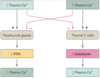
Regulation of PTH and Calcitonin Secretion

A

** primary signal for PTH regulation is plasma free Ca++

** simple negative feedback regulation- no nervous or other hormonal influence

12
Q

What is this? What happens when it is activated?

A

Extracellular calcium sensing receptor. This is how changes in plasma Ca++ concentration are detected. It is expressed on parathyroid cells and C cells in the thyroid gland, in the kidney, osteoblasts in bone, hematopoietic cells in bone marrow, placenta, GI mucosa, squamous epithelial cells of the oesophagus. (calcium acting as a hormone has direct effects on the function of many cell types)

  1. activation of phospholipase C, which leads to generation of the second messengers diacylglycerol and inositol triphosphate
  2. Inhibition of adenylate cyclase, which suppresses intracellular concentrations of cyclic AMP
13
Q

What is Vitamin D considered? Why?

A

* a hormone (aka calcitriol)- as it can be produced in the skin from a precursor related to cholesterol by sunlight and is subsequently released into the blood to act at a distant target site.

** vitamin D must be activated via 2 sequential additions of an -OH group in the liver and then the kidneys

** Vitamin D’s major target is the mucosa of the SI. In the proximal SI it increases active transcellular transport of Ca++ and in the distal part the transport of phosphorous.

14
Q

Difference in the role of vitamin D and PTH

A

Vitamin D acts to ensure retention of sufficient mineral ions for mineralisation of bone matrix; whereas PTH maintains the proper ratio of calcium to phosphorous in extracellular fluids.

* a small amount of vitamin D is needed to permit PTH to exert its action on bone

15
Q

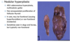
What is PTH hypersecretion?

A

* occurs in domestic animals in response to a poor Ca++ diet e.g. all meat diet in carnivores

* can also result from a hyper secreting tumour of the parathyroid gland but it is rare

* Consequences can include: reduced excitability of muscle and nervous tissue which leads to muscle weakness and neurological disorders; excessive mobilisation of Ca++ and PO4 from skeletal stores results in thinning of bones, skeletal deformities and increased fracture risk; increase incidence of Ca++ containing kidney stones from excessive Ca++ filtered through kidneys

16
Q

What are the consequences of a Vitamin D deficiency?

A

* major cause is impaired intestinal absorption of Ca++, under these conditions PTH maintains plasma Ca++ at the expense of bones. Improper mineralization can lead to soft and deformed bones- rickets in young and osteomalacia in adults

17
Q
A

Parturient Paresis (Milk fever, parturient hypocalcaemia)- most common metabolic disorder affecting high producing dairy cattle. Inability of dairy cow to mobilize adequate amounts of calcium results in characteristic symptoms usually obvious 72 hours post-parturition: of restlessness, anxiety, anorexia, uncoordination, and lack of interest in a calf. Incidence increases with age and yield.

** without intervention– cows progress to second stage which is manifest by recumbency– body temp drops– eventually dullness to coma

18
Q

How does hypocalcaemia in dogs manifest?

A

Excitement (eclampsia) with restlessness, panting, trembling, muscular tetany and convulsive seizures. Disorder occurs in bitches of small breeds of dogs during lactation.

19
Q

Why do dogs and cows react differently with hypocalcaemia?

A

* cows- paresis, dogs- tetany

* differences in functions of neuromuscular junction– the release of acetylcholine and transmission of nerve impulses across the NMJ are blocked by hypocalcaemia in cows (but not in dogs)

* tetany occurs in dogs as a result of spontaneous repetitive firing of motor nerve fibres- owing to the loss of stabilising membrane-bound calcium, nerve membranes become more permeable to ions and require stimulus of a less magnitude to depolarise

20
Q
A
21
Q

What are the two main hormones secreted by the thyroid? Which is the most abundant (>90%)? Which is more potent?

A

* Thyroxine (T4) (most abundant)
* Tri-iodothyronine (T3)– may be directly synthesized or formed from T4, 3-4xmore potent than T4

22
Q

What are the effects of thyroid hormones?

A
  1. Increase basal metabolic rate in all tissues (except brain, spleen, testes, and lung)– increased mitochondrial size and number, increased protein turnover (both catabolism and synthesis), increased carbohydrate turnover (both catabolism and synthesis), increased lipolysis
  2. Promote growth in young animals (acting with growth hormone)- thyroid hormone particularly important for normal neurological and musculoskeletal development
  3. Promote hyperglycaemia through glycolysis, gluconeogenesis and intestinal glucose absorption
  4. Stimulatory effects on cardiac function- both chronotropic (increases heart rate) and inotropic (increases heart contraction strength)
23
Q

Regulation of thyroid secretion

A

* Thyrotropin-releasing hormone (TRH) is released from the anterior pituitary–> TRH stimulates secretion of thyroid- stimulating hormone (TSH) by anterior pituitary–> thyroid secretes T3 and T4

* TSH secretion is suppressed by T4 and somatostatin with minor effects of glucocorticoids and sex hormones

24
Q
A

Ectopic thyroid tissue- common incidental finding in ventral neck, mediastinum, and heart base. May become neoplastic.

25
What is this? Who is it common in? What is the most likely cause? What occurs?
Congenital hypothyroidism \* Common in lambs, other species rarely affected - typically due to iodine deficiency - usually weak or stillborn and may have goiter \* Congenital cases may display certinism (severely stunted mental and physical growth) - disproportionate dwarfism with long limbs, short bodies, and domed heads with shortened muzzle - mental impairment common - retention of juvenile hair coat (fine hair)
26
What is its? Causes? Effects?
Adult-onset hypothyroidism \* Common in dogs, rare in other species Causes: 1. lymphocytic thyroiditis- bilateral invasion and destruction of thyroid tissue by lymphocytes, unknown cause -- affected animals often develop antibodies directed against thyroglobulin and other thyroid hormones 2. Idiopathic follicular atrophy- may represent end stage of lymphocytic thyroiditis but inflammation usually minimal-- thyroid follicles are small and sparsely distributed with adipose tissue infiltration \* Vague and insidious course \* Decreased metabolic rate - mental dullness - exercise intolerance - cold intolerance - weight gain other: - poor coat quality, hyperpigmentation, alopecia, seborrhea and hyperkeratosis, secondary pyoderma and otitis externa, myxoedema- non-pitting oedematous thickening of the forehead, eyelids, and skin folds of the face and neck-- "tragic facial expression-- due to subcutaneous accumulation of hydrophyllic glycosaminoglycans \* may be infertile, megaoesophagus due to neuropathy affecting oesophageal innervation, bradycardia with absence of thyroid homrone (severe cases hypotension, hypothermia, become comatose), hyperlipidaemia prediposes to atherosclerosis (severe cases may develop infarction, corneal lipidosis are common)
27
28
29
Causes of adult-onset hypothyroidism
\* Decreased thyroid functional tissue- aplasia, hypoplasia (rare), thyroid atrophy, typically due to decreased TSH stimulation \* Thyroid destruction: immune mediated thyroiditis, neoplasia (destructive thyroid carcinoma), iatrogenic (e.g. thyroidectomy to remove thyroid neoplasia, overdose of thyroid hormone inhibitor) \* Impaired thyroid hormone production (usually associated with goiter) - dietary iodine deficiency, dietary iodine excess, enzyme defects in thyroid hormone synthesis, exposure to goitrogenic toxins which interfere with thyroid hormone synthesis
30
What is goiter generally?
\* Non-inflammatory and non-neoplastic thyroid enlargement \* typically associated with hypothyroidism (nodular hyperplasia is the exception) \* Occur sporadically in calves, lambs, kids and pups due to inability to synthesize thyroid hormone
31
What are the three forms of goiter?
1. Hyperplastic goiter- reflects hypersecretion of TSH and hypertrophy and hyperplasia of thyroid tissue without adequate thyroid hormone production-- inadequate T3/T4 so feedback does not work and just continues secretion of TRH and TSH--\> hypertrophy & hyperplasia--\> thyroids are diffusely enlarged, firm, dark red with a "meaty" texture due to increased cellularity and blood flow-- animals may be weak poorly haired, and display myxoedema \*\* most cases reversible 2. Colloid goiter: reflects the resolution stage of hyperplastic goiter once thyroid hormonal function has been restored- restoration of normal T3/T4--\> decreased TSH stimulation of thyroid--\> follicular atrophy and accumulation of colloid within follicles. Thyroids are diffusely enlarged pale tan coloured with a waxy appearance on cut surface. 3. Nodular hyperplasia- idiopathic hyplasia of thyroid tissue, common in older cats, dogs and horses. Mat be associated with hyperthyroidism inc ats, but not typically an indicator of hypothyroidism and often incidental findings
32
33
34
35
36
37
What are the causes of Goiter?
\* iodine deficiency (usually seen in mountain and inland areas- seawater is rich in iodine), causes hyperplastic goiter, rarely observed now due to supplementation \* iodine excess- may also cause hyperplastic goiter as excess iodine impairs uptake by thyroid gland and thyroid hormone synthesis, usually associated with overzealous supplementation, neonates and fetuses more sensitive due to concentration of iodine in palcenta and milk. \* Goitrogenic toxins- some toxins interfere with the SYNTHESIS of THYROID HORMONES (e.g. goitrin, derived from glucosinolates in Brassica plant spp.) causing hyperplastic goiter \* Congenital dyshormonogenetic goiter: hyperplastic goiter due to genetic defects in ENZYMES responsible for thyroid hormone synthesis
38
hyperplastic goiter
39
hyperplastic goiter
40
41
42
\* Clinical features: increased metabolic rate (weight loss, polyphagia, hyperactivity/ irritability); skin (unkempt hair coat/ patchy alopecia); Cardiac effects (arrhythmias, secondary hypertrophic cardiomyopathy, hypertension)
43
44
45
Causes of hyperthyroidism
46
47
48
What are the parathyroid glands? What cells are they composed of? What other cell near by are associated with the function of the parathyroid?
\* Four glands in most species that are associated with thyroids: one pair within the thyroid parenchyma and an external pair \* composed of chief cells which secrete parathyroid hormone (PTH) \* C Cells- interstitial cells within the thyroid secrete calcitonin-- usually individually cells or small clusters that sit between thyroid follicles
49
What are the effects of calcitonin and PTH?
\* Opposing effects on serum calcium - PTH: provides general regulation of serum calcium concentrations, secretion induced by decrease in serum calcium as well as phosphate, promotes renal calcium retention and phosphorous excretion, increases osteoclastic bone resorption, promotes synthesis of active vitamin D3 - Calcitonin: provides emergency regulation in the event of hypercalcaemia, secretion induced by increase in serum calcium, promotes renal calcium and phosphorous excretion, inhibits osteoclastic bone resorption
50
What is primary hypoparathyroidism?
Occasionally seen in small animals, rare in other species \* Causes: due to lymphocytic plasmacytic parathyroiditis (presumed autoimmune parathyroid destruction), occasionally induced by thyroidectomy \* Effects: reflect decreased PTH secretion and hypocalcaemia, clinical effects typically develop suddenly and are episodic, animals develop tremors/ tetany, hyperpnoea and are often hyperexcitable- seizures may occur in severe cases, muscle weakness and arrhythmia may also develop
51
What is parturient hypocalcaemia?
\* Complex metabolic disease in cattle (milk fever/ parturient paralysis) and sometimes in dogs (eclampsia) \* Cause: high dietary calcium intake during pregnancy suppresses the PTH secretion and increases calcitonin secretion, leading to decreased calcium mobilization--\> following parturition, calcium demand increases markedly due to lactation, while calcium intake is decreased due to inappetance and post-partum GI stasis--\> PTH unable to respond rapidly to changes in calcium supply and demand--\> hypocalcaemia develops Effects- downer cows, dogs show overstimulation
52
Parathyroids
53
Parathyroid gland
54
C cells- interstitial cells within the thyroid that secrete calcitonin
55
How does the body maintain calcium homeostasis?
56
57
What are some causes of the primary disease in this picture?
Hyperparathyroidism- Causes: older dogs, caused by excessive PTH, functional parathyroid neoplasia (adenoma (B) or carcinoma (M))
58
What are some causes of the secondary RENAL disease seen in this picture?
Secondary RENAL hyperparathyroidism: \* Caused by hypocalcaemia or hyperphosphataemia- renal tubular defects causing increased calcium excretion and phosphate retention (phosphate binds calcium), decreased vitamin D synthesis \* Parathyroid hyperplasia and PTH hypersecretion secondary to hypocalcaemia in renal failure \* all parathyroids enlarged (due to hyperplasia stimulated by calcium imbalance, in contrast to parathyroid neoplasia which affects a single gland)
59
Other than secondary renal hyperparathyroidism, what is the other kind of secondary hyperparathyroidism?
Secondary Nutritional hyperparathyroidism \* Parathyroid hyperplasia and PTH hypersecretion secondary to low dietary calcium and/or dietary phosphorous (phosphate binds calcium) \* Commonly induced by: all meat diets (dogs, reptiles), high concentrate diet especially bran (horses), diets high in oxalates (oxalates bind calcium) \* all parathyroids enlarged (due to hyperplasia stimulated by calcium imbalance, in contrast to parathyroid neosplasia which affects a SINGLE GLAND)
60
What do you see with hyperparathyroidism?
\* osteoporosis due to osteoclastic bone resporption and demineralization \* susceptible to pathological fracture- folding fractures common, animals may develop shifting lameness due to microfractures or paressis due to spinal fracutres \* Facial bones most severely affected -- fibrous tissue may result in firm swellings ("big head" in horses) \*\* Metastatic MINERALIZATION- only occurs in primary hyperparathyroidism due to hypercalcaemia (because secondary is a response to low calcium and so calcium levels are typically low)-- lungs, stomach, kidneys, and blood vessels (including endocardium) most commonly affected
61
Could be a result of what?
Osteoporosis from hyperparathyroidism
62
Could be a result of what?
Fibrous osteodystrophy- from hyperparathyroidism
63
Could be a result of what?
Fibrous osteodystrophy- hyperparathyroidism
64
Could be a result of what?
Rubber jaw- hyperparathyroidism
65
What is pseudohyerparathyroidism? Causes, effects?
Aka hypercalcaemia of malignancy \* secretion of parathyroid hormone- related protein (PTHrP) by some neoplasms - anal sac apocrine adenocarcinoma, Lymphoma (especialy T cell) \* Causes soft tissue mineralization- renal, gastric, mammary carcinomas, SCC (especially gastric SCC in horses) Effects: similar to primary hyperparathyroidism but less bone resorption; major effects are hypercalcaemia and metastatic mineralization
66
What is Thryoid C Cells Neoplasia?
\* Rare but in horses and old bulls \* Adenomas are circumscribed, encapsulated, firm, cream to grey- tan, one or many \* Carcinomas may be unilateral or bilateral, locally invasive, may metastasize to LNs and lungs \* In bulls, C cell tumours may be present concurrently with pheochromocytoma +/- pituitary adenomas \*\* C cell tumours in bulls may be functional and cause vertebral spondylosis and osteopetrosis- hypocalcaemia is typically mild \*\* C Cells in horses is usually solitary and asymptomatic
67
Pathology of the Endocrine Pancrease
\* uncommon, seen in dogs and ferrets; islet of langerhans produce insulin, glucagon, somatostatin, and others. 1. Insulinoma- episodic seizures, weakness and collapse, reflecting CNS glucose depletion, triggered by fasting, exercise or excitement, episodes are brief due to counter-regulatory hormones (catecholamines, glucagon, cortisol, and growth hormone) 2. Gastrinoma- not normally produced by islets of langerhan but may be in neoplasm-- causes Zollinger-Ellison syndrome- increased gastric acid production therefore gastric and duodenal erosions and ulcerations; anorexia, melaena and/or haematemesis 3. Glucagonoma- rare, hyperglycaemic (may develop diabetes mellitus due to insulin resistance) and display vacuolar hepatopathy, superficial necrolytic dermatitis (aka hepatocutaneous syndrome)-- bilaterally symmetrial hyperkeratotic crusting, lesions of muzzle, lips, periocular skin, pinnal margins, distal extremities, ventrum, pressure points (hocks), external genital mucocutaneous margins. Footpads are hyperkeratotic.
68
What ultimately occurs with hypoglycaemia?
The CNS cannot synthesize, store or concentrate glucose-- hypoglycaemia ultimately leads to coma and death.
69
What is meants by the exocrine and endocrine functions of the pancrease?
Exocrine= glands that produce and secrete substances onto an epithelial surface by way of a duct, in the case of the pancreas this refers to its function in the GIT Endocrine= Secretes hormones using ducts. In the case of the pancreas, this refers to the Islets of Langerhans
70
What are the four main hormones the islets of Langerhans secrete? Who secretes them? What do they do?
\* Insulin, glucagon, somatostatin, pancreatic polypeptide \* GlucAgon- catabolic hormone secreted by ALPHA cells-- increased insulin & increased somatostatin secretion-- Role is to maintain NORMOGLYCAEMIA \* Insulin- anabolic hormone secreted by BETA cells-- decreased glucagon secretion-- ALLOWS the body to use carbs as energy sources and to store nutrients. Causes tissue uptak and sequestration of glucose, fatty acids, and amino acids with resultant DECREASE IN PLASMA LEVELS. \* Somatostatin- secreted by delta cells-- inhibits digestive processes and other pancreatic hormones-- decreased insulin & decreased glucagon secretion-- INHIBITS glucagon and insulin \* Pancreatic polypeptide- secreted by F cells-- increases gut motility and gastric emptying
71
Hormones of the pancreas are secreted where? What is the innervation of the pancreas?
\*\* Hormones are secreted into pancreatic vein--\> portal vein--\> preferentially affects liver \* innervated by the sympathetic and parasympathetic NS
72
What is insulin secretion stimulated by? What is insulin inhibited by?
1. High blood glucose (most important stimulant) 2. Blood amino acid (arginine and alanine) 3. Gastrointestinal hormones e.g. cholycystokinin 4. Parasympathetic stimulation Insulin secretion is inhibited by: 1. Low plasma glucose (fasting, exercise) 2. Somatostatin sympathetic stimulation 3. Sympathetic- alpha adrenergic activity
73
What is meant by insulin secretion is biphasic?
1. In the acute phase (10 minutes) release of preformed insulin occurs 2. In the chronic phase (rises over next hour) release of newly formed insulin occurs. \*\* from beta cells of the pancreas directly into the portal vein which ensures the liver receives high concentrations 1/2 life of insulin in blood is 5-8 minutes \*\* metabolized by the kidney and liver by the enzyme inulinase that splits disulphide bonds
74
What does insulin do?
Allows the body to use carbohydrates as energy sources and store nutrients. Causes tissue uptake and sequestration of glucose, fatty acids, and amino acids with resultant DECREASE in plasma levels. Toward hypoglycaemia because it is storing!!!
75
What happens with glucoregulatory failure?
Caused by either inadequate insulin secretion or inappropriate insulin action on target tissues and therefore hyperglycaemia and can contribute to Type I or Type II diabetes mellitus.
76
What causes insulin to be secreted?
1. Glucose influences Beta islet cells electrical activity via metabolically inudced changes in the activity of ATP-sensitive K+ channels 2. These channels open when glucose is low and close when glucose is high 3. ATP blocks K+ channel activity, whereas ADP stimulates K+ channel opening. (common target for anti-diabetic drugs) 4. Depolarizing cell membrane opens voltage- gated Ca2+ channels and results in both synthesis of insulin and exocytosis of insulin granules leading to insulin secretion
77
What are the intracellular actions of glucose?
\* Insulin binding alpha and beta subunits of tyrosin kinase--\> allowing the receptor to phosphorylate tyrosine residues --\> ACTIVATION OF PIP2 pathways and MAP kinase pathways--\> OVERALL a result in a range of GENES and ENZYMES activatied which have important roles in increased glucose uptake and storage, protein synthesis and lipogenesis.
78
What are the metabolic actions of insulin?
\* Promotes glycolysis (liver, pyrvate+ lactate--\> glucose), lipogenesis, and inhibits glycogenolysis (liver, glucose output) with major targets being the liver, adipose tissue, and muscle. \* Muscle--\> stimulates glucose uptake via carrier system, Glut4, stimulates glycolysis, stimulates glycogen synthesis, promotes protein synthesis \* Adipose tissue--\> stimulates glucose uptake via Glut4, glycolysis, some effect on protein metabolism, stimulates fat deposition, regulates lipoprotein lipase which promotes breakdown of triglycerides in blood (VLDL & chylomicrons) to free glycerol and free fatty acids
79
What is glucagon?
Essentially opposite to insulin-- the role is to maintain normoglycaemia. Secreted by the ALPHA cells of the pancreas. Half life of 6 minutes in the blood. 50% extracted by the liver on single pass. 1. Secreted in response to low plasma glucose 2. Stimulated by protein meal 3. Inhibited by insulin and somatostatin MOST IMPORTANT ACTION= maintenance of hepatic glucose output \* little effect on glucose utilisation \*\*\*\* insulin and glucagon control the relative rates of gluconeogenesis and glycolysis by altering the hepatic fructose 2,6-biphosphate levels plus other key enymes involved in glycolysis
80
What is somatostatin?
\* Stimuli for secretion: 1. Glucose, amino acids, free fatty acids, gastrointestinal hormones, glucagon & autonomic stimuli 2. Inhibition of secretion: autonomic stimuli & insulin Actions: 1. inhibition of insulin and glucagon 2. reduces assimilation rate of all nutrients from GIT. Prevents rapid nutrient overload.
81
Glucagon and insulin compare and contrast
82
What is Diabetes Mellitus?
A condition in which large amounts of glucose are excreted in the urine instead of supplying the muscles and organs. Untreated suffer from periodic energy supply crises- severe weakness and occasionally diabetic coma. The paradox is that animals are not malnourished and indeed contain an abundance of biological fuel which cannot be "burnt." \*\* Type 1-- underproduction of the hormone insulin, which the function of insulin is to open the glucose "gates" of cells for storage, and in the absence, the gates remain shut and cell starved of glucose.
83
Diabetes Mellitus type 1 vs. type 2? Clinical signs and symptoms?
\*PU/PD, high glucose in urine, excretion in ketone bodies, acetone smelt on breath, etc. \* e.g. normal blood glucose is about 4mM, but in diabetic dogs and cats it can exceed 8mM-- renal threshold is 7mM and is excreted in high amounts
84
What are the two parts to the primary problem in all forms of diabetes mellitus?
\* Chronic disorder of carboyhydrate metabolism. results in hyperglycaemia due to absolute or relative insulin insufficiency or reduced sensitivity of target cells. \* The primary problem in all forms of diabetes mellitus is the inability to conduct glycolysis despite an abundance of blood glucose for 2 reasons 1. In the absence of normal insulin responsiveness, glucose cannot enter cells at a normal rate and therefore blood glucose cannot be utilized. Brain and liver are exempt from this problem in diabetes because uptake of glucose does NOT depend on insulin. Brain can utilize ketone bodies. Insulin's effect on muscle cells is to promote expression of gates called Glut4-- this doesn't happen without insulin. 2. Glucagon, present in normal levels in diabetics promotes gluconeogenesis and glycogen degradation in the liver. IN heart muscle and other tissue glucagon promotes glycolysis. The overall effect of this hormonal imbalance in the diabetic is for the liver to degrade valuable glycogen to glucose which is released into the blood and finally voided in the urine. Gluconeogenesis in the liver is inappropriately promoted and glycolytic intermediates are diverted from the production of oxaloacetate via the anaplerotic reactions.
85
What is type 1 diabetes in dogs?
Also called insulin dependent diabetes mellitus (IDDM) The most common type but seems to develop mainly in middle age or in older animals. \* Dogs manufacture littler or no active insulin due to an immune attack. \* Susceptiblity is inherited in most cases, the trigger is unknown. \* Pancreatitis is often seen as well \* generally managed by SQ admin of insulin
86
What is type 2 diabetes mellitus?
\* Non-insulin dependent diabetes mellitus (NIDDM) \* most common type in humans and in cats \* it also occurs in dogs but less commonly \* Burmese cats especially \* Typified by a lower sensitivity to low doses of insulin than type 1 disease. Primarily due to reduced production of insulin and increase insulin resistance. Loss of sensitivity to insulin; often the problem is that insulin receptors are under-expressed. Sometimes this is linked to OVEREATING!! \*\* Diabetes in cats normally has degenerative lesions due to amyloid deposition within the islets of Langerhans, whereas the remainer of the pancreas appears to be normal-- possibly by increased glucose toxicity or underlying infectious/ inflammatory process \* Treatment- glargine (tradename lantus): this is the preferred insulin cats-- exercise and diet changes
87
How is Diabetes Mellitus a disease of carbohydrate metabolism?
88
Diabetes mellitus and fatty acid metabolism
89
Why isn't Diabetes Mellitus immediately lethal?
The body has the ability to use amino acids-- therefore a high protein diet is often recommended. However, large amounts of urea are produced which must be excreted along with commensurate amounts of water.
90
How does Lipaemia occur in Diabetes Mellitus?
\* Under normal circumstances glucagon promote the release of fatty acids from triglycerides stored in lipoproteins into the bloodstream; the primary enzyme which glucagon actives is called hormone sensitive lipase. Insulin inhibits this enzyme. Thus the absence of insulin causes excessive release of fatty acids into the blood. Additionally, insulin promotes transfer of triglycerides from blood lipoproteins to vascular endothelial cells; the key enyme here is called lipoprotein lipase. THEREFORE absence of insulin = FAT ACCUMULATING IN THE BLOOD
91
Why is there polyuria/polydipsia in DM?
\* Osmotic diuresis- water following the glucose--\> dehydration--\> Polydipsia
92
If this is the pancreas, what cells are the arrows pointing to? What are the other cells?
93
What is the most important regulator of insulin?
Glucose! Beta cells monitor levels of glucose, leucine, and alanine
94
Basic: Insulin effect on liver
\* Promotes storage of glucose as glycogen as well as conversion of glucose to triglycerides
95
Basic: Insulin on muscle
1. Insulin stimulates Glut4 in muscle and adipose 2. Enhances conversion glucose to glycogen 3. Increases glucose breakdown oxidation phosphofructokinase pyruvate dehydrogenase 4. Stimulates the synthesis of protein in skeletal muscle and slows the degradation of existing proteins
96
Amyloidosis of islets of langerhans in a cat, what disease process likely?
Type II Diabetes Mellitus
97
What is ultimately happening in diabetic ketoacidosis?
\* Glucose unavailable for use--\> fatty acids used for ATP production \* Ketone bodies are produced--\> accumulate--\> Ketone acidosis--\> increased H+ = brain dysfunction and contributes to coma and death
98
Cataracts-- as blood glucose increases some tissues convert the glucose into sorbitol-- sorbitol does not cross cell membranes and therefore accumulates intracellularly producing osmotic swelling, hypoxia and damage--\> Reduced transparency of the lens (cataracts) \*\* Neurons, kidneys, blood vessels and the lens f the eye have aldose reductase which means glucose can be converted to sorbitol in these cells
99
What are long-term complications of DM?
100
What's the typical caue of hyperthyroidism in what aged cats?
\* Cats \> 8 yo, usually autonomous release of T4 and T3, neoplasia is uncommon but can occur-- would be functional adenocarcinoma if resulting in hyperthyroidism \*\* in 90% of cats palpable mass, 70% bilateral too
101
When would we suspect hyperthyroidism clinically?
102
What changes would we expect on the haemogram (CBC) with hyperthyroidism?
103
Heinz body from endogenous oxidative stress- damage to hemoglobin from ROS- leading to premature cell lysis. Can indicate hyperthyroidism, paracetamol and onions by cats, dogs.
104
What changes will we see in biochemistry with hyperthyroidism?
\* Mild to moderate increases in liver enzymes are the most common alternations-- elevated ALT (secondary liver disease), elevated ALP, +/- AST (up to 90% have at least one liver enyme abnormality) \* Azotaemia (20%)- concurrent renal disease, dehydration \* Elevated UPC (75%)- proteinuria is common due to hypertension \* Hyperglycaemia (5%)- stress- related or concurrent diabetes mellitus
105
How can we test for feline hyperthyroidism?
\* Serum total T4 (TT4) (90% elevated, 10% within reference) - initial screening test of choice \* If normal but still suspect hyperthyroidism, REPEAT TT4 at later date if signs are mild--\> measure free T4 (higher sensitivity; less affected by non-thyroidal illness; especially useful when TT4 is high normal) \*\* You can also use T3 suppression test- no change in T4 lelve with hyperthyroidism as TSH is already suppressed OR Thyroid scintigrpahy
106
So minimum diagnositc testing if you suspect hyperthyroidism?
\* CBC, serum biochemistry, urinalysis (looking for high protein due to hypertension), serum total T4 concentration (TT4), thoracic imaging, measure blood pressure, then if TT4 borderline high, repease at a later date or measure free T4
107
What in Non-thyroidal illness syndrome?
Renal disease can mask hyperthyroidism & Hyperthyroidism can mask chronic kidney disease (via increase renal perfusion) and also exacerbate renal disease (via renal hypertension) \*\* assessment of renal parameters is recommended before using definitive treatment for hyperthyroidism e.g. start with oral anti-thyroid drugs that are reversible before surgical thyroidectomy or radioactive iodine
108
Hypothyroidism \* Impaired production and secretion of thyroid hormones resulting in a decreased metabolic rate Primary hypothyroidism (95%)- thyroid gland dysfunction e.g. lymphocytic thyroiditis or thyroid atrophy Secondary hypothyroidism (5%)- pituitary dysfunction (impaired TSH secretion) e.g. congenital-- pituitary dwarfism, drugs, neoplasia \*\* rare in cats but can occur iatrogenic post treatment for hyperthyroidism
109
Clinical signs of hypothyroidism
110
What changes would you expect to see on a CBC or haemogram in hypothyroidism?
\* Mild normocytic normochromic non-regenerative anaemia (30%)- likely due to decreased erythrocyte production \* Codocytes can be seen- reduced quantity of intracellular hemoglobin in hypothyroid patients
111
Changes to serum biochemistry with hypothyroidism
\* hypercholesterolaemia (75%)- hypertriglyceridaemia is also common (decreased hepatic LDL receptor activity, therefore decreased cholesterol uptake) \* Mild elevations in CK, AST, ALT (hypothyroid myopathy) \*\* atherosclerosis- rare in dogs but can occur with hypothyroidism
112
Testing for canine hypothyroidism
\*\* Do all 3 TT4, Free T4, and endogenous TSH to maximize sensitivity and specificity!! \* Serum TT4- basal level of T4 is usually low in hypothyroidism-- a normal baseline of TT4 usually excludes hypothyroidism \* Free T4- decreased in hypothyroidism (NTIS normally have normal FT4 levels) \* Endogenous TSH-- Primary would be elevated (lack of thyroid hormone removes negative feedback-- tests lack sensitivity- 25% of hypothyroid would be normal), Secondary decreased to undetectable levels, NTIS normally have normal TSH levels \*NOTE: TT4 Tests lack sensitivity and specificity-- in low numbers of hypothyroid caes, anti-T4 antibodies may cross-react with the assay (false negative); plus there are many other common non-thyroidal causes of decreased TT4 (fals positive)-- such as non-thyroidal illness syndrome, some drugs (prednisone, sulphonamides, clomipramine, carprofen, phenylbutazone), normal sight hounds have lower TT4 than other dog breeds
113
Screening for lymphocytic thyroiditis
(early hypothyroidism) \* Thyroglobulin auto-antibodies (TgAA) - detected in 50% of hypothyroid dogs- leakage of Tg into circulation \*\* positive test implies THYROIDITIS-- may increase suspicion of hypothyroidism in dogs with clinical signs but equivocal TT4 levels-- antibodies may be found before the development of clinical hypothyroidism-- not all cases develop it though (may be negative with end stage disease)
114
Minimum diagnostic testing when suspecting hypothyroidism
\* CBC, Serum biochemistry profile, urinalysis, serum TT4, if TT4 is low then test free T4 (if low), endogenous TSH (and if high)--\> hypothyroidism \*\* NOTE: withdrawal corticosteroids or thyroxine for 6-8 weeks prior to testing for hypothyroidism
115
What are the 3 fractions of calcium? What can affect proportions?
116
What are some causes of hypercalcaemia?
HARDIONS \* hyperparathyroidism (primary) \* Addisons (hypoadrenocorticism) \* Renal Failure (esp horses, some dogs/cats) \* Vitamin D toxicosis (plants, rodenticide) \* Idiopathic (hypercalcaemia of cats) \* Osteolysis (granulomatous disease, MM (multiple myeloma)) \* Neoplasia (humoral hypercalcaemia of malignancy) \* Spurious- lab error e.g. lipaemia, icterus, haemoconcentration, hyperproteinaemia
117
Why does hypercalcaemia cause PU/PD?
\* Impaired renal tubular response to ADH-- results in lack of aquaporin 2 water channel expression collecting ducts \* Reduced renal tubular sodium resorption- resutling in osmotic diuresis & lack of medullary concentration gradient \* Renal damage- metastatic mineralisation-- if Ca x Phos \> 70 mg/dL or \> 5.6 mmol/L
118
Why does hypercalcaemia so often occur with hypoadrenocorticism?
\* Multifactorial, decreased GFR--\> increased intestinal and renal tubular absorption, hyperproteinaemia
119
What species is renal failure a common cause of hypercalcaemia?
Horses- calcium retention, as the kidney eliminates large amounts of calcium in the horse (and rabbit) in health \* Dogs and cats normally have NORMAL Ca with renal failure BUT acute kidney insufficiency caused by grape/ raisin toxicity can cause hypercalcaemia --\> lab findings: Increased total calcium, increased phosphate( due to decreased GFR)-- plus azotaemia, USG isosthenuric
120
How does renal secondary hyperparathyroidism come about?
\*\* ultimately low serum Ca++ therefore increased PTH
121
What is Vitamin D toxicosis?
An excess of vitamin D causes high blood calcium, which causes overcalcification of bones, soft tissues, heart and kidneys. \* Due to plants containing calcitiol glycosides- Cestrum, Solanum, instesinal absorption, rodenticides containing cholecalciferol, granulomatous disease e.g. balstomycosis (infection with parasitic fungi- Blastomyces dermatitidis), histoplasmosis \* Lab findings: Increased total Ca, increased phosphorous
122
What is the most common cause of hypercalcaemia of malignancy?
\* Neoplasia-- Lymphoma, MM, thymoma, melanoma, many carcinomas ( NOT bone tumours ironically) \* Caused by tumour tissue secretion of PTHrp: induces osteoclast- mediated bone resorption, stimulates tubular resorption of calcium, decreases renal tubular phosphate resorption \* Lab findings: high total calcium, low phosphorous, high PTHrp (very labile, requires special collection), PTH low to undetectable
123
Differentials for hypocalcaemia
124
How does hypoalbuminaemia result in hypocalcaemia?
Due to decreased protein binding- normally a large portion of calcium is protein bound \* ionized calcium values remain unchanged \* In the presence of hypoalbuminaemia: a mild low TCa is unlikely to indicate a calcium disorder, a high normal TCa may indicate hypercalcaemia
125
Total calcium = ?
Complexed Calcium (e.g. with phosphate, bicarbonate) + Protein Bound Calcium (albumin) + Ionized Calcium (biologically active form)
126
Why is hypoparathyroidism?
Uncommon endocrine disorder from iatrogenic (thyroidectomy), immune mediated destruction or decreased Mg Clinical signs: seizures, muscle tremors, rear leg cramping, restless, aggressive, hypersensitive, weight loss, inactivity \* leading to hypocalcaemia and hyperphosphataemia \* Lab findings: decreased ionized Ca, decreased PTH, increased phosphorous
127
128
What are some causes of hypocalcaemia?
\* Pacreatitis- calcium precipitates into soluble soaps via saponification of fatty acids (cats) \*Parturient paresis and eclampsia \* Ethylene glycol toxicity- calcium oxalate crystals form, metabolic acidosis \* Hypomagnesaemia- Magnesium is an important cofactor for PTH secretion and function- ruminants more prone \* Spurious hypocalcaemia- e.g. EDTA contamination of sample.. what else would be present if this was the case? MARKED HYPERKALAEMIA
129
What is the role of lipids? What are the three clinically important types of lipids?
\*\* cell membrane components and other roles but also used to store energy 1. Triglycerides- most common form of stored energy in mammals, derived from dietary sources or endogenous hepatic production 2. Cholesterol- the main sterol in animal tissues 3. Fatty acids
130
How are lipids transported?
Because lipids are insoluble in water, they cannot be transported in aqueous solutions such as blood plasma. Therefore triglycerides and cholesterol are transported as macromolecular complexes called lipoproteins. Spherical structures consisting of a lipid core and an amphophilic outer layer of phospholipids, free cholesterol, and proteins. Fatty acids are transported bound to albumin.
131
What happens to lipids following digestion?
HYdrolysis products (fatty acids and monoglycerides) are converted to triglycerides in the intestinal mucosal cells. They are then combined with phospholipids, cholesterol, and apolipoprotein to form chylomicrons which are transported to the liver and other tissues. \*\* cholesterol and triglycerides are transported as VLDL and cholesterol as LDL
132
What is lipoprotein lipase?
Triglycerides are released by lipoprotein lipase- it is expressed on the endothelium in many different tissues for the uptake as free fatty acids into e.g. fat or muscle Triglycerides--\> (lipoprotein lipase)--\> free fatty acids
133
What is the rate limiting step in the breakdown of fat?
\* Hormone sensitive lipase: under sensitive regulation by hormones- fatty acids are released from stored triglycerides in fat cells by HSL (they are then transported as NEFA (non-esterified fatty acids) bound to albumin where they are taken up principally by the liver, which has a very efficient uptake process
134
What is the hormonal control of fat metabolism?
\* Insulin- Promotes fat storage. Promotes esterification into triglycerides & Ups activity of lipoprotein lipase = fatty acids into adipose tissue \* Glucagon- promotes glycolysis and increase in plasma fatty acids, which have a negative feedback on glucagon \* Cortisol and ACTH- increased rate of lipolysis in adipocytes by stimulating HSL. Fat may become redistributed to the liver and abdomen, which may contribute to pot bellied appearance \* Catecholamines- adrenaline- activate HSL- energy for fight or flight \* Thyroid hormones- causing lipolysis, thyroid hormones also tend to reduce plasma cholesterol levels, via increased cell uptake of LDLs and increased cholesterol degradation
135
What are adipokines?
Adipose derived hormones \*not just a storage site for fat but also a rich source of hormone mediators (and inflammatory cytokines in obesity) 1. Leptin- informs the brain of levels of stored fat (present in serumin direct proportion to the amount of adipose tissue) 2. Adiponectin- glucose regulation and fatty acid catabolism 3. Apelin- participates in the control of blood glucose 4. Visfatin- expressed in visceral fat, serum levels correlate with obesity 4. Visfatin 5. Cytokines: Resistin; TNF alpha; IL-6. These cytokines are pro-inflammatory and may contribute to insulin resistance.
136
What is obesity?
Dogs in cats when BW exceeds optimum by 15%. 4 or 5 out of 5
137
Causes of obesity in dogs and cats
\* Dietary carbohydrate storage- when circulating glucose is high the body goes into storage mode. (Insulin)... when it is is low (glucagon) gets glucose out into the blood stream-- also during periods of high blood glucose, the metabolic pathways required for burning fat (e.g. lipolysis) are shut down \* Leptin resistance- never satieted as hypothalamus is less responsive \* Speying- loss of circulating sex hormones following spaying of female dogs appears to affect satiety centre in the brain and slow the metabolism \* Also high fat diets, environment, breed disposition, genetic polymorphisms
138
Consequences of obesity
\* Reduced life span, osteoarthritis, exercise intolerance, tracheal collapse, hyperadrenocorticism, urethral sphincter mechanism incompetence, hypothyroidism, diabete mellitus, hyperlipidaemia, mammary neoplasia, heat intolerance, increased anaesthetic risk
139
How does obesity result in insulin resistance? What happens in fat and muscle? Liver?
\* As adipocytes swell with fat, the cellular changes are associated with a decreased number of insulin receptors and postreceptor failure to activate tyrosine kinase \* Also inflammatory cytokines (TNFalpha and resistin) may contribute \*\* insulin resistance in muscle and fat cells reduces glucose uptake \*\* insulin resistance in liver cells results in reduced glycogen synthesis and storage and failure to suppress glucose production and release into the blood
140
What is obesity in cats often related to? What about in ponies and some breeds of horses?
Insulin resistance and the pancreas trying to release insulin to compensate-- eventually pancreas becomes exhausted and type II diabetes mellitus may result (weight loss alone may be enough) \*\* Ponies -- Equine Metabolic Syndrome (EMS)-- insulin resistance and obesity-- grazing on high sugar content grass-- high plasma insulin levels, risk for acute laminitis--\> regional adiposity e.g. fat on nuchal crest
141
What are some causes of hyperlipidaemia in dogs?
\* Hypothyroidism, DM, Hyperadrenocorticism, liver disease, protein losing nephropathy, (high fat meal) \*\* Mostly associated with insulin resistance, leading to impairment of lipoprotein lipase and hypercholesterolaemia
142
Equine hyperlipidaemia
\* Negative energy balance in obese animals- metabolic rate slows to decrease consumption of glucose- glucagon secretion increases and insulin decreases-- net effect is catabolic with gluconeogenesis, glycogenolysis and peripheral lipolysis occurring--\> SHIFT towards using fatty acids as the primary energy source. LEPTIN release increases. Physiological stress from surgery or illness causes cortisol and catecholamine levels to rise, upregulating HSL activity and lipolysis. \*\* ponies and donkeys are more insulin resistant \* HSL is the rate limiting step in the breakdown of fat, if not regulated effectively then excess FFAs are released, thereofre hyperlipidaemia if the liver cannot process them via the oxidative pathway (KETONE BODY FORMATION IS NOT WELL DEVELOPED IN HORSES)--\> triglycerides in plasma overwhelms their clearance by peripheral tissues, lipidosis of other organs such as kidneys occurs = organ failure \* Can be fatal
143
Treatment of hyperlipidaemia in dogs and horses.
Dogs- low fat diet, supplement with fish oil and omega 3 fatty acids which inhibit TG synthesis, decrease VLDL production/ secretion, and increase VLDL metabolism \* ponies and donkeys- high carbohydrate feed such as oats with high fructose corn syrup. Parenteral nutrition can be required. You are trying to correct negative energy balance here. Insulin therapy is also used, despite insulin resistance-- insulin will suppress HSL activity and activate LPL. Heparin has also been found to stimulate LPL activity and promote triglyceride use.
144
What is the link between glucose and fat metabolism?
145
Equine Metabolic Syndrome
146
Hepatic Lipidosis
Could be from hyperlipidaemia; could be Equine Metabolic Syndrome in donkeys or ponies
147
What is medullary tonicity? What is medullary washout?
\* The medulla of the kidney has increased Na, Cl, and urea to enable passive water resorption from renal tubules. \* Medullary washout can results from polyuria due to decrease in those solutes, therefore an inability to resorb water from the filtrate
148
What does renal insensitivity mean?
\* Polyuria results from reduced renal tubular response to ADH- many causes such as pyelonephritis- endotoxin mediated, Chronic renal disease, hypercalcaemia (failure of aquaporin 2 water channel expression), hypokalaemia, hyperadrenocorticism/ glucorticoid therapy, hyperthyroidism
149
What is isosthenuria? Hyposthenuria? Hypersthenuria?
150
What are levels of normal water intake (ml/kg/d)? Urine output (ml/kg/d)? Therefore what is considered PD? PU?
151
What regulates urine concentration and thirst?
\* Blood prssure and water balance are regulated by the RAAS, hypothalamus, and pituitary gland - key regulators are renin, angiotensin, aldosterone, and ADH \* ADH promotes urine concentration via regulating water resorption in the renal collecting ducts (= increases water resorption)-- RENAL MEDULLARY MUST BE HYPERTONIC to attract the water (more than 1/3 of the nephron population must be functional) \* ADH secretion from the pituitary is stimulated via: - osmoreceptors sensing increased serum osmolality (high osmalility= need to let some of the electrolytes+ other molecules out, more water) - baroreceptors sensing reduction in blood pressure - juxtaglomerular apparatus sensing decreased blood volume- RAAS activation and generation of angiotensin II
152
What causes primary polyuria?
\* ADH deficiency: Central Diabetes Insipidus-- pituitary disorders (rare, failure of ADH production and secretion) \* Reduced tubular response to ADH: Nephrogenic Diabetes Insipidus-- primary/ congenital (rare, failure of ADH acting on renal tubules), seconday/ acquired (common, reduced renal tubular response to ADH) \* Osmotic diuresis- increased solutes in renal filtrate (e.g. glucose or urea) \* Medullary washout- hypotonicity due to reduced interstitial Na, Cl, urea \* Primary Polydipsia: Psychogenic polydipsia (rare)
153
What is Central Diabetes Insipidus?
\* Caused by lack of ADH production & release from pituitary \* Most cases of CDI are idiopathic, but may result from: pituitary neoplasia, trauma, congenital pituitary lesion \* PU/PD is usually severe \* USG usually hyposthenuric-- if after routine diagnostics and endocrine tests still UNK consider CDI \*specialized testing: lack of response after water deprivation test; positive response after administration of exogenous ADH
154
What is Nephrogenic Diabetes Insipidus?
1. Primary NDI is rare e.g. renal congenital structural or functional defect 2. Secondary (acquired) NDI is common-- PU results from reduced renal tubular response to ADH (renal insensitivity) and has many causes: Infectious diseae: pyometra, pyelonephritis, Metabolic disease: hypercalaemia (reduced medullary tonicity), hypokalaemia (reduced medullary tonicity) 3. Endocrine Disease: Hyperadrenocorticism: renal insensitivity, also inhibits ADH secretion and decreased medullary tonicity; Hyper thyroidism: same as hyperA; Chronic Kidney Disease (same as above); Iatrogenic e.g. diuretic or other drugs e.g. glucocorticoids, thyroxine, anticonvulsants
155
What are some causes of osmotic diuresis?
\* Glucosuria: DM, Fanconi's syndrome (congenital- Basenji), Primary Renal Glucosuria \* Increased Urea: Postobstructive diuresis-- renal damage
156
When can medullary washout occur?
Any persistent polyuric condition or diuretic therapy (lack of medullary tonicity)-- due to decreased tubular resorption of solute (Na, Cl, urea) \* With Marked hypochloridemia and hyponatraemia- e.g. GIT loss \* Hypoadrenocorticism- lack of aldosterone results in sodium wasting, hypoosmolality inhibits ADH secretion from pituitary \* Hepatic failure: decreased urea production, hepatic encephalopathy may increase thirst
157
What is Psychogenic polydipsia?
Primary polydipsia \* Compulsive water drinking - decreases ADH release from pituitary - reduces medullary tonicity \* Psychology, physiological or pathological mechanisms-- reaction to stress, fever or pain; encephalopathy/ neurologic diseae; damage to thirst centre (neoplasia, trauma) \*\* only consider if ruled all else out-- special testing: positive response after water deprivation test, lack of response to exogenous ADH
158
Steps to clinical approach to PU/PD
1. Confirm polydipsia- measuring water intake (avg. over a few days best); confirmed \> 100 ml/kg/d 2. Physical exam and history 3. Urinalysis- USG, dipstick, sediment exam, +/- UPC (urine protein: creatinine ratio) 4. CBC & biochemistry including electrolytes 5. Further testing: imaging, endocrine function tests e.g. TT4, ACTH stim/ LDDST, water deprivation test, ADH response test
159
Primary differentials to hyposthenuria USG \< 1.008?
Central Diabetes Insipidus (CDI), Nephrogenic Diabetes Insipidus- (hyperCa, hypoK, hyperA, hyperT4, pyometra, pyelonephritis), PPD (psychogenic polydipsia) and liver disease \*\* rules out CKD because kidneys are able to alter the glomerular filtrate-- isosthenuria can indicate CKD
160
What are the differentials for lack of appropriate concentration? Isosthenuria?
1.008- 1.030/35 \* Can occur with most of the differentials: Central Diabetes Insipidus (CDI), Nephrogenic Diabetes Insipidus- (hyperCa, hypoK, hyperA, hyperT4, pyometra, pyelonephritis), PPD (psychogenic polydipsia) and liver disease \*\* CDI, PPD less likely
161
Differentials with hypersthenuria?
\*\* OSMOLALITY ELEVATED!!! \* USG \> 1.030/35 With confirmed PU/PD may indicate osmotic diuresis
162
What are you looking for in a urine biochemistry exam with PU/PD?
\* Glucosuria (DM), proteinuria (renal disease), pyuria & culture (pyelonephritis, pyometra, UTI)
163
If you have PU/PD and you have an inflammatory leukogram, what could this mean? How about a lack of a stress leukogram?
\* Inflammatory- Pyelonephritis, pyometra \* Lack of stress leukogram- hypoA
164
If you have PU/PD and you have elevated liver enzymes, what could you have?
\* Hepatic disease, DM, hyperT, hyperA
165
If you have PU/PD and serum osmolality is elevated, what is a possible ddx?
Osmotic diuresis
166
1 Confirm PU/PD 2 Perform physical exam 3 Baseline diagnostics: Urinalysis, CBC, Biochem
167
What potential mechanisms are at work here? With high haemoglobin, high PCV, high WBC, left shift, neutrophilia, lymphocytopeania, monocytosis... And biochem: high urea, high creatinine, high phosphorous, hypernatraemia, TP= high, Globulin is high, ALT is high Urinalysis: occult blood from cystocentesis Estimated serum osmolality= 325 mmol/L (high) Na: K= 40.3
\* Decreased tubular response to ADH (secondary NDI) \* Osmotic diuresis \* Medullary washout \* Primary PD (psychogenic) \* Primary CDI (ADH deficiency) \* Primary NDI (congenital)