Vocab Flashcards

1
Q

Components of the central nervous system

A

brain and spinal cord

2
Q

Structural way to think of the nervous system

A

central and peripheral nervous system

3
Q

functional way to think of the nervous system

A

somatic and autonomic nervous system

4
Q

lobes of cerebrum

A

frontal, parietal, occipital, temporal

5
Q

where does name change from brainstem to spinal cord

A

foramen magnum

6
Q

what are the layers of the meninges

A

dura, arachnoid, pia

7
Q

what emerges from between two adjacent vertebrae

A

spinal nerve through the intervertebral foramen

8
Q

spinal cord organization

A

white matter outside (myelinated) and gray matter inside; ascending and descending tracts

9
Q

peripheral nervous system

A

nerve fibers and cell bodies outside of the CNS

10
Q

neuron

A

nerve cell specialized for rapid communication

11
Q

neuroglia

A

support cells

12
Q

nerve fiber

A

axon and its coverings

13
Q

myotome

A

muscle fibers innervated by a single spinal nerve

14
Q

transverse temporal gyri

A

primary cortical areas of the auditory system inferior to the insula (the insula is deep to the lateral fissure)

15
Q

pre occipital notch

A

intersection of occipital lobe frontal lobe and cerebellum

16
Q

cerebral aquaduct

A

connect third and fourth ventricle

17
Q

calcimine sulcus

A

divides the occipital lobe into the cuneus gyrus and lingual gyrus

18
Q

septum pellucidum

A

covers the lateral ventricle

19
Q

substantia nigra

A

area in the midbrain involved with movement! assoc with parkinsons disease bc dopamine neurons of SN die.

20
Q

diencephalon

A

thalamus and hypothalamus mammillary bodies infundibular stalk optic tract, optic chiasm, optic nerve

21
Q

fornix

A

C-shaped bundle of nerve fibers in the brain that acts as the major output tract of the hippocampus

22
Q

the columns of the fornix ends in the…

A

mammillary bodies

23
Q

the crura of the fornix lead in to the

A

hippocampus

24
Q

what nerves are found in medulla

A

9,10,11,12

25
Q

what nerves are found in pons

A

5,6,7,8

26
Q

what are the two key aspects of the medulla

A

pyramids and olives

27
Q

what produces purkinje cells in the cerebellum?

A

ventricular zone progenitors

28
Q

rhombomere 1

A

generates cerebellum

29
Q

isthmus organizer

A

area between midbrain and hindbrain

30
Q

rhombomeres

A

developmental units of the embryonic hindbrain

31
Q

pontine flexure

A

generates 4th ventricle

32
Q

cervical flexure

A

formed between brain stem and spinal cord by week 5

33
Q

cephalic flexure

A

pushes mesencephalon upwards

34
Q

Chiari I malformation

A

ectopic cerebellar tonsils; associated spinal cavitation

35
Q

syringomyelia

A

cystic cavity within central canal of spinal cord

36
Q

Chiari II malformation

A

herniation of low lying cerebellar vermis and tonsils through foramen magnum; associated with myelomeningocele

37
Q

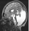
Dandy Walker syndrome

A

genesis of cerebellar vermis; cystic enlargement of 4th ventricle; 70-80% cases associated with hydrocephalus
38
Q

what types of neurons are affected in huntingtons disease

A

basal ganglia (cuadate/ putamen)

39
Q

what types of neurons are affected in Alzheimers

A

hippocampus, cortex

40
Q

What types of neurons are affected in parkinsons

A

substantia nigra within the midbrain

41
Q

chondroitin sulfate proteoglycans

A

growth inhibitory substance produced by reactive astrocytes

42
Q

White matter territories

A

Dorsal (posterior) Funiculus

Lateral Funiculus

Ventral (anterior) Funiculus

Anterior White Commissure

43
Q

Fasciulus gracilis

A

present at all cord levels

aspect of the dorsal colum medial lemniscus pathway

carries info from lower extremities to the inferior poriton of the medial lemniscus

44
Q

Fasiculus Cuneatus

A

present only from T6-C1

part of dorsal column medial lemniscus pathway

carries info from the upper extremities to superior medial lemniscus

45
Q

Gray matter territories

A

dorsal/posterior horn

ventral/anterior horn

46
Q

stereognosis

A

object identification by touch; done by the dorsal column medial lemniscus system

47
Q

syrinx

A

fluid collection in the spinal cord that expands the central canal; compressess and ultimatel destroys the nervous tissue in the affected area

48
Q

Type IV anderson’s disease

A

glycogen storage disease; poorly branched and insoluble

due to deficieincy in transglucosidase (branching enzyme)

no epilepsy!

49
Q

type VII tauri’s disease

A

muscle specific glycogen storage disease; no phosphofructokinase leads to incr G6P which allosterically activates glycogen synthase

therefore incr synthase to branhcing ration –> accumulation of poorly branched polyclucosan

50
Q

lateral geniculate nucleus

A

receives information from retina, visual input

51
Q

medial geniculate nucleus

A

receives auditory info from inferior colliculous

52
Q

locations of neuropathies

A

mononeuropathy = focal

mononeuropathy multiplex = multifocal

polyneuropathy = generalized

53
Q

Intermediate/ lateral horn

A

enlargement seen on the lateral aspect of the thoracic gray matter in the spinal cord

location of the autonomic nerves

54
Q

denticulate ligament

A

separates dorsal from ventral roots; anchors the dura

55
Q

which are the fast sensors?

A

pacinian, meissner, hair

56
Q

which are the slow sensors

A

merkel, ruffini, free nerve ending

57
Q

ruffini

A

stretch

58
Q

meissner

A

pressure

59
Q

pacinian

A

vibration

60
Q

merkle

A

light touch

61
Q

what do we mean when we say we are looking at duration?

A

whether something is fast or slow adapting

62
Q

two ways to measure intensity

A

population code and frequency code

63
Q

frequency code

A

how many AP per unit time (depends on intensity of stimulus)

64
Q

population code

A

how many are activated

65
Q

periaqueductal gray

A

around the cerebral aqueduct in the midbrain

rostral ventral medulla - serotonergic (5HT) neurons of raphe nuclei project to spinal cord

Neuropeptide substance P - sent by RVM to locus ceruleus

66
Q

hyperalgesia

A

increased sensitivity to an already painful stimulus

67
Q

allodynia

A

normally non painful stimuli are felt as painful

68
Q

3 models for how neuroectoderm differentiates into epidermis v. neurons

A

1: epidermal factor –> epidermis + neuronal factor –> neurons

Epidermal factor = BMP-4 (promotes epidermal fate and inhibits neuronal fate)

69
Q

corticospinal tract

A

PYRAMIDS

70
Q

MLF

A

pontine tract; medial in the rostral medulla

71
Q

vestibular nuclei locaiton

A

near the inferior cerebellar peduncal in the rostral medulla

72
Q
A
73
Q

upper motor neuron syndrome

A

inital spinal shock/hypotonia, followed by spasticity, clonus, hyporeflexia, muscle paresis and positive babinski sign

74
Q

what does a positive or upgoing babinsci sign indicate

A

fanning of toes/extensor plantar response indicates upper motor neuron syndrome

75
Q

why does UMN leison cause spasticity

A

inputs from brainstem and cerebral cortex have SUPPRESSIVE effect on reflexes; so when you lose the suppression, you get spasticity = hyperreflexia and hypertonia in muscles

however!!! you also get hyporeflexia for superficial reflexes

76
Q

filopedia

A

actin filaments growing at end of oligodendrocyte process

77
Q

lamellipodia

A

widening filopedia; formed as microtubules migrate into membrane protrusions, bind actin and stabilize filaments

78
Q

NOGO

A

usually sequestered to the nucleus, but released to the ER upon damage to the oligodendrocyte; when released, it binds neuron and inhibit growth –> aka growth is a no-go!

79
Q

steps for oligodendrocyte

A
  1. differentiation
  2. process extension
  3. contact formation and initial wrapping
  4. myelin compaction/formation
80
Q

lipid rafts

A

specialized microdomains at leading edge of oligodendrocyte process that accumulate as oligodendrocyte differentiate

contain: glycosphingolipids, cholesterol, integrins , NCAM120

81
Q

alternating hemiplesia

A

contralateral trunk and extremity and ipsilateral CN motor n issue

82
Q

macula

A

receptive element of uticle and saccule; located within membranous labryinth

83
Q

endolymph composition

A

sodium poor

potassium rich

84
Q

perilymph composition

A

potassium poor

sodium rich

85
Q

otolythic membrane

A

gelatinous mass overlying the macula (which is the receptive element for uticle and saccule )

86
Q

what are stereocilia of the ear

A

large microvilli

87
Q

what are kinocilium

A

true cilia

88
Q

crista

A

sensory receptive element of the semicircular canals; has a population of hair cells, which also have stereocilia and kinocilia that extend upwards into the cupula

89
Q

in head turning, oculomotor does

A

medial rectus

90
Q

abducens

A

lateral rectus

91
Q

key area in human brain for processing vestibular information

A

parieto-insular region

92
Q

fasiculation

A

LMN!

93
Q

decorticate rigidity

A

rubrospinal and vestibulospinal tracts are active

lesion above midbrain

arms flexed (point toward head), legs extended, rigid

94
Q

decerebrate rigidity

A

vestibulospinal tract active

lesion below midbrain

arms and legs extended, rigid

95
Q
A
96
Q

where are mirror neurons found?

A

Premotor cortex, inferior parietal lobule

97
Q

primary gustatory cortex

A

anterior region of insular lobe and “the frontal operculum” located on the inferior frontal gyrus of the frontal lobe

98
Q

what nerves control taste of tongue

A

CNX (Back)

CN IX (mid)

CN VII (front)

99
Q

What nerves control sensation of tongue

A

CNX (back)

CNIX (mid)

CNV3 (front)

100
Q

what is the majority of the blood supply to the lateral geniculate nucleus?

A

posterior cerebral artery

101
Q

magnocellular pathway

A

dorsal pathway, the where pathway for vision;

goes to superior parietal lobe

responsible for action associated with objects

102
Q

parvocellular pathway

A

the what pathway, important for object identification, goes to the inferior temporal lobe

103
Q

Retrolenticular fibers

A

carry information from the inferior aspect of the visual field; take that information through the parietal lobe to the cuneus of the occipital lobe

104
Q

Sublenticular fibers

A

carry information from the superior aspect of the visual field; go through the TEMPORAL lobe to get to the lingula of the occipital lobe

105
Q

plexiform layers of the eye

A

where the synapses between layers of cells occur

106
Q

glaucoma

A

lose peripheral vision and have tunnel vision in advanced cases.

patterns of the visual outcomes of glaucoma and ARMD are opposite or complementary.

107
Q

What is outlined in red line?

A

Pretectal area; if stimulated will get bilateral activation of the parasympathetics of the CN III

108
Q

posterior parietal lobe role in vision

A

detection of motion and location and also vision for action

(part of the where pathway, magnocellular)

109
Q

temporal cortex role in vision

A

comprehension of form & color

part of what parvocellular pathway

110
Q

how do you know its inferior colliculi?

A

theyre dark

111
Q

when you see inferior colliculi what should you think

A

egg and nest –> trochlear nerve and MLF

Trochlear nerve only found in inferior midbrain!!!!

112
Q

rubrospinal

A

flexors (mainly upper extremities)

starts in red nucleus

on in decorticate rigidity but off in decerebrate rigidity (b/c below midbrain)

113
Q

An individual with neurologic disease exhibits a generalized increase in muscle tone, whereas deep tendon reflexes are normal. Which one of the following can account for this finding?

A

Abnormal increase in excitatory drive to static gamma motor neurons.

114
Q

tegmentum of pons contains

A

spinal nucleus

trigeminal spinal tract

VTT

ALS

medial lemniscus

115
Q

Horner’s syndrome where would lesion be

A
  • lateral spinal cord
  • sympathetic chain
116
Q

Where is this

A

Cuadal medulla

117
Q

Identify A

A

solitary nucleus (for taste) in the caudal medulla

118
Q

Identify B

A

Contains ALS and spinal tract of V in caudal medulla

119
Q

“locked-in” syndrome

A

bilateral lesion base of the upper pons

120
Q

What is the arrow pointing to

A

left optic tract

121
Q

What is the arrow pointing to

A

Optic nerve

122
Q

What is the arrow pointing to

A

lateral geniculate nucleus

123
Q

where is the fovea

A

most medial aspect of the temporal hemiretina

124
Q

medial reticulospinal

A

pons

125
Q

lateral reticulospinal

A

medulla!

126
Q

where are the corticospinals located in the pons

A

base

127
Q

whats here?

A

corticospinal

128
Q

vestibular spinals

A

Provide powerful anti gravity drive

129
Q

What is the orange delinating

A

the efferent aspect of the corneal blink reflex

130
Q

middle cerebellar peduncle

A

CN V afferent

efferent –> muscles of mastication

131
Q

interpedunclar fossa

A

where oculomotor comes out ; level of midbrain

132
Q

cerebellopontine

A

7 exits, smile ! at pons medulla junciton

133
Q

What cranial nerve could be affected
by an aneurysm of the posterior
communicating artery?

A

CN III

134
Q

inner retina

A

central retina

135
Q

chondrotion sulfate proteoglycan

A

made by reactive astrocytes

136
Q

posterior ciliary supplies

A

inner and outer segments containing photoreceptors, outer nuclear layer and outer plexiform layer

137
Q

Whats this

A

a lesion to the edinger westphal nucleus bilaterally (issues with pupillary light reflex & accomodation bilaterally)

138
Q

whats in purple

A

internal capsule

139
Q

whats in red

A

lgn

140
Q

whats this in red

A

parvocellualr - inferior temporal lobe ; posterior cerebral

141
Q

what artery comes to mind for horner’s syndrome?

A

PICA!

142
Q

true vertigo

A

spinning or sensation of movement

143
Q

vestibular neuritis

A

nystagmus is horizontal, follows alexanders law and does not change direction

h-HIT corrective saccade

144
Q

vestibular nystagmus or direction chanigng nystagmus

A

sign of stroke

145
Q

h-HIT

A

head impuse test; corrective saccade observed in vestibular neruitis but not stroke

146
Q

CGRP

A

calcitonin gene related peptide; target of migraine med

147
Q
A
148
Q

CSPG

A

chondroitin sulfate proteoglycans –> prodcued by reactive astrocytes