Visual Systems Flashcards
which visual field information remains ipsilateral?
which visual field information crosses to the contralateral side of the optic chiasm?
nasal
temporal
identify 1 and 2


Upper Visual Field is controlled by which part?
Lower Bank of Calcarine Sulcus
Lower Visual Field is controlled by which part?
Upper Bank of Calcarine Sulcus
what is the circuit followed by signals in vision?
Optic Nerve
Optic Chiasm
Optic Tract
LGN
Visual Cortex
what does the visual cortex receive?
fibers from the LGN
what happens in the LGN?
it divides into upper and lower
where does the optic tract go?
sends info from the left/right to the LGN
optic tract, ipsilateral nasal field fibers will project where?
contralateral temporal field fibers will project where?
layers 2,3,5 of the LGN
layers 1,4,6 of the LGN
all fibers that the LGN receives will then project where?
into layer 4 of the visual cortex
the LGN has two types of neurons, what are these?
Parvocellular: small cells which receive input from small ganglion cells in the retina.
Magnocellular: large cells, receiving input from large ganglion cells.
identify the areas

- Optic Nerve
- Lateral Optic Chiasm
- Central Optic Chiasm
- Optic Tract
- Meyer’s Loop (lower part of the Geniculocalcarine tract)
- Upper part of Geniculocalcarine tract
- Visual cortex
what do you get if the optic nerve is damaged?
what may cause optic nerve damage?
unilateral blindness
trauma and optic neuritis
what happens if you damage the lateral part of the Optic chiasm?
what may cause bilateral damage to the lateral optic chiasm?
what is the most common cause for damage to the lateral part of the optic chiasm?
binasal hemianopia
internal carotid aneurysm
calcified internal carotid arteries
what do you get if there is damage to the central part of the optic chiasm?
what may cause damage to the central part of the optic chiasm?
bitemporal hemianopia
pituitary tumor and craniopharyngioma
what happens if you get damage to the optic tract?
right or left homonymous hemianopia
what happens is there is damage to Meyer’s loop?
what happens if there is damage to the upper geniculocalcarine tract?
what happens if there is damage to the Visual cortex?
upper quadrantinopia
lower quadrantinopia
homonymous hemianopia with macular sparing
if there is damage to the visual cortex, there is macular sparing due to what?
because of anastomosis of the calcarine and middle cerebral arteries in the most posterior region of the visual cortex, which receives macular fibers.
what is this called?
what can lead to it?

constricted field
end stage glaucoma or conversion disorder
what is this called?
what can lead to it?

central scotoma
optic neuritis, MS
what is this?
what can cause it?

upper altitudinal hemianopia
bilateral damage to the lingual damage
what is this?
what can cause it?

lower altitudinal hemianopia
bilateral damage to cuneus gyrus
what is the blood supply to the following areas:
- optic nerve
- optic chiasm
- optic tract
- LGN
- geniculocalcarine tract
- visual cortex
- circle of willis
- anterior cerebral and internal carotid
- PCA and anterior choroidal
- MCA, anterior choroidal and calcarine
- calcarine and MCA
calcarine artery is a branch off which other artery?
Anterior choroidal artery is a branch off what artery?
PCA
internal carotid
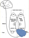
what are the 6 eye muscles and the CN that controls each?
- CN 3
- superior rectus
- inferior rectus
- medial rectus
- inferior oblique
- CN 4
- superior oblique
- CN 6
- lateral rectus
in what direction will each muscle turn the eye
- superior rectus
- inferior rectus
- medial rectus
- inferior oblique
- lateral rectus
- superior oblique
- up
- down
- adduct
- up and abduct
- abduct
- down and abduct
how do you test for superior and inferior rectus muscles?
how do you test for superior and inferior oblique?
tell patient to look 23 degrees lateral and then move up and down
Ask the patient to look medially 23 degrees and then move up and down
a lesion of CN 3,4,6 is at what level?
damage to the MLF is at what level?
if horizontal or vertical gaze damage is at what level?
nuclear level
internuclear
supranuclear
if horizontal gaze is damaged what part is affected?
what about vertical gaze?
frontal eye field
pontine tegmentum
Damage to CN VI will result in?
Lack of abduction, medial deviation at rest (strabismus)
Horizontal diplopia only, when looking in direction of lesion.
what is 1 and a half syndrome?
Damage to abducens nucleus may only affect the lateral rectus itself; it may also affect the connections of abducens nucleus to the contralateral, CN III (lateral/horizontal conjugate gaze, reviewed in a few slides).
can result from damage to pons: leading
- Ispilateral CN VI (lateral rectus).
- MLF connection to ispilateral CN III (medial rectus).
- MLF connection to contralateral CN III (medial rectus).
what do you get with this lesion?

1 and a half syndrome
Control of voluntary conjugate movements includes which structures?
frontal eye fields, parietal lobe, PPRF, and rostral
MLF.
rostral MLF damage can cause what?
what happens if you damage the Frontal Eye Fields?
paralysis of vertical (upward) gaze (Parinauds)
there is no conjugate gaze to the lesioned side and inability of the eyes to look voluntarily to the opposite side.
Damage to frontal eye fields, or PPRF, can be considered as?
supranuclear opthalmoplegia
what is MLF Syndrome?
demyelination of the medial longitudinal fasciculus interrupts connections between CN III, IV, VI, and VIII.
basically Disconnection of CN III and VI interferes
with the lateral conjugate gaze.
Parietal eye field is superior parietal lobule; damage here results in what?
decreased eye movements towards the opposite side and neglect
how will someone with CN 6 walk?
- dejected appearance: Patient will tilt head forward to compensate for difficulties in depressing the eye.
- Patient will tilt head to the side to compensate
for rotational problems (torsional diplopia). Head tilt is towards a nucleus, and away from a nerve, lesion.
what is Aniscoria?
what is scotoma?
asymmetric pupils
blind spots in visual field
what is a Argyl Robertson Pupil?
what may cause it?
an absence of direct and consensual light reflexes. Miotic reaction to accommodation/convergence is still intact
(doesnt react to light but do constrict in conversion)
syphilis, diabetes, and lupus.
how does the puppillary reflex work?
- Light stimulates the afferent component of CN II; these axons synapse in both pretectal areas.
- Neurons in the pretectal areas send axons to the Edinger-Westphal nucleus of CN III (bilaterally).
- Efferents from CN III innervate the pupillary sphincter via the ciliary ganglion, causing pupillary constriction.
how many layes does the superior colliculus have?
what are they?
what is the main function of each?
what is the overall function of the superior colliculus?
what happens if there is damage here?
Three:
- Superficial: receives info from retina and visual association areas (18, 19).
- Middle: receives frontal eye field info.
- Deep: gets ascending sensory info.
- big role in reflexive eye movements,
- nothing happens