Imaging Flashcards
what do you look for a pathology in imaging?
midline, shift, asymmetry
is this MRI, CT, XRay…
at what level?

CT
left: lateral ventricles so it is very superior
right: brainstem level (the eyeballs are present)
which will be anatomical: T1 or T2?
anatomical = white matter is white, grey matter is grey
T1 is anatomical
Is this T1 or T2?

T-1
is this T1 or T2?

T 1
is this T1 or T2?

T2
Which machine uses T1/T2?
MRI
identify how the things look in the image


answer


answer

1) anterior commissure
2) interthalamic adhesion
3) third ventricle
4) spleneium
5) cingulate gyrus
answer


What is Flow Void?
In which imaging can you see it and how does it look?
Flowing blood (not bleeding) appears dark on a T2 but not on CT/T1.
answer


a bleed is always what color in all CT and MRI?
infarcts, tumors and MS plaque are dark in which?
MRI T1, MRI T2, CT
bright
T1 and CT
is this T1, T2, CT?
what is the pathology?

CT
epidural hematoma
is this T1, T2, CT?
What is the pathology?

T1
sub-arachnoid bleed
is this T1 or T2, or CT?
pathology?

T1
shaken baby = right is old blood, left is new blood
what 2 enhancement can you use for T2?
what do they do?
- Proton density (T2-PD)
- FLAIR
better resolution near the ventricles
what enhancements can be used for T1?
when do you use it?
what enhancements can we use for strokes?
just GAD for tumors
DWI and PWI
what is the pathology?
is this CT or MRI?
hemorrhage
CT
if someone has a hemmorrhage, what imaging is the best?
If someone has an infarct, is CT good for that?
CT
no
what is the pathology here?
what imagin is being used?

subdural hematomas
CT
what type of image?
what is the pathology?
what enhancement is used?

CT
stroke
DWI
what type of image is this?
how do you know?
what is the enhancement?

T2
CSF is very bright
none
what is the type of image?
what type of enhancement?
what is the pathology?
which artery would be damaged?

T2
FLAIR
infarct on left lower side (spots)
Left MCA
what does PWI measure?
what does DWI measure?
change in cerebral blood flow
extent of diffusion of water in short distance
what enhancement is used?

DWI and a FLAIR
DWI and PWI are good for measuring what?
extent of an infarct
what is this imaging?
what is the pathology?

angiogram
right internal carotid occlusion

which area is underperfused?
what should you do?

the right side in blue
give clot buster
what is the penumbra?
the bigger the penumbra, the ……
the area at risk of further infarct damage
given by using DWI and PWI
more reason to give a clot buster
what imaging enhancement is used in MS?
FLAIR and T2-PD
what is the MS criteria?
3 lesions:
1 on the side of lateral ventricle
1 infrantentorial
lesions must be 5mm big
using T1 GAD in MS will help by doing what?

shows new lesions
what are these elongated extensions off the axis fo the ventricles?

Dawson’s fingers
In what disease do you see dawson’s fingers?
MS
what individuals are susceptible to progressive multifocal encephalopathy?
what part of the brain does it affect mostly?
what enhancement doesn’t work well with this disease?
some patients will present only with a leasion localized where?
immunocompromised
white matter
GAD
cerebellum or midbrain
name the disease?

Progressive Multifocal Leukoencephalopathy
what is Diffuse Axonal Injury?
they lose what?….
damage to cerebral hemispheres, brainstem, and possibly cerebellar white matter because of multidirectional trauma
(acceleration/deceleration with rotational forces)
you tear white matter and the brain is twisted
consciousness
how does a CT of a patient who has suffered diffuse axonal injury look like?
why?
normal
because edema is usually minor initially and they are non-haemorrhagic
what is the pathology?

multiple lesions due to diffuse axonal injury
(multi-car accident)
tumors are viewed best with what enhancement and what machine?
MRI T1-GAD
what is GAD?
contrast agent administered into blood circulation peripherally and since tumors like blood..they suck it up and light up in the MRI
identify the type of image and the pathology

what is the type of image and what is the pathology?


This machine measures the changes in blood flow by detecting positrons injecting them into the body?
PET scan
which machine detects radiopharmaceuticals?
this machines is bad for what patients?
PET scans
diabitus!
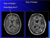
what is happening in the picture?

1 - resting
2 - looking at something
what is going on in the picture?

1- resting
2- moving or hearing
3- thinking, cognitive task (math)
what is going on in the picture?

1 - moving an arm and thinking about it
2 - visual memory task (hippocampus and occipital lobe)
what neuropsychiatric condition is this?

schizophrenia

L-DOPA is given to a patient and placed in a PET-MRI…what condition is this? which side in abnormal?


what type of imaging is this?

PET-CT
what is moya moya disease?
severe bilateral stenosis or occlusion of the arteries around the Circle of Willis with a lot of collateral circulation.
what is this disease?

moya moya
