Visual Histo Flashcards
Identify the blanked out structures or cell types
![](https://s3.amazonaws.com/brainscape-prod/system/cm/128/533/415/q_image_thumb.png?1659502461)
Acinar Cells
Centroacinar cells
Intercalated duct
![](https://s3.amazonaws.com/brainscape-prod/system/cm/128/533/415/a_image_thumb.jpg?1659446192)
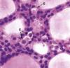
What do the arrows indicate?
![](https://s3.amazonaws.com/brainscape-prod/system/cm/128/533/609/q_image_thumb.jpg?1659502461)
Islets of langerhans
What are three cell types present in the light zone?
What type of pancreatic tissue is this?
![](https://s3.amazonaws.com/brainscape-prod/system/cm/128/533/653/q_image_thumb.jpg?1659502461)
Alpha cells at the periphery
Beta cells at the core of the islet
Delta cells at the periphery
Endocrine tissue
Identify the structures indicated.
![](https://s3.amazonaws.com/brainscape-prod/system/cm/128/533/853/q_image_thumb.png?1659502462)
Light - Posterior Lobe, Neurohypophysis
Dark - Anterior Lobe, Adenohypophysis
![](https://s3.amazonaws.com/brainscape-prod/system/cm/128/533/853/a_image_thumb.jpg?1659446193)
Identify the indicated structures.
![](https://s3.amazonaws.com/brainscape-prod/system/cm/128/534/676/q_image_thumb.png?1659502470)
AP: anterior pituitary
PP: posterior pituitary
P: pituitary stalk (infundibulum)
H: hypothalamus
V: ventricle
O: optic chiasm
![](https://s3.amazonaws.com/brainscape-prod/system/cm/128/534/676/a_image_thumb.jpg?1659446203)
What structure is this tissue from?
![](https://s3.amazonaws.com/brainscape-prod/system/cm/128/535/637/q_image_thumb.jpg?1659502471)
Pineal Gland. Recognize by the brain sand. (S)
What is this tissue from? Is it active?
![](https://s3.amazonaws.com/brainscape-prod/system/cm/128/538/094/q_image_thumb.jpg?1659502475)
Thyroid. Is active
What cells are shown around the colloid structure?
![](https://s3.amazonaws.com/brainscape-prod/system/cm/128/538/114/q_image_thumb.jpg?1659502476)
Inactive Follicular epithelial cells of the thyroid
What do the indicated cells secrete?
![](https://s3.amazonaws.com/brainscape-prod/system/cm/128/538/296/q_image_thumb.jpg?1659502476)
Calcitonin
These are parafollicular cells. Recognize them by small pale staining clusters.
Where are these tissue samples taken from?
![](https://s3.amazonaws.com/brainscape-prod/system/cm/128/538/956/q_image_thumb.png?1659502483)
Parathyroid. Recognize from the presence of adipocytes.
Identify the portions of this tissue. What is this gland?
![](https://s3.amazonaws.com/brainscape-prod/system/cm/128/539/280/q_image_thumb.png?1659502483)
Adrenal Gland
![](https://s3.amazonaws.com/brainscape-prod/system/cm/128/539/280/a_image_thumb.jpg?1659446219)
Cap: Capsule
C: Cortex
M: Medulla
Identify the layers of the Adrenal gland shown.
![](https://s3.amazonaws.com/brainscape-prod/system/cm/128/539/772/q_image_thumb.png?1659502484)
•Capsule (Cap)
•Cortex
–Three zones:
•Zona glomerulosa (G)
•Zona fasciculata (F)
•Zona reticularis (R)
•Medulla (M)
![](https://s3.amazonaws.com/brainscape-prod/system/cm/128/539/772/a_image_thumb.jpg?1659446220)
Where is this from? What layers are shown?
![](https://s3.amazonaws.com/brainscape-prod/system/cm/128/540/180/q_image_thumb.jpg?1659502488)
adrenal gland
Z. Reticularis
Medulla