UWSA1-1 Flashcards
(224 cards)
What is this?

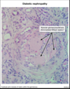
Diabetic nephropathy
When the glomerular sclerosis is nodular, it is described as a Kimmelstiel-Wilson (KW) lesion and is pathognomonic for DN. KW lesions consist of dense hyaline deposits and commonly arise from the glomerular arterioles. As the disease progresses, increased mesangial matrix proteins can lead to global glomerulosclerosis.
Diabetic glomerulosclerosis
pathophys
Diabetic nephropathy (DN) is a common complication of both type 1 and type 2 diabetes mellitus. Prolonged hyperglycemia in diabetes results in glycosylation of endothelial proteins, inflammation, and increased production of growth factors, collagen, and fibronectin. Over time, this results in mesangial expansion, glomerular basement membrane thickening, and glomerular sclerosis.
The first clinical sign of DN is albuminuria, which can progress to overt nephrotic syndrome (eg, peripheral edema, heavy proteinuria, fatty casts) and renal failure. The urine sediment is typically bland (ie, no red or white blood cells or casts).

Focal Segmental Glomerulosclerosis Histology
Focal segmental glomerulosclerosis causes nephrotic syndrome and is associated with foci of sclerosis and hyalinosis in some, but not all, glomeruli.

IgA Nephropathy Presenting symptoms
IgA nephropathy presents with recurrent hematuria associated with upper respiratory infections (synpharyngitic hematuria).
IgA Nephropathy Histology
Light microscopy demonstrates mesangial proliferation, and immunofluorescence demonstrates mesangial immune deposits composed of IgA.

Minimal Change Disease Presenting Features
Minimal change disease causes nephrotic syndrome and typically occurs in children.
Minimal Change Disease Histology
Light microscopy demonstrates normal glomeruli, but electron microscopy demonstrates diffuse effacement of podocytes.

Rapidly Progressive glomerulonephritis presenting features
Rapidly progressive glomerulonephritis causes nephritic syndrome and can occur as an end stage in multiple diseases (eg, Goodpasture syndrome, systemic lupus erythematosus).
Rapidly progressive Glomerulnephiritis Histology
light microscopy demonstrates glomerular crescents composed of proliferating parietal and inflammatory cells.

Cardiac Conduction Velocity Fastest to slowest

Paget Disease Pathophysiology
- Osteoclast dysfunction
- Excessive & disordered bone formation
Paget disease begins with the lytic phase, in which normal bone is resorbed by osteoclasts that are more numerous, are larger, and have many more nuclei (up to 100) than normal osteoclasts (5-10 nuclei). Bone turnover rates increase to as much as 20 times normal.
The second phase, the mixed phase, is characterized by rapid increases in bone formation from numerous osteoblasts. Although increased in number, the osteoblasts remain morphologically normal. The newly made bone is abnormal, however, with collagen fibers deposited in a haphazard fashion rather than linearly, as with normal bone formation. As the osteoclastic and osteoblastic activities of bone destruction and formation repeat, a high degree of bone turnover occurs.
In the final phase of Paget disease, the sclerotic phase, bone formation dominates and the bone that is formed has a disorganized pattern (woven bone) and is weaker than normal adult bone. This woven bone pattern allows the bone marrow to be infiltrated by excessive fibrous connective tissue and blood vessels, leading to a hypervascular bone state.
Paget Disease Clinical manifestations
- Often asymptomatic
- Bone pain & deformity
- Skull: headache, dizziness, hearing loss
- Spine: spinal stenosis, radiculopathy
- Long bones: bowing, focal enlargement, arthritis of adjacent joints
- Elevation in alkaline phosphatase
- X-ray: lytic or mixed lytic/sclerotic lesions; cortical & trabecular thickening
Paget disease of bone is characterized by excessive and disordered bone formation. It commonly involves the skull (eg, headache), spine (eg, radiculopathy, spinal stenosis), or long bones (eg, bone pain, bowing, arthritis of adjacent joints). Long-term complications of Paget disease include fracture, hearing loss, and malignant transformation leading to osteosarcoma.
Paget Disease Complications
- Fracture
- Excessive blood flow: high-output heart failure, vascular steal
- Malignant transformation (eg, osteosarcoma)
Cardiac stress test

coronary steal syndrome

Paget Disease AKA
Osteitis Deformans
Radiologic findings of Osteosacrcoma
Radiographic findings of osteosarcoma include destruction of the normal trabecular bone pattern, mixed radiodense (sclerotic) and radiolucent (lytic) areas, and periosteal new bone formation with lifting of the periosteum (Codman triangle). Adjacent soft tissue often demonstrates ossification in a “sunburst” pattern.

Avascular necrosis caused by
Avascular necrosis can be caused by long-term glucocorticoid use, autoimmune disorders (eg, systemic lupus erythematosus), or conditions that affect blood viscosity (eg, sickle cell disease).
Avascular necrosis radiologic findings
Radiographic findings include the pathognomonic crescent sign, which indicates subchondral collapse at the femoral head
Osteoarthritis ragiographic findings
Osteophytes and joint-space narrowing
Osteoporosis Causes
Osteoporosis is common in older adults, and the risk is increased in patients with corticosteroid exposure (including inhaled corticosteroids)
Osteoporosis radiographic findings
X-ray would show trabecular thinning and possible fracture, rather than lytic/sclerotic lesions and new bone formation.
Secondary Hyperparathyroidism in CKD mechanism
Hypocalcemia and hyperphosphatemia in chronic kidney disease stimulate parathyroid hormone production (secondary hyperparathyroidism).
The resulting effects on bone, known as renal osteodystrophy, can include osteomalacia, osteitis fibrosa cystica, and adynamic bone disease. Patients usually have a known history of advanced renal disease.
Risendronic acid AKA
Risendronate
Risendronate indications
- help prevent osteoporosis
- treat Pagets disease of the bone


