UW7 Flashcards

1
Q

SLE Antibodies

A

Antinuclear Antibodies (ANA) sensitive

Anti-DSDNA antibodies & Anti-Smith antibodies (specific)

2
Q

Lupus nephritis is caused by

A

type III hypersensitivity reaction, immune-complexes deposition in the mesangium

3
Q

Ankylosing spondylitis description

A

Ankylosing spondylitis is a chronic inflammatory arthritis involving the spine and sacroiliac joints. It presents with low back pain and stiffness. Common associated features include enthesitis, dactylitis, and uveitis

4
Q

Question

A

This patient is pregnant with twins of different sexes which can only occur in dizygotic twins due to the fertilization of 2 oocytes by 2 different sperm. Dizygotic twins are almost always dichorionic/diamniotic (eg, 2 chorions and 2 amnions) but may falsely give the appearance of being monochorionic/monoamniotic if the chorions and amnions fuse due to proximity of implantation sites.

5
Q

Dizygotic twins

A

Dizygotic twins are almost always dichorionic/diamniotic (eg, 2 chorions and 2 amnions) but may falsely give the appearance of being monochorionic/monoamniotic if the chorions and amnions fuse due to proximity of implantation sites

6
Q

Monozygotic twins

A

monozygotic twins develop from division of a single zygote after fertilization of a single oocyte. They are of the same sex, are genetically identical, and are similar in appearance. The type of placentation in monozygotic twins depends on when zygote division occurs during embryonic development.

Early division (days 0-4) can result in monozygotic twins with 2 chorions and 2 amnions (eg, dichorionic/diamniotic), which may or may not be fused. If the sexes are the same, it may be difficult to distinguish whether the twins are monozygotic or dizygotic until later in the pregnancy.

Division between days 4-8 is the most common outcome in monozygotic twins and results in 1 chorion (eg, shared placenta) but 2 amnions (eg, monochorionic/diamniotic twins)

Late division (8-12 days) results in 1 chorion and 1 amnion. A monochorionic/monoamniotic pregnancy is associated with a high fetal fatality rate, due primarily to the increased risk of umbilical cord entanglement

Division occurring after 13 days can result in monochorionic/monoamniotic conjoined twins

7
Q

Monozygotic twins: Early division (days 0-4)

A

Early division (days 0-4) can result in monozygotic twins with 2 chorions and 2 amnions (eg, dichorionic/diamniotic), which may or may not be fused. If the sexes are the same, it may be difficult to distinguish whether the twins are monozygotic or dizygotic until later in the pregnancy.

8
Q

Monozygotic twins: Division between days 4-8

A

Division between days 4-8 is the most common outcome in monozygotic twins and results in 1 chorion (eg, shared placenta) but 2 amnions (eg, monochorionic/diamniotic twins)

9
Q

Monozygotic twins: Late division (days 8-12)

A

Late division (8-12 days) results in 1 chorion and 1 amnion. A monochorionic/monoamniotic pregnancy is associated with a high fetal fatality rate, due primarily to the increased risk of umbilical cord entanglement

10
Q

Monozygotic twins: Division > or = 13 days

A

Division occurring after 13 days can result in monochorionic/monoamniotic conjoined twins

11
Q

Question

A

C. polymorphic ventricular tachycardia (torsade du pointes)

Relatively young patients with symptomatic paroxysmal atrial fibrillation are often treated with a rhythm-control strategy to restore and maintain normal sinus rhythm and to eliminate symptoms. Antiarrhythmic drugs commonly used for rhythm control of atrial fibrillation include class IC (eg, flecainide, propafenone) and class III (eg, ibutilide, dofetilide) agents.

Dofetilide and ibutilide selectively block the rapid component of the delayed-rectifier potassium current. This slows repolarization and prolongs action potential duration and the effective refractory period in cardiomyocytes, suppressing the electrical foci that lead to atrial fibrillation. As a result, the QT interval is prolonged, leading to an increased risk of torsade de pointes, a form of polymorphic ventricular tachycardia that can cause syncope and sudden cardiac death. To minimize this risk, dofetilide or ibutilide therapy is started only in the hospital with temporary cardiac monitoring.

(Choice A) Adenosine can trigger bronchospasm, likely by stimulating the release of histamine and leukotrienes from mast cells. Class III antiarrhythmics are not associated with bronchospasm.

(Choice B) Beta blockers (via inhibition of beta-1 receptor–driven chronotropy) and nondihydropyridine calcium channel blockers (via blockade of L-type atrioventricular [AV] node calcium channels) slow AV node conduction and in overdose can cause complete AV block. Digoxin (via enhanced vagal tone) and adenosine can also cause AV block. Although some class III antiarrhythmics (ie, sotalol, amiodarone) have negative chronotropic effects and may slow AV node conduction, dofetilide and ibutilide have no significant effect on AV node conduction.

(Choice D) Amiodarone can disrupt thyroid function and lead to hyper- or hypothyroidism, but dofetilide and other class III antiarrhythmics do not affect thyroid function. Compared with other class III antiarrhythmics, amiodarone is associated with a lower risk of torsade de pointes.

(Choice E) Most antiarrhythmic drugs, including class III, are not associated with an increased risk of venous thromboembolism.

(Choice F) Digoxin toxicity can cause visual disturbances consisting of blurred or yellow vision, but visual disturbance is not an adverse effect of class III antiarrhythmics.

Educational objective:
Dofetilide and ibutilide are class III antiarrhythmics that treat atrial fibrillation by blocking the rapid component of the delayed-rectifier potassium current to slow repolarization and increase the effective refractory period. As a result, the QT interval is prolonged, creating an increased risk of polymorphic ventricular tachycardia (torsade de pointes).
12
Q

Question

A

E.

13
Q

How to organize a table into standard format

A
14
Q

Question

A

B. Effect modification

Effect modification occurs when the effect of an exposure on an outcome is modified by another variable. It can be identified using stratified analysis (analyzing the cohort as different subgroups), as the different strata will have different measures of association. In this scenario, smoking status modified the effect of the new estrogen receptor agonist (exposure) on deep vein thrombosis (DVT) incidence (outcome). Using stratified analysis by smoking status:

Among smokers, there was a statistically significant association between taking the new estrogen receptor agonist and risk of developing DVT with a relative risk of >1, indicating higher risk, and a p-value of <0.05, indicating statistical significance.

In contrast, among nonsmokers, there was no statistically significant association between taking the medication and risk of DVT (p-value >0.05).

Effect modification is not a bias (Choices D and E), as it is not due to flaws in the design or analysis phases of the study. It is a natural phenomenon that should be described, not corrected.

Effect modification is most easily confused with confounding (Choice A), but stratified analysis can help distinguish between these 2 scenarios. With effect modification, the different strata will have different measures of association, as seen in this example of the association between taking the estrogen receptor and the risk of DVT among smokers compared to nonsmokers. In contrast, with confounding, stratification usually reveals no significant difference between the strata. For instance, in an analysis of primary school students (of all grade levels), age can be a confounder that muddies the association between shoe size and intelligence. Children with bigger shoe sizes may appear to be more intelligent on initial analysis. However, this association is likely not due to shoe size but rather to age because older children tend to have both bigger feet and more intelligence. When older and younger children are analyzed separately (stratification based on the confounder), the association between shoe size and intelligence disappears.

(Choice C) The latent period is the time required for an exposure to begin having an effect. However, there is no information on how latency was handled in this study.

Educational objective:
Effect modification is present when the effect of the main exposure on the outcome is modified by the presence of another variable. Effect modification is not a bias.

15
Q

Question

A

C. Neisseria Meningitidis

Paroxysmal nocturnal hemoglobinuria (PNH) is caused by a mutation in hematologic stem cells that eliminates production of an anchoring protein that attaches surface molecules to the cell membrane. Loss of this membrane anchor prevents erythrocytes from expressing the complement-inactivating surface proteins CD55 and CD59. Without these surface proteins, autoactivated components of the alternative complement cascade cannot be disabled. This leads to spontaneous complement amplification, the generation of membrane attack complexes (MACs) on the red cell membrane, and subsequent complement-mediated hemolysis.

Treatment with a monoclonal antibody (eg, eculizumab) that targets C5, the first complement protein that contributes to the formation of MACs, can drastically reduce hemolysis and improve symptoms (eg, fatigue) in patients with PNH. However, the generation of MACs is crucial for defense against encapsulated organisms (eg, Neisseria meningitidis, Streptococcus pneumoniae) because these pathogens are resistant to other host defense mechanisms (eg, immunoglobulin binding, phagocytosis).

Patients receiving anti-C5 therapy require vaccination against encapsulated pathogens (N meningitidis, S pneumoniae) and appropriate antibiotic prophylaxis (eg, penicillin) to prevent fulminant infection.

(Choice A) Nontypable strains of Haemophilus influenzae are not encapsulated; therefore, these strains are susceptible to phagocytosis and immunoglobulin-mediated opsonization. Control is not primarily mediated by the formation of MACs.

(Choice B) Host defense to Mycobacterium tuberculosis depends more on T-cell and antigen-presenting cells than on complement activation and MAC formation.

(Choices D and E) Staphylococcus aureus and Streptococcus pyogenes often secrete factors that disable the complement cascade. Therefore, they are primarily countered by other components of the immune system (eg, neutrophils, humoral immunity).

Educational objective:
Paroxysmal nocturnal hemoglobinuria leads to the formation of membrane attack complexes on erythrocytes. Treatment with monoclonal antibody against C5, the first component of the membrane attack complex, can improve symptoms. However, it also increases risk for encapsulated bacterial infection. Therefore, patients require vaccination and antibiotic prophylaxis against Neisseria meningitidis and Streptococcus pneumoniae.

16
Q

Paroxysmal nocturnal hemoglobinuria Pathophysiology

A

Paroxysmal nocturnal hemoglobinuria (PNH) is caused by a mutation in hematologic stem cells that eliminates production of an anchoring protein that attaches surface molecules to the cell membrane. Loss of this membrane anchor prevents erythrocytes from expressing the complement-inactivating surface proteins CD55 and CD59. Without these surface proteins, autoactivated components of the alternative complement cascade cannot be disabled. This leads to spontaneous complement amplification, the generation of membrane attack complexes (MACs) on the red cell membrane, and subsequent complement-mediated hemolysis.

17
Q

Paroxysmal nocturnal hemoglobinuria Tx

A

Treatment with a monoclonal antibody (eg, eculizumab) that targets C5, the first complement protein that contributes to the formation of MACs, can drastically reduce hemolysis and improve symptoms (eg, fatigue) in patients with PNH. However, the generation of MACs is crucial for defense against encapsulated organisms (eg, Neisseria meningitidis, Streptococcus pneumoniae) because these pathogens are resistant to other host defense mechanisms (eg, immunoglobulin binding, phagocytosis).

Patients receiving anti-C5 therapy require vaccination against encapsulated pathogens (N meningitidis, S pneumoniae, typeable H. flu) and appropriate antibiotic prophylaxis (eg, penicillin) to prevent fulminant infection.

18
Q

Question

A

Vitamin D is derived from food and generated in the skin due to sunlight exposure (ie, conversion of 7-dehydrocholesterol to cholecalciferol). Endogenous production from sunlight accounts for approximately half of daily requirements (~200-400 IU/day [required: 600]). The dietary share is usually obtained primarily from fortified dairy/dairy-alternative foods; oily fish (eg, salmon, herring) is also an abundant source. Other foods contain vitamin D but are often insufficient to meet requirements.

Vitamin D deficiency reduces intestinal absorption of calcium. Parathyroid hormone secretion increases to mobilize calcium from bone and maintain plasma calcium levels but causes renal phosphate wasting. The resulting hypophosphatemia causes impaired mineralization of bone. In children, this manifests as rickets, characterized by excessive unmineralized osteoid matrix at the epiphyseal (growth plate) cartilage.

Clinical manifestations of rickets include frontal bossing, craniotabes (softened skull bones), and costochondral widening from cartilage overgrowth (rachitic rosary). Dental enamel hypoplasia can also be seen. Weight-bearing children may show lower extremity bowing (genu varum). Radiographs of growth plates (eg, distal ulna) often reveal metaphyseal plate widening and cupping.

(Choice A) Glucocorticoids (eg, prednisone) cause osteoporosis because they inhibit proliferation and differentiation of osteoblast precursors and increase apoptosis of mature osteoblasts. Fragility fractures may occur, but this patient’s deformities are more consistent with rickets.

(Choice B) The early osteolytic phase of Paget disease of bone is characterized by proliferation of abnormally large, hypernucleated osteoclasts. Paget disease causes bone pain and deformity (eg, frontal bossing, bowing of long bones) but findings are typically focal and asymmetric and usually occur in adults age >50.

(Choice C) Replacement of bone with disorganized fibrous connective tissue (fibrous dysplasia) can occur in a single bone (eg, femur) or multiple bones (polyostotic). Polyostotic fibrous dysplasia (McCune-Albright syndrome) is typically associated with café au lait macules and pituitary endocrine disorders (eg, precocious puberty, thyrotoxicosis, acromegaly).

(Choice D) Osteosarcoma, the most common malignant bone tumor in children, is characterized by excessive production of mineralized bone. The lesions are typically single and would not cause short stature, frontal bossing, or enamel defects.

Educational objective:
Vitamin D deficiency decreases intestinal absorption of calcium. Parathyroid hormone secretion increases to maintain plasma calcium, which causes renal phosphate wasting and impaired bone mineralization. In children, this causes rickets characterized by excessive unmineralized osteoid matrix at the epiphyseal cartilage.

19
Q

Physical manifestations of Ricketts

A
20
Q

Question

A

E. interchain cross-links involving lysine
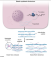
This patient’s emphysema is likely due to alpha-1 antitrypsin deficiency. Neutrophil-secreted elastase is an endogenous proteolytic enzyme that hydrolyzes elastin within alveolar walls. The liver synthesizes alpha-1 antitrypsin, a protein that inhibits neutrophil elastase and prevents alveolar wall degradation, particularly in the lower airways. Patients with alpha-1 antitrypsin deficiency consequently develop excessive alveolar elastin degradation, which clinically manifests with early-onset, lower lobe–predominant emphysema.

Elastin is a fibrous connective tissue protein that provides elasticity to the skin, blood vessels, and pulmonary alveoli. The fibers can stretch to several times their length and recoil back to their original size once stretching forces are withdrawn. Elastin assembly is closely related to that of collagen. Similar to collagen, elastin is synthesized as a large polypeptide precursor (tropoelastin) composed of about 700, mostly nonpolar, amino acids (eg, glycine, alanine, valine) (Choice D). Elastin also contains proline and lysine residues; however, in contrast to those found in collagen, few of these amino acids are hydroxylated (Choice C).

After tropoelastin is formed, it is secreted into the extracellular space where it interacts with microfibrils (fibrillin) that function as a scaffold. Next, lysyl oxidase, a copper-dependent enzyme, oxidatively deaminates some of the lysine residues of tropoelastin, facilitating the formation of desmosine cross-links between neighboring polypeptides that hold the elastin molecules together. These cross-links, along with the high content of nonpolar (hydrophobic) amino acids, account for the rubber-like properties of elastin.

(Choices A and B) Disulfide bridges are formed during collagen, not elastin, synthesis. After post-translational hydroxylation and glycosylation of procollagen molecules, disulfide bond formation between the C-terminal propeptide regions of 3 alpha chains brings the chains into a favorable alignment for triple helix assembly.

Educational objective:
The rubber-like properties of elastin are due to high content of nonpolar (hydrophobic) amino acids and extensive cross-linking between elastin monomers facilitated by lysyl oxidase. Patients with alpha-1 antitrypsin deficiency can develop early-onset, lower lobe–predominant emphysema due to excessive alveolar elastin degradation.

21
Q

Question

A

D. Tyrosine to fumarate

Alkaptonuria is an autosomal recessive disorder of tyrosine metabolism. Deficiency of homogentisic acid dioxygenase blocks homogentisic acid metabolism, preventing the conversion of tyrosine to fumarate. Homogentisic acid accumulates in the body and is excreted in the urine, imparting a black color to the urine if allowed to sit and undergo oxidation. In patients with alkaptonuria, the retained homogentisic acid selectively binds to collagen in connective tissues, tendons, and cartilage. This leads to “ochronosis,” a blue-black pigmentation most evident in the ears, nose, and cheeks, and ochronotic arthropathy, which typically manifests during adulthood.

(Choice A) Leucine is a branched-chain amino acid that is elevated in maple syrup urine disease. Isoleucine and valine are also increased. Impaired metabolism of these amino acids leads to cerebral edema, seizures, and a sweet smell of the urine.

(Choice B) Conversion of phenylalanine to tyrosine is defective in phenylketonuria and usually occurs due to a defect in phenylalanine hydroxylase. Undiagnosed and untreated phenylketonuria results in significant intellectual disability not seen in this patient.

(Choice C) Impaired renal cystine (a homodimer of cysteine) transport leads to cystinuria, a disease characterized by flank pain, hematuria, and renal stones in childhood or adolescence.

(Choice E) Sickle cell anemia results from the substitution of valine for glutamic acid due to a single-nucleotide polymorphism. This mutation leads to loss of red cell elasticity, polymerization of sickle hemoglobin, and sickling of red blood cells, which results in vasoocclusive crises.

Educational objective:
Alkaptonuria is an autosomal recessive disorder in which the lack of homogentisic acid dioxygenase blocks the metabolism of tyrosine, leading to an accumulation of homogentisic acid. Clinical features include a black urine color when exposed to air, a blue-black pigmentation on the face, and ochronotic arthropathy.

22
Q

Alkaptonuria pathophysiology

A

autosomal recessive disorder of tyrosine metabolism.

Deficiency of homogentisic acid dioxygenase blocks homogentisic acid metabolism, preventing the conversion of tyrosine to fumarate.

Homogentisic acid accumulates in the body and is excreted in the urine, imparting a black color to the urine if allowed to sit and undergo oxidation.

In patients with alkaptonuria, the retained homogentisic acid selectively binds to collagen in connective tissues, tendons, and cartilage. This leads to “ochronosis,” a blue-black pigmentation most evident in the ears, nose, and cheeks, and ochronotic arthropathy, which typically manifests during adulthood.

23
Q

Question

A

A. Expanded tri-nucleotide repeats

This child with intellectual disability has weakness and slow muscle relaxation, findings suggestive of myotonic dystrophy (DM). DM is an autosomal dominant muscular dystrophy due to an expansion of CTG trinucleotide repeats within the dystrophia myotonica protein kinase (DMPK) gene. Increased repeat length occurs with successive generations and is associated with earlier onset of more severe disease (ie, anticipation); this patient’s parent with mild muscle weakness is most likely due to adult-onset DM.

In the childhood form of DM, intellectual disability and behavioral problems are often the predominant features. Other key findings characteristic of classic DM may be present or develop over time:

Weakness, atrophy, and pain of facial and distal limb musculature

Myotonia, or delay in muscle relaxation, such as sustained hand contraction after attempted grip release or sustained thumb abduction after thenar eminence percussion (as seen here)

Cardiac abnormalities (eg, conduction disturbances, cardiomyopathy)

Gastrointestinal tract disturbances (eg, dysphagia, constipation)

Nonmuscular involvement such as frontal hair loss, cataracts (lens opacities), insulin resistance, and daytime somnolence

(Choice B) Mitochondrial myopathies are often maternally inherited and characterized by muscle weakness (eg, exercise intolerance) and lactic acidosis in more severe cases. Cataracts may be present, but myotonia does not occur.

(Choice C) Partial chromosome deletion can be seen with inherited causes of intellectual disability (eg, cri-du-chat syndrome) but would not explain this patient’s myotonia or cataracts.

(Choice D) Uniparental disomy is the mode of inheritance for Prader-Willi and Angelman syndromes, which are associated with cognitive dysfunction and behavioral disturbances but not myotonia.

(Choice E) X-linked frameshift mutation in dystrophin gene can cause both Duchenne and Becker muscular dystrophy. These conditions present with muscle weakness, but the proximal lower extremities are typically affected first and cataracts and myotonia are not seen.

Educational objective:
Myotonic dystrophy is an autosomal dominant condition caused by a trinucleotide repeat expansion; successive generations typically have an increased number of repeats, resulting in earlier and more severe disease (ie, anticipation). In children, cognitive/behavioral issues may be the initial findings before development of muscle weakness and myotonia.

24
Q

Clinical presentation of Myotonic Dystrophy in childhood

A

In the childhood form of myotinc dystrophy (DM), intellectual disability and behavioral problems are often the predominant features. Other key findings characteristic of classic DM may be present or develop over time:

Weakness, atrophy, and pain of facial and distal limb musculature

Myotonia, or delay in muscle relaxation, such as sustained hand contraction after attempted grip release or sustained thumb abduction after thenar eminence percussion (as seen here)

Cardiac abnormalities (eg, conduction disturbances, cardiomyopathy)

Gastrointestinal tract disturbances (eg, dysphagia, constipation)

Nonmuscular involvement such as frontal hair loss, cataracts (lens opacities), insulin resistance, and daytime somnolence

25
Q

Myotonic Dystrophy AKA

A

DM

26
Q

Myotonic dystrophy pathophysiology

A

DM is an autosomal dominant muscular dystrophy due to an expansion of CTG trinucleotide repeats within the dystrophia myotonica protein kinase (DMPK) gene. Increased repeat length occurs with successive generations and is associated with earlier onset of more severe disease (ie, anticipation)

Autosomal dominant trinucleotide (CTG) repeat expansion in DMPK gene

Untranslatable mutant mRNA disrupts gene expression:

Disorganized T tubulesweakness & type I myofiber atrophy

Skeletal muscle chloride channel dysfunction → delayed muscle relaxation (myotonia)

27
Q

Question

A

D. increased fetal hemoglobin synthesis

Patients with sickle cell disease (SCD) have a missense mutation in the beta globin gene that leads to the substitution of valine for glutamic acid in the 6th position of the beta globulin chain. This altered form of hemoglobin is called hemoglobin S (Hb S); unlike normal adult hemoglobin (Hb A), Hb S polymerizes when deoxygenated or dehydrated, leading to erythrocyte membrane damage (ie, intravascular hemolysis) and clogging of small vessels (ie, painful vasoocclusive crises, ischemia).

Heterozygotes for Hb S are usually asymptomatic because there is enough Hb A to prevent extensive polymerization of Hb S, but homozygotes typically have severe disease. These individuals often require repeated blood transfusions for symptomatic anemia and treatment with hydroxyurea, an antimetabolite that inhibits the ribonucleotide reductase enzyme. Although inhibition of ribonucleotide reductase leads to the myelosuppressive effects of hydroxyurea (it causes cell cycle arrest in rapidly dividing hematopoietic cells), hydroxyurea also shifts globin gene transcription from the beta globin locus to the gamma globin locus (via unclear mechanisms), thereby increasing circulating levels of fetal hemoglobin (HB F).

HB F is unaffected by the sickle cell mutation (because it is not composed of beta chains) and has physiologic properties similar to hemoglobin A; therefore, hydroxyurea is often used in the treatment of SCD to improve oxygen delivery, reduce vasoocclusive crises, and lessen need for transfusions.

(Choice A) The calcium-dependent (Gardos) potassium channel regulates the transport of potassium and water through the erythrocyte membrane; blocking this channel could help prevent erythrocyte dehydration but has not been shown to improve symptoms.

(Choice B) Voxelotor directly inhibits polymerization of Hb S by binding to the alpha chain of the molecule and causing an allosteric change that results in increased oxygen binding.

(Choice C) Hb A2 is a variant of normal adult hemoglobin that has 2 delta chains instead of 2 beta chains. Because it does not contain the abnormal beta chains, increased hemoglobin A2 could reduce sickle cell crises. However, hydroxyurea increases gamma globulin expression, not delta globulin expression.

(Choice E) L-glutamine, a precursor to the antioxidant glutathione, reduces oxidative stress in erythrocytes with HB S. It is used for patients with SCD who have persistent vasoocclusive pain or those who cannot tolerate hydroxyurea.

(Choice F) Stimulation of erythrocyte production (eg, erythropoietin) would likely worsen vasoocclusive crises by increasing circulating levels of Hb S. Hydroxyurea inhibits (not stimulates) the production of erythrocytes, leukocytes, and platelets by blocking ribonucleotide reductase; however, patients with SCD still benefit from administration due to stimulated HB F production.

Educational objective:
Hydroxyurea is used in patients with sickle cell disease to increase expression of gamma globin chains, which leads to increased circulating fetal hemoglobin (Hb F) concentration. This reduces vasoocclusive crises and symptomatic anemia episodes.

28
Q

Hydroxyurea MOA in Sickle Cell Disease

A

hydroxyurea, an antimetabolite that inhibits the ribonucleotide reductase enzyme. Although inhibition of ribonucleotide reductase leads to the myelosuppressive effects of hydroxyurea (it causes cell cycle arrest in rapidly dividing hematopoietic cells), hydroxyurea also shifts globin gene transcription from the beta globin locus to the gamma globin locus (via unclear mechanisms), thereby increasing circulating levels of fetal hemoglobin (HB F).

HB F is unaffected by the sickle cell mutation (because it is not composed of beta chains) and has physiologic properties similar to hemoglobin A; therefore, hydroxyurea is often used in the treatment of SCD to improve oxygen delivery, reduce vasoocclusive crises, and lessen need for transfusions.

29
Q

Question

A

D. Increased peripheral conversion of testosterone to estradiol

Gynecomastia is the abnormal development of glandular breast tissue in males. It is characterized by ductal epithelial hyperplasia with fibrosis of the surrounding stroma. Breast growth is inhibited by androgens and promoted by estrogens; conditions that cause increased estrogen/androgen ratio can lead to gynecomastia.

This patient, who did not respond to first-line therapy (ie, tamsulosin, an alpha-adrenergic antagonist) for benign prostatic hyperplasia, has likely been prescribed a 5-alpha reductase inhibitor (eg, finasteride, dutasteride). These medications block conversion of testosterone to dihydrotestosterone, which has a higher affinity for the testosterone receptor and mediates most testosterone effects (including prostatic hyperplasia).

However, the excess testosterone is then converted to estrogen (eg, estradiol) by aromatase in various tissues (eg, adipose, bone), leading to gynecomastia. Both the beneficial effects and potential gynecomastia of 5-alpha reductase inhibitors develop slowly and are generally not apparent in the first several months of therapy.

(Choice A) Androgen receptor antagonists (eg, flutamide, bicalutamide) are competitive inhibitors that block the binding of androgens in target tissues; these agents are widely used in metastatic prostate cancer but not for treating BPH.

(Choices B and C) Ketoconazole is an antifungal agent that inhibits enzymes in the steroid hormone biosynthetic pathway, reducing testosterone synthesis. Spironolactone is an aldosterone receptor antagonist that also decreases testosterone synthesis in addition to cross-inhibition of the androgen receptor. Both drugs also can displace estrogen from plasma binding proteins and raise free estrogen levels, but neither is used in BPH.

(Choice E) Pituitary prolactin secretion is under negative regulation by dopamine. Dopamine receptor antagonists (eg, antipsychotic medications) facilitate increased prolactin release and can occasionally cause galactorrhea. The resulting hyperprolactinemia can also suppress release of FSH and LH, leading to central hypogonadism (and rarely gynecomastia).

Educational objective:
5-alpha reductase inhibitors (eg, finasteride, dutasteride) are used in the treatment of benign prostatic hyperplasia. They block the conversion of testosterone to dihydrotestosterone; the excess testosterone is then available for conversion to estrogens (eg, estradiol) by aromatase, which can lead to gynecomastia.

30
Q

anti-andgrogen receptor drugs

A

Flutamide

Bicalutamide

31
Q

Question

A

A bursa is a fluid-filled synovial sac that serves to alleviate pressure and friction at bony prominences and ligamentous attachments throughout the body. Bursae are vulnerable to injury from acute trauma or chronic repetitive pressure and may also become inflamed due to infection (septic bursitis), crystalline arthropathy (eg, gout), or autoimmune conditions (eg, rheumatoid arthritis). Because bursae are located in prominent and exposed positions, the pain of bursitis may be exquisite, and point tenderness is typical. Other features of bursitis may include swelling and erythema, particularly with more superficial bursae. Active range of motion is often decreased or painful, but passive motion is usually normal as it results in less pressure on the inflamed bursa.

This patient with acute pain and localized tenderness associated with repetitive anterior knee trauma from kneeling has typical features of prepatellar bursitis, sometimes called “housemaid’s knee.” The prepatellar bursa is located between the patella and the overlying skin. Other occupations associated with prepatellar and infrapatellar bursitis include carpet layers, mechanics, and plumbers.

(Choice A) Anserine bursitis presents with pain along the medial knee and well-defined tenderness approximately 4 cm distal to the anteromedial joint margin of the knee. It frequently results from obesity or overuse in athletes.

(Choices B and D) Popliteal (Baker) cysts are caused by swelling of the gastrocnemius or semimembranosus bursa. They often form due to extrusion of synovial fluid from the knee joint into the bursa in patients with osteoarthritis or inflammatory joint disease.

(Choice E) The suprapatellar bursa is located anteriorly between the distal femur and quadriceps. Bursitis here is most often caused by a direct blow to the distal thigh or prolonged/repetitive quadriceps activity (eg, running).

Educational objective:
A bursa is a fluid-filled synovial sac that serves to alleviate pressure from bony prominences and reduce friction between muscles and tendons. Acute trauma or chronic repetitive pressure can cause injury, leading to localized pain and tenderness. Prepatellar bursitis causes anterior knee pain and is usually due to repetitive or prolonged kneeling.

32
Q

Question

A

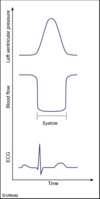
The blood flow curve shows a cyclical variation in flow during the cardiac cycle, with maximum flow occurring during diastole and minimal flow occurring during ventricular systole. This pattern is unique for the left ventricular myocardium. The great majority of blood flows through the vascular beds of the left ventricle during diastole, when the coronary vessels are not compressed by the high ventricular wall pressures created by myocardial contraction. This systolic reduction in blood flow is greatest in the subendocardial myocardium (where wall pressures are the highest), making it the region most prone to ischemia and myocardial infarction.

(Choices A, B, D, E, and G) In all other organs, blood flows continuously throughout the cardiac cycle down the pressure gradient from the arterial source into the venous circulation.

(Choice F) During systole, pressures in the right ventricle are much lower than in the left ventricle (only ~25 mm Hg compared to ~120 mm Hg). As a result, coronary perfusion pressure is able to overcome right ventricular wall pressure throughout the cardiac cycle, leading to relatively constant blood flow to the right ventricular myocardium.

Educational objective:
During ventricular systole, the coronary vessels supplying the left ventricle are compressed by the surrounding muscle. As a result, the majority of left ventricular blood flow occurs during diastole. The systolic reduction in coronary blood flow is greatest in the subendocardial region, making this portion of the left ventricle most prone to ischemia and infarction.

33
Q

Question

A

E.

34
Q

The capacity to refuse treatment requires

A

the ability to express a choice, understand the relevant medical information, appreciate the potential consequences of treatment options, and offer a rationale for the decision.

Patients with Alzheimer disease in the mildly affected range often retain decision-making capacity.

35
Q

Question

A

C.

This patient has an early-peaking systolic murmur and wide and fixed splitting of S2. These findings are characteristic of an atrial septal defect (ASD), a common congenital heart lesion involving an abnormal connection between the right and left atria.

After birth, pulmonary vascular resistance decreases and systemic vascular resistance increases, creating a left-to-right shunt through the ASD. As a result, the right atrium and ventricle receive increased blood volume (ie, venous return to the heart plus blood delivered via the shunt) as compared to normal. Increased blood flow into the pulmonary arteries across the pulmonic valve creates an early-peaking, midsystolic ejection murmur best heard over the left second intercostal space (ie, pulmonic flow murmur). In addition, increased blood flow through the tricuspid valve may cause a diastolic rumble best heard at the lower left sternal border (not present in this patient). It is notable that the blood flow through the ASD itself is low velocity and does not typically cause a murmur.

Increased blood flow across the pulmonic valve also accounts for the characteristic ASD finding of a widely split, fixed S2. Under normal conditions, S2 is split only during inspiration due to increased venous return to the heart, resulting in delayed pulmonic valve closure (P2) relative to aortic valve closure (A2). With ASD, increased right ventricular volume further delays P2, and the communication between the left and right atria maintains high right-sided blood volume throughout the respiratory cycle, eliminating respiratory variation of splitting.

(Choice A) Aortic stenosis causes a crescendo-decrescendo systolic murmur over the right second intercostal space. A2 may be delayed until after P2 (paradoxical splitting) due to the increase in left ventricular ejection time.

(Choice B) Ventricular septal defect creates a holosystolic murmur along the lower left sternal border due to a high-pressure, left-to-right shunt from the left to the right ventricle. Splitting of S2 may be widened but is usually not fixed.

(Choice D) Aortic insufficiency is characterized by regurgitant blood flow through the aortic valve, resulting in a decrescendo diastolic murmur along the sternal border. Splitting of S2 is usually not affected.

Educational objective:
Atrial septal defects result in increased right-sided blood flow due to left-to-right shunting and characteristic findings of a systolic ejection murmur (ie, pulmonic flow murmur) and widely split, fixed S2.

36
Q

Question

A

Sudden upward jerking of the arm at the shoulder can cause injury to the lower trunk of the brachial plexus. The lower trunk carries nerve fibers from the C8 and T1 spinal levels that ultimately contribute to the median and ulnar nerves. Together, these nerves innervate all of the intrinsic muscles of the hand (eg, lumbricals, interossei, thenar, hypothenar).

Injury to the lower trunk of the brachial plexus may cause paralysis of all the intrinsic hand muscles (Klumpke’s palsy). Weakness of lumbricals causes impaired flexion of the metacarpophalangeal joints and impaired extension of the interphalangeal joints. Relative sparing of the extrinsic flexors and extensors of the hand contributes to the total claw hand deformity seen in this condition. Sensory loss can also occur over the medial aspect of the hand/forearm. Involvement of the T1 nerve root proximal to the sympathetic trunk may cause concomitant Horner’s syndrome (eg, ipsilateral ptosis, miosis, anhidrosis).

(Choice A) Shoulder dislocations or fractures of the proximal humerus can cause axillary nerve injury, which results in weakness of the deltoid and teres minor muscles.

(Choice B) Long thoracic nerve injury classically occurs during axillary lymph node dissection and results in paralysis of the serratus anterior muscle, leading to winging of the scapula.

(Choice D) Injury to the musculocutaneous nerve causes weakened elbow flexion because the nerve innervates the biceps brachii and brachialis muscles.

(Choice E) Fractures at the midshaft of the humerus commonly cause radial nerve injury. Patients may experience posterior forearm and dorsolateral hand numbness with paralysis of the wrist and finger extensors (wrist drop).

(Choice F) The suprascapular nerve innervates the supraspinatus and infraspinatus muscles. Nerve injury may result in problems with abduction and lateral rotation of the arm, respectively.

Educational objective:
The lower trunk of the brachial plexus carries nerve fibers from the C8 and T1 spinal levels that are responsible for innervating all of the intrinsic muscles of the hand (via the median and ulnar nerves). Sudden upward stretching on the arm at the shoulder can damage the lower trunk, resulting in finger clumsiness with total claw hand deformity.

37
Q

Identify

A
38
Q

Question

A

D. Piriformis muscle

The sciatic foramen is a pelvic opening serving as the major pathway for pelvic neurovascular structures to the lower limbs. It is divided into greater and lesser sciatic foramina by the sacrospinous ligament (Choice E). The greater sciatic foramen is bordered anterolaterally by the greater sciatic notch of the ilium, inferiorly by the ischial spine and sacrospinous ligament, superiorly by the anterior sacroiliac ligament, and posteromedially by the sacrotuberous ligament (Choice F).

The piriformis originates on the anterior aspect of the sacrum and occupies most of the space in the greater sciatic foramen. It inserts on the greater trochanter of the femur and acts to externally rotate the thigh when extended and abduct the thigh when flexed. Structures running above the piriformis include the superior gluteal vessels and superior gluteal nerve. Structures crossing below the piriformis include the inferior gluteal vessels, internal pudendal vessels, and multiple nerves (most notably the sciatic nerve). Muscle injury or hypertrophy can compress the sciatic nerve to cause sciatica-like symptoms (eg, pain, tingling, and numbness in the buttocks and along the nerve distribution) known as piriformis syndrome. The muscle can be tender with deep palpation or on stretching with adduction and internal rotation.

(Choice A) The coccygeus muscle is a pelvic floor muscle anterior to the sacrospinous ligament and combines with the levator ani to form the pelvic diaphragm.

(Choice B) The gluteus minimus is the smallest of the 3 gluteal muscles and is immediately beneath the gluteus medius. It works with the gluteus medius to stabilize the hip and abduct the thigh when the limb is extended.

(Choice C) The obturator internus is a fan-shaped muscle originating from the medial surface of the obturator membrane, ischium, and pubic rim. It exits the pelvis through the lesser sciatic foramen and inserts on the greater trochanter of the femur, where it functions similarly to the piriformis.

Educational objective:
The piriformis passes through the greater sciatic foramen and is involved with external hip rotation. Muscle injury or hypertrophy can compress the sciatic nerve in the foramen, causing piriformis syndrome.

39
Q

Question

A

B. Decreased tear production on right side of face

This patient’s acute hemifacial weakness is consistent with Bell palsy, an idiopathic mononeuropathy affecting the facial nerve (CN VII). CN VII is a mixed nerve, consisting of the following:

Motor output to the muscles controlling movement of the face (including eye closure)

Somatic sensation afferents from portions of the pinna and external auditory canal

Special sensation afferents for taste from the anterior two-thirds of the tongue

Parasympathetic innervation of the submandibular and lacrimal glands

Motor innervation of the stapedius muscle, which causes sound dampening

Patients with Bell palsy typically have a sudden onset (within hours) of unilateral facial paralysis. Specific findings include impaired eye closure; eyebrow sagging; inability to smile, frown, or purse lips on the affected side; disappearance of the nasolabial fold; and lips drawn to the nonaffected side. Patients with Bell palsy may also have decreased tear production, hyperacusis, and/or loss of taste sensation over the anterior two-thirds of the tongue.

(Choice A) Trigeminal nerve (CN V) somatic afferents are responsible for providing general sensation over the face; the motor branch of CN V (V3) innervates the muscles of mastication and the tensor tympani (sound dampening).

(Choice C) The levator veli palatini elevates the soft palate during swallowing (and when saying “ah”). Palate elevation is mediated by the pharyngeal branch of the vagus nerve (CN X).

(Choice D) Horner syndrome occurs due to interruption of sympathetic innervation and classically presents with hemifacial ptosis, miosis, anhydrosis, and enophthalmos. Postsynaptic sympathetic axons travel in a perivascular plexus along the carotid arteries to reach and innervate the face.

(Choice E) Taste sensation from the posterior one-third of the tongue is provided by the glossopharyngeal nerve (CN IX).

Educational objective:
Because of the diverse functions of the facial nerve (CN VII), patients with Bell palsy may experience decreased tear production, hyperacusis, and/or loss of taste sensation over the anterior two-thirds of the tongue, in addition to unilateral facial weakness.

40
Q

Bell Palsy Pathophysiology

A

acute hemifacial weakness is consistent with Bell palsy, an idiopathic mononeuropathy affecting the facial nerve (CN VII).

41
Q

Bell Palsy Clinical Features

A

Mononeuropathy affecting the facial nerve (CN VII)

CN VII is a mixed nerve, consisting of the following:

  • Motor output to the muscles controlling movement of the face (including eye closure)
  • Somatic sensation afferents from portions of the pinna and external auditory canal
  • Special sensation afferents for taste from the anterior two-thirds of the tongue
  • Parasympathetic innervation of the submandibular and lacrimal glands
  • Motor innervation of the stapedius muscle, which causes sound dampening

Patients with Bell palsy typically have a sudden onset (within hours) of unilateral facial paralysis. Specific findings include impaired eye closure; eyebrow sagging; inability to smile, frown, or purse lips on the affected side; disappearance of the nasolabial fold; and lips drawn to the nonaffected side. Patients with Bell palsy may also have _decreased tear produ_ction, hyperacusis, and/or loss of taste sensation over the anterior two-thirds of the tongue.

42
Q

Question

A

B. Hookworm infection

This patient, who developed a serpiginous, pruritic rash shortly after walking barefoot on a beach, likely has hookworm infection. Hookworms spread when egg-containing feces are deposited into shady, warm, moist sand or soil. Eggs hatch into larvae and are transmitted when they come into direct contact with human skin (eg, when walking barefoot).

Dermal penetration is usually marked by a pruritic maculopapular lesion (“ground itch”) at the site of larval entry (eg, foot, toe webs). Spread of larvae through the adjacent dermal tissue can lead to the formation of migrating, reddish-brown, serpiginous tracks. Deeper spread may occur, depending on the species of infecting hookworm, as follows:

Human hookworms (eg, Necator americanus, A duodenale) penetrate the basement membrane and spread from the dermis to the bloodstream. They rupture into the alveoli and are coughed up and swallowed into the small intestine. There, larvae mature into adult hookworms that feed on blood from the duodenal mucosa (often causing iron deficiency anemia) and shed up to 10,000 eggs/day, thereby spreading the infection to others.

Cat and dog hookworms (eg, Ancylostoma braziliense, A caninum) cause a dermal eruption in humans but are unable to penetrate the cutaneous basement membrane and spread to deeper tissues. Therefore, larvae do not mature into adult hookworms or spread from human-to-human (humans are incidental hosts).

(Choice A) A brown recluse spider bite usually appears as a red plaque or papule with central pallor; it may develop a dark central eschar that ulcerates due to necrosis. Pain is often severe.

(Choice C) Mycobacterium marinum lesions usually form a few weeks after exposure to contaminated fresh/salt water (eg, swimming pool, fish tank). Lesions begin as solitary papules or nodules that eventually ulcerate and scar. Serpiginous tracts would be uncommon.

(Choice D) Sporothrix schenckii, a dimorphic fungus, is usually transmitted when contaminated organic material (eg, moss, soil) is inoculated into the skin or subcutaneous tissue. A papule forms at the inoculation site and usually ulcerates; additional lesions form along the proximal route of lymphatic drainage.

(Choice E) Staphylococcus aureus is the most common cause of purulent cellulitis. Lesions tend to develop over days and are characterized by painful areas of fluctuance with surrounding erythema.

(Choice F) Vibrio vulnificus is a free-living bacterium that grows in brackish water and marine environments. It can contaminate wounds and cause mild cellulitis. More severe manifestations (eg, myositis, necrotizing fasciitis, sepsis) can develop in patients with certain underlying comorbidities (eg, liver disease, hemochromatosis).

Educational objective:
Hookworm infections are transmitted via direct contact between human skin and contaminated soil/sand (eg, walking barefoot). Dermal penetration is often characterized by an intensely pruritic papule that may form serpiginous tracks due to the subcutaneous migration of hookworm larvae.

43
Q

Question

A

D. Impairment of Protein Synthesis

Corynebacterium diphtheriae are nonmotile, unencapsulated, gram-positive rods. On microscopic examination, they are often found in clumps (classically said to resemble Chinese characters) or joined in V- or Y-shaped chains. Their cytoplasm contains metachromatic granules that stain with aniline dyes (eg, methylene blue).

C diphtheriae produce classic 2-subunit AB exotoxin. The B (binding) subunit binds specifically to the heparin-binding epidermal growth factor receptor on cardiac and neural cells, which explains the toxin’s affinity for heart and brain tissue. The B subunit induces endocytosis of the toxin, and the subsequently released A (active) subunit inhibits host cell protein synthesis by catalyzing the ADP-ribosylation of protein elongation factor 2 (EF-2). EF-2 is necessary for tRNA to insert new amino acids into the growing protein chain during translation. Therefore, the toxin causes cell death by inhibiting cell protein synthesis.

(Choice A) Certain virulent enteric bacteria (eg, Campylobacter jejuni, Vibrio cholerae) release AB exotoxins that activate electrolyte transport. These toxins have 5 B subunits responsible for cell binding and toxin endocytosis and 1 A subunit that stimulates a G protein to increase intracellular cAMP concentrations. As a result, active efflux of sodium and chloride ions occurs, and the subsequent cell water loss into the gut lumen leads to watery diarrhea and dehydration.

(Choice B) Botulinum toxin is a neurotoxin that blocks exocytosis of acetylcholine-containing presynaptic vesicles from peripheral nerve terminals, causing cranial and peripheral nerve palsies, muscle weakness, and respiratory paralysis. Tetanus toxin inhibits the release of the inhibitory neurotransmitters gamma-aminobutyric acid and glycine. Classic manifestations of tetanus include muscle spasms and autonomic hyperactivity.

(Choice C) Clostridium perfringens releases a cytolytic toxin (alpha-toxin, or phospholipase C) that degrades cell membrane phospholipids, causing cell destruction.

(Choice E) Superantigens (eg, staphylococcal and streptococcal toxic shock toxins) bind nonspecifically to T cell receptors and major histocompatibility complex class II molecules on antigen-presenting cells, leading to widespread T cell stimulation and inflammation. However, they are classically implicated in toxic shock syndrome related to tampon use or nasal packing, not upper respiratory infections.

Educational objective:
Diphtheria exotoxin inhibits host cell protein synthesis by catalyzing the ADP-ribosylation of host cell elongation factor-2.

44
Q

Diphtheria Toxin MOA

A
  • Diphtheria toxin is an AB exotoxin that inhibits protein synthesis by ADP-ribosylation of EF-2
  • Toxin acts locally, causing respiratory cell necrosis with formation of fibrinous, coagulative exudates
45
Q

Corynebacterium diphtheriae appearance

A

Corynebacterium diphtheriae are nonmotile, unencapsulated, gram-positive rods. On microscopic examination, they are often found in clumps (classically said to resemble Chinese characters) or joined in V- or Y-shaped chains. Their cytoplasm contains metachromatic granules that stain with aniline dyes (eg, methylene blue)

46
Q

Question

A

This patient has adenomyosis, a disorder caused by an abnormal collection of endometrial glands and stroma within the uterine myometrium. Adenomyosis is common in multiparous women, and prior uterine surgery (eg, cesarean delivery) is a risk factor.

Although the exact pathogenesis is unclear, adenomyosis may occur due to endometrial invagination into the myometrium during periods of myometrial weakening or changes in vascularity at the endomyometrial interface (eg, pregnancy, uterine surgery). The clinical features of adenomyosis reflect its pathophysiology:

endometrial gland proliferation and cyclic bleeding within the myometrium leads to dysmenorrhea and uterine tenderness

abnormal myometrial hyperplasia and hypertrophy results in a concentric, uniformly enlarged uterus

uterine enlargement and subsequently increased endometrial surface area causes regular, heavy menstrual bleeding

Definitive therapy is with hysterectomy, which allows for histologic diagnosis.

(Choice A) Leiomyomas (ie, uterine fibroids) are benign myometrial smooth muscle cell tumors. Although fibroids can cause regular, heavy menses (also due to increased endometrial surface area), the uterus is typically nontender and irregularly enlarged rather than tender and uniformly enlarged.

(Choice B) An ectopic pregnancy most commonly occurs due to abnormal blastocyst implantation in the fallopian tube. Although prior tubal surgery (eg, bilateral tubal ligation) is a risk factor, ectopic pregnancy is unlikely in this patient with a negative urine β-hCG. In addition, uterine enlargement would not be seen.

(Choice D) Endometrial polyps are benign, intracavitary, focal hyperplastic growths of endometrial tissue. In contrast to adenomyosis, endometrial polyps cause painless intermenstrual bleeding rather than painful, cyclic, heavy menses. There is also no associated uterine tenderness or enlargement.

(Choice E) Patients with endometrial hyperplasia have unregulated endometrial gland proliferation with increased gland-to-stroma ratio; the thickened endometrial lining may slightly increase uterine size and cause heavy menses. However, endometrial hyperplasia does not typically cause dysmenorrhea or uterine tenderness. In addition, the most common risk factor is unopposed estrogen from chronic anovulation and/or obesity; this patient has regular menses and a normal BMI.

Educational objective:
Adenomyosis is the abnormal presence of endometrial glands and stroma within the uterine myometrium. Affected patients are typically multiparous women with dysmenorrhea, heavy menses, and a uniformly enlarged uterus.

47
Q

Botulinum toxin MOA

A

Botulinum toxin is a neurotoxin that blocks exocytosis of acetylcholine-containing presynaptic vesicles from peripheral nerve terminals, causing cranial and peripheral nerve palsies, muscle weakness, and respiratory paralysis. Tetanus toxin inhibits the release of the inhibitory neurotransmitters gamma-aminobutyric acid and glycine. Classic manifestations of tetanus include muscle spasms and autonomic hyperactivity.

48
Q

C. perfringens toxin MOA

A

Clostridium perfringens releases a cytolytic toxin (alpha-toxin, or phospholipase C) that degrades cell membrane phospholipids, causing cell destruction.

49
Q

Superantigens MOA

A

Superantigens (eg, staphylococcal and streptococcal toxic shock toxins) bind nonspecifically to T cell receptors and major histocompatibility complex class II molecules on antigen-presenting cells, leading to widespread T cell stimulation and inflammation. However, they are classically implicated in toxic shock syndrome related to tampon use or nasal packing, not upper respiratory infections.

50
Q

Question

A

A. Antiphagocytic D-glutamate capsule

Bacillus anthracis is a large, nonmotile, nonhemolytic, gram-positive rod that forms colonies in culture with multiple curled extensions at the edges that resemble a “Medusa head.” Because the organism proliferates in soil contaminated with manure, pasture-grazing herbivores such as sheep, cattle, and horses are at greatest risk for infection. However, transmission to humans occasionally occurs when B anthracis spores are inhaled, ingested, or inoculated into the skin.

Inhalation of spores into the alveoli causes pulmonary anthrax. Alveolar macrophages phagocytose the spores and bring them to mediastinal lymph nodes, where the spores germinate into vegetative rods and multiply. The vegetative form of the bacteria contains a polypeptide capsule composed of D-glutamic acid, which is antiphagocytic. B anthracis rods also produce a potent exotoxin complex composed of edema factor, lethal factor, and protective antigen, which enter host cells and cause cell death.

Initial manifestations of infection are nonspecific (eg, myalgia, fever, malaise), but proliferation of the bacteria in the mediastinum eventually causes hemorrhagic mediastinitis (often visible on chest x-ray as a widened mediastinum). Subsequent dissemination through the bloodstream causes shock, brain edema/hemorrhage, and death. Although pulmonary anthrax is quite rare, it can be seen in individuals who handle contaminated hides or animal hair (“woolsorter’s disease”) or those exposed to aerosolized biologic weapons (eg, opening contaminated mail).

(Choice B) Many bacteria (eg, Streptococcus pneumoniae) are resistant to phagocytosis due to the presence of a polysaccharide capsule. However, B anthracis has a polypeptide, not polysaccharide, capsule.

(Choice C) Staphylococcus aureus produces protein A, which binds the Fc portion of IgG. This inhibits opsonization and complement-mediated killing of the bacteria.

(Choice D) Intracellular polyphosphate granules are characteristic of Corynebacterium diphtheriae; they can be visualized on microscopy after growing the pathogen on Loeffler medium and staining it with methylene blue.

(Choice E) Peritrichous flagella (flagella distributed uniformly over the entire surface of a bacterial cell) are not present on the surface of B anthracis as this organism is nonmotile. Peritrichous flagella are characteristic of highly motile organisms such as Proteus mirabilis.

Educational objective:
Bacillus anthracis is a large, sporulating, gram-positive rod that is encased in an antiphagocytic polypeptide capsule composed of D-glutamic acid. Inhalation of B anthracis spores can cause pulmonary anthrax, which is usually characterized by nonspecific symptoms followed by hemorrhagic mediastinitis, shock, and death.

51
Q

Question

A

C. Gastric Hypersecretion

Gastric acid secretion by parietal cells in the fundus and body of the stomach is stimulated by:

Histamine binds H2 receptors and increases intracellular cyclic AMP (cAMP) concentration.

Acetylcholine binds M3 muscarinic receptors and leads to an increase in intracellular calcium.

Gastrin binds to the cholecystokinin B receptor and increases the intracellular calcium concentration. It also stimulates histamine synthesis and release by enterochromaffin-like cells in the stomach.

Intracellular calcium and cAMP activate protein kinases and lead to increased transport of H+ by H+/K+ ATPase into the gastric lumen.

In systemic mastocytosis, clonal mast cell proliferation occurs in the bone marrow, skin, and other organs. Mast cell proliferation often is associated with mutations in the KIT receptor tyrosine kinase. These cells are characterized by prominent expression of mast cell tryptase. Excessive histamine release from degranulation of mast cells mediates many of the symptoms of the disease, such as syncope, flushing, hypotension, pruritus, and urticaria. In addition, histamine induces gastric acid secretion, which can lead to gastric ulceration. The excess acid also inactivates pancreatic and intestinal enzymes, causing diarrhea. Other gastrointestinal symptoms include nausea, vomiting, and abdominal cramps.

(Choices A, B, and F) Gastric mucosal atrophy often results from colonization with Helicobacter pylori. It can also be caused by autoimmune gastritis, which may lead to pernicious anemia due to loss of intrinsic factor from parietal cells. Symptoms of atrophic gastritis may include nausea, indigestion, and epigastric discomfort, but this patient’s skin symptoms are more consistent with mastocytosis.

(Choice D) Gastric hypomotility (gastroparesis) occurs with diabetes mellitus, uremia, hypothyroidism, and other metabolic disorders. Gastroparesis causes constipation, early satiety, and food stasis with vomiting.

(Choice E) Pancreatic endocrine tumors may secrete gastrin (Zollinger-Ellison syndrome), insulin (hypoglycemia), glucagon (hyperglycemia, rash), somatostatin (diarrhea, cholelithiasis, hyperglycemia), or vasoactive intestinal peptide (watery diarrhea, hypokalemia, achlorhydria). These disorders would not be associated with excess mast cells in the skin.

Educational objective:
Systemic mastocytosis is characterized by the abnormal proliferation of mast cells and increased histamine release. Histamine causes hypersecretion of gastric acid by parietal cells in the stomach as well as a variety of other symptoms (eg, hypotension, flushing, pruritus).

52
Q

Pathophysiology of Mastocytosis

A

In systemic mastocytosis, clonal mast cell proliferation occurs in the bone marrow, skin, and other organs. Mast cell proliferation often is associated with mutations in the KIT (CD 117) receptor tyrosine kinase. These cells are characterized by prominent expression of mast cell tryptase. Excessive histamine release from degranulation of mast cells mediates many of the symptoms of the disease, such as syncope, flushing, hypotension, pruritus, and urticaria. In addition, histamine induces gastric acid secretion, which can lead to gastric ulceration. The excess acid also inactivates pancreatic and intestinal enzymes, causing diarrhea. Other gastrointestinal symptoms include nausea, vomiting, and abdominal cramps.

53
Q

Question

A

D. aromatization

This obese patient with postmenopausal bleeding most likely has endometrial hyperplasia or cancer. The most common risk factor for endometrial hyperplasia/cancer is chronic unopposed estrogen exposure, which is associated with obesity, early menarche, late menopause, and chronic anovulation.

Postmenopausal women have decreased ovarian function (ie, low estrogen and progesterone production), resulting in cessation of endometrial proliferation and subsequent amenorrhea. However, small amounts of androgens continue to be produced in the ovaries and adrenal glands after menopause. In obese women, these androgens (eg, androstenedione) are readily converted to estrogens (eg, estrone) by the aromatase enzyme found in adipose tissue.

The increased peripheral production of estrogens in adipose tissue and the decrease in sex-hormone binding globulin levels (that normally occurs with menopause) lead to elevated free estrogen levels (Choice C). However, ovarian progesterone production remains low; therefore, the unopposed estrogen causes unregulated endometrial proliferation and subsequent endometrial hyperplasia or cancer, which often presents as postmenopausal or abnormal uterine bleeding.

(Choice A) Hyperprolactinemia due to autonomous pituitary secretion of prolactin (ie, pituitary adenoma) causes negative feedback to the hypothalamus. This results in decreased hypothalamic GnRH secretion, decreased pituitary FSH and LH secretion, and decreased estrogen and progesterone levels. Therefore, premenopausal women often have signs of hyperprolactinemia (eg, galactorrhea) and hypoestrogenism (eg, amenorrhea). In postmenopausal women, the most common symptom is tumor mass effect (eg, headache, bitemporal hemianopsia).

(Choice B) Endometriosis, the ectopic implantation of endometrial glands and stroma, typically causes painful menses (ie, dysmenorrhea) due to inflamed tissue on the peritoneum. Endometriosis is uncommon after menopause because it requires high premenopausal estrogen levels that do not occur even in obese women.

(Choice E) Suppression of hypothalamic GnRH secretion (ie, hypothalamic hypogonadism) most commonly occurs in women with severe caloric restriction (eg, anorexia), excessive energy expenditure (eg, female athlete triad), or stress. The decreased GnRH secretion results in decreased pituitary FSH and LH secretion and subsequent decreased ovarian estrogen and progesterone production. Therefore, patients typically have amenorrhea rather than postmenopausal bleeding.

Educational objective:
Endometrial hyperplasia and cancer often present with postmenopausal bleeding. A common risk factor for endometrial cancer is obesity due to the peripheral aromatization of androgens to estrogens in adipose tissue, which leads to chronic unopposed estrogen exposure and uncontrolled endometrial tissue proliferation.

54
Q

Question

A

E. the barrier is formed by type I pneumocytes

This patient who developed hypoxia, pulmonary edema (bilateral pulmonary infiltrates), and protein-rich fluid in the alveoli had acute respiratory distress syndrome (ARDS). ARDS is a severe inflammatory reaction that occurs in the lungs and results in respiratory failure with hypoxemia and noncardiogenic pulmonary edema. Risk factors include sepsis, severe trauma (eg, long-bone fractures with fat embolism, extensive tissue injury), and transfusion of blood products, all of which can initiate pulmonary injury and trigger a cascade of inflammation:

In response to injury, alveolar macrophages release multiple proinflammatory cytokines that recruit neutrophils to the lungs (Choice A).

Upon arrival to pulmonary tissue, the neutrophils release inflammatory mediators (eg, proteases, free radicals). This increases inflammation and leads to damage to the pulmonary endothelium and alveolar pneumocytes.

The alveolar-capillary barrier, which is formed by the endothelial cells and type I pneumocytes, is then destroyed, allowing fluid and red blood cells to escape the vascular and interstitial spaces and pour into the alveoli. There, they combine with material from necrotic cells and form a thick proteinaceous fluid and a hyaline membrane.

Destruction of type II pneumocytes, which normally produce surfactant and proliferate in response to injury, leads to alveolar collapse (but does not cause destruction of the alveolar capillary barrier) (Choice F).

The net effect is impaired alveolar gas exchange and respiratory failure, with affected patients having a high mortality rate.

(Choices B and D) Ciliated epithelium and goblet cells line the larger airways. Goblet cells secrete mucus that traps particulate matter (eg, dust, bacteria), while ciliated epithelial cells assist in its elimination by sweeping the mucus up the bronchi and trachea to the pharynx, where it is swallowed. These cells are not prominently involved in ARDS.

(Choice C) Club cells (formerly called Clara cells) are nonciliated, secretory constituents of the terminal respiratory epithelium. They secrete protein and surfactant components and help detoxify inhaled substances (eg, tobacco smoke). They are not prominently involved in ARDS.

Educational objective:
Acute respiratory distress syndrome is a severe inflammatory reaction that occurs in the lungs and results in hypoxemia and noncardiogenic pulmonary edema. Pulmonary injury leads to an inflammatory response resulting in breakdown of the capillary-alveolar barrier (created by type I pneumocytes and endothelial cells), increased capillary permeability, intraalveolar fluid accumulation, and hyaline membrane formation.

55
Q

Question

A

E. Neutrophils

This patient with dyspnea, productive cough, and an obstructive pattern on pulmonary function testing—in the setting of a prolonged smoking history—likely has chronic obstructive pulmonary disease (COPD). COPD is characterized by chronic airway inflammation, which results in both parenchymal destruction (emphysema) and remodeling of the airways (chronic bronchitis). The primary cell lines that are increased in COPD are neutrophils, macrophages, and CD8+ T lymphocytes. These cells release enzymes and proteases, such as neutrophil elastase, that cause alveolar damage, reduced ciliary motion, and increased mucus secretion by goblet cells. In addition, the inflammatory cells show impaired ability to phagocytize bacterial pathogens, possibly contributing to increased risk of respiratory infections such as community-acquired pneumonia.

(Choice A) The involvement of B lymphocytes in COPD pathogenesis is controversial; however, they do not appear to have a major role, and their increased numbers may actually be protective against bacterial colonization.

(Choice B) Although CD4+ T lymphocytes are slightly increased in COPD, their numbers and pathologic role are much less significant than those of CD8+ T lymphocytes. However, CD4+ T lymphocytes have been implicated in asthma pathogenesis.

(Choice C) Eosinophils play an important role in asthma but do not appear to have a major impact in COPD. Their increased presence in patients with COPD may indicate coexisting asthma.

(Choice D) Patients with mast cell disorders can have flushing, abdominal discomfort, and respiratory symptoms such as dyspnea and wheezing due to bronchoconstriction. However, mast cells do not play an important role in the pathogenesis of COPD.

Educational objective:
Neutrophils, macrophages, and CD8+ T lymphocytes are the primary mediators of disease in chronic obstructive pulmonary disease. They secrete enzymes and proteases that cause and perpetuate both the alveolar destruction of emphysema and the mucus hypersecretion found in chronic bronchitis

56
Q

Question

A

E. Medulloblastoma

Gait instability and limb ataxia are symptoms of cerebellar involvement. The mass described indicates that a brain tumor is causing the cerebellar symptoms in this child. Because both pilocytic astrocytomas and medulloblastomas arise in the cerebellum, microscopic findings must be relied on to determine which type of tumor this patient has. Sheets of primitive cells with many mitotic figures described in the vignette indicate a medulloblastoma, not a pilocytic astrocytoma. Medulloblastomas are part of a group called “primitive neuroectodermal tumors” (PNETs). PNETs are composed of sheets of small cells with deeply basophilic nuclei and scant cytoplasm (small, round, blue cells). Abundant mitoses are also seen. Medulloblastomas are undifferentiated and aggressive tumors.

The cerebellar vermis is the most common location of a medulloblastoma. Symptoms include signs of increased intracranial pressure (morning headaches, vomiting, and lethargy). Cerebellar dysfunction occurs as the tumor compresses adjacent structures.

(Choice A) Pilocytic astrocytoma is the most common brain neoplasm of childhood. Although both pilocytic astrocytomas and medulloblastomas are often located in the cerebellum, they can be differentiated by histology. On microscopic examination of a pilocytic astrocytoma, pilocytic astrocytes and Rosenthal fibers are seen. Pilocytic astrocytomas are low-grade tumors and have a better prognosis than medulloblastomas.

(Choice D) Ependymomas are the third most common brain neoplasm found in children. They arise in the walls of the ventriculi and can hamper CSF flow and cause hydrocephalus. On microscopic examination, ependymal cells form gland-like structures called “rosettes.”

Educational Objective:
Medulloblastoma is the second most common brain neoplasm of childhood. It is located in the cerebellum, often at the vermis, and consists of sheets of small, blue cells. Like other “PNET” tumors, medulloblastomas are poorly differentiated and have a bad prognosis.

57
Q

Pilocytic astrocytoma location and histology

A

often located in the cerebellum

On microscopic examination of a pilocytic astrocytoma, pilocytic astrocytes and Rosenthal fibers are seen. Pilocytic astrocytomas are low-grade tumors

58
Q

Ependymomas location & histology

A

Ependymomas are the third most common brain neoplasm found in children.

They arise in the walls of the ventriculi and can hamper CSF flow and cause hydrocephalus.

On microscopic examination, ependymal cells form gland-like structures called “rosettes.”

59
Q

Medulloblastoma location and histology

A

It is located in the cerebellum, often at the vermis

consists of sheets of small, blue cells (Sheets of primitive cells with many mitotic figures).

Like other “PNET” tumors, medulloblastomas are poorly differentiated and have a bad prognosis.

60
Q

Question

A

F. Serum TSH

Amiodarone is a class III anti-arrhythmic agent used to suppress life-threatening rhythm disturbances. Because it is 40% iodine by weight, amIODarone can cause a number of alterations in thyroid function. It can cause hypothyroidism due to decreased production of thyroid hormone. Individuals with pre-existing autoimmune thyroid disease are at greatest risk and so should be screened for subclinical hypothyroidism with a serum TSH assay prior to initiating amiodarone therapy. Amiodarone can also cause hyperthyroidism due to increased thyroid hormone synthesis or destructive thyroiditis with release of preformed thyroid hormone.

(Choices A and B) Amiodarone is not associated with altered function of the adrenal cortex or medulla.

(Choice C) The oral glucose tolerance test is used to screen for diabetes mellitus, primarily in pregnant women. Amiodarone is not associated with significant alterations in insulin sensitivity or blood glucose levels.

(Choice D) Release of prolactin is primarily regulated by inhibitory effects of dopamine from the hypothalamus. Release is stimulated to a small degree by thyrotropin-releasing hormone and therefore may be mildly affected by amiodarone. However, these effects would not be clinically significant.

(Choice E) Amiodarone can cause erectile dysfunction, which may be related to its alpha and beta adrenoreceptor blocking effect. Amiodarone does not significantly alter serum testosterone levels.

Educational objective:
Amiodarone is 40% iodine by weight. It can cause hypothyroidism due to decreased production of thyroid hormone. Amiodarone can also cause hyperthyroidism due to increased thyroid hormone synthesis or destructive thyroiditis with release of preformed thyroid hormone.

61
Q

Amiodarone MOA

A

Amiodarone is a class III antiarrhythmic that blocks potassium channels.

62
Q

Major adverse effects of amiodarone

A
63
Q

AmIODarone

A

amiodarone is 40% iodine by weight and can induce hypo or hyperthyroidism

64
Q

Question

A

A.

This infant has a harsh systolic ejection murmur that disappears during an episode of cyanosis, an expected finding with tetralogy of Fallot (ToF). The major defects in ToF are right ventricular outflow tract (RVOT) obstruction and a ventricular septal defect (VSD); the murmur is created by blood flow through the narrowed RVOT, not the VSD.

The degree of RVOT obstruction is the major driver of symptoms in ToF because it determines the direction and magnitude of shunting through the VSD. A certain degree of RVOT obstruction is fixed, but some degree is dynamic and can change based on activity (eg, feeding, squatting) that alters the underlying hemodynamics. Many patients have left-to-right or neutral shunting at rest (and are acyanotic), but they develop cyanosis (“tet” episodes) when a dynamic increase in RVOT obstruction (eg, precipitated by crying or feeding) leads to increased right-to-left shunting through the VSD.

During such an episode, decreased blood flow through the RVOT causes the systolic murmur to disappear. In addition, oxygen saturation (SaO2) in the left ventricle is reduced (eg, <90%) due to mixing with deoxygenated blood from the RV. The SaO2 in the left atrium (LA) remains normal as the LA continues to receive oxygenated blood from the pulmonary veins.

(Choice B) These values show a step-down in SaO2 at the LA (pulmonary vein SaO2 > LA SaO2), consistent with a right-to-left shunt through an atrial septal defect (ASD). Right-to-left shunting through an ASD (ie, Eisenmenger syndrome) takes years to develop and is not an expected cause of episodic cyanosis in infants.

(Choice C) These values show a step-up in SaO2 at the RV (RV SaO2 > right atrium SaO2), consistent with left-to-right shunting through a VSD, as occurs during acyanotic periods in patients with ToF.

(Choice D) These values reflect the SaO2 distribution in a normal heart.

(Choice E) These values show a step-up in SaO2 at the pulmonary artery (pulmonary artery SaO2 > RV SaO2), consistent with left-to-right shunting through a patent ductus arteriosus. Left-to-right shunts do not cause cyanosis.

Educational objective:
Tetralogy of Fallot involves right ventricular outflow tract (RVOT) obstruction and a ventricular septal defect (VSD). Cyanotic (“tet”) episodes are commonly precipitated by a dynamic increase in RVOT obstruction that increases right-to-left shunting through the VSD. These episodes involve decreased blood flow through the RVOT, causing disappearance of the associated systolic murmur, and reduced oxygen saturation of the left ventricular blood being pumped into the systemic circulation.

65
Q

Defects of Tetralogy of Fallot

A
66
Q

Eisenmenger syndrome

A

over time elevated pulmonic pressures exceed aortic pressures and a right to left shunt occurs across VSD

67
Q

Question

A

D. Reduction in glomerular hyperfiltration

This patient has incipient diabetic nephropathy with moderately increased albuminuria (ie, 30-300 mg/g). Although patients with more advanced diabetic kidney disease typically have reduced glomerular filtration (rising serum creatinine), in earlier stages, filtration is normal to increased and serum creatinine may be normal.

In the kidneys, glucose is freely filtered in the glomerulus and is normally reabsorbed with sodium by sodium-glucose cotransporter-2 (SGLT2) in the proximal tubule. In patients with diabetes mellitus, the increased filtered glucose load leads to the following pathophysiologic changes:

Reabsorption of the excess tubular glucose causes increased co-reabsorption of sodium by SGLT2.

This results in decreased tubular sodium delivery to the macula densa, stimulating renin secretion by the juxtaglomerular apparatus and, ultimately, increasing angiotensin II levels.

Angiotensin II induces relative vasoconstriction in the renal efferent arterioles, increasing hydrostatic pressure in the glomerular capillaries, contributing to glomerular hyperfiltration.

SGLT2 inhibitors (eg, empagliflozin) lower blood glucose levels by decreasing the reabsorption of glucose (and sodium) in the proximal tubule, causing urine glucose wasting and increased sodium excretion. The concurrent increase in tubular sodium delivery to the macula densa leads to decreased renin secretion, lower glomerular pressure, reduced hyperfiltration, and delayed progression of nephropathy.

(Choice A) Glucose is absorbed in the intestine primarily via SGLT1 (not SGLT2), which is not significantly affected by empagliflozin.

(Choices B and C) SGLT2 inhibitors lower blood glucose levels, leading to decreased secretion of insulin by pancreatic beta cells. Most patients taking SGLT2 inhibitors experience modest weight loss, not gain, due to urinary glucose wasting and lower circulating insulin levels.

(Choice E) SGLT2 inhibitors are associated with increased, not suppressed, glucagon secretion, possibly due to lower blood glucose and insulin levels or a direct effect on pancreatic alpha cells. Glucagon-like peptide-1 agonists (eg, exenatide) lower blood glucose levels by increasing glucose-dependent insulin secretion and suppressing glucagon secretion.

Educational objective:
In patients with diabetes mellitus, excess glucose in the proximal tubule causes increased concurrent reabsorption of sodium by sodium-glucose cotransporter-2 (SGLT2). This leads to decreased sodium delivery to the macula densa and increased renin secretion, which increases glomerular filtration pressure and promotes glomerular hyperfiltration. SGLT2 inhibitors increase sodium delivery to the macula densa, decreasing renin production and reducing hyperfiltration.

68
Q

Question

A

H. Pastuerella multocida

This patient has a skin and soft-tissue infection (SSTI) that developed at the site of a dog bite relatively soon following the bite. Pasteurella multocida is an organism found in the mouths of dogs and is responsible for the majority of acute skin infections following a dog bite. Infection typically occurs within 24 hours of inoculation and has a characteristic mouse-like odor (indole-positive species). P multocida SSTI can also occur following a cat bite. Other organisms associated with dog bites include staphylococci, streptococci, and Capnocytophaga canimorsus. Management includes wound care and antibiotics (eg, amoxicillin-clavulanate).

(Choice A) Bartonella henselae is associated with cat-scratch disease, which can develop following inoculation from an infected cat and presents with lymphadenopathy that is often self-limiting. It is not associated with dog bites.

(Choices B and I) Campylobacter jejuni typically causes gastrointestinal (diarrheal) illness, and Proteus mirabilis is a common cause of urinary tract infection.

(Choices C and E) Clostridium perfringens is an anaerobe that can cause necrotizing SSTI, which occurs most commonly after a traumatic wound and leads to myonecrosis and gas gangrene. Erysipelothrix rhusiopathiae is a gram-positive rod (not a gram-negative coccobacillus) that causes erysipeloid.

(Choices D and G) Coxiella burnetii can cause Q fever, a mild form of pneumonia. Fusobacterium is part of the anaerobic oral flora potentially causing aspiration pneumonia or pharyngitis (Lemierre disease).

(Choice F) Francisella tularensis causes tularemia, a zoonotic infection that can occur following contact with lagomorphs and rodents (eg, rabbits, beavers, squirrels). The presentation is variable (eg, severe febrile pulmonary infection, ulcerative disease at inoculation site).

Educational objective:
Pasteurella multocida is a cause of soft-tissue infection that develops within 24 hours following a dog or cat bite. Management includes wound care and antibiotics targeted against this organism.

69
Q

Question

A

E. inhibition of osteoclast-mediated bone resorption

Bisphosphonates (eg, alendronate, risedronate) treat osteoporosis by inhibiting osteoclast-mediated bone resorption. They have a chemical structure similar to that of pyrophosphate and attach to hydroxyapatite binding sites on bony surfaces. Osteoclasts that resorb the bone take up the bisphosphonate and are unable to adhere to the bony surface to continue resorption. Bisphosphonates also decrease osteoclast proton production, induce osteoclast apoptosis, and decrease development/recruitment of osteoclast precursor cells. The net result is to slow the rate of bone loss, and some patients may experience a small increase in bone mineral density.

(Choice A) Sclerostin is a glycoprotein produced by osteocytes that inhibits osteoblast bone formation. Monoclonal antibody preparations (eg, romosozumab) that bind sclerostin reverse this effect and facilitate increased osteoblast activity.

(Choice B) Denosumab is a monoclonal antibody that decreases bone resorption by binding to the receptor activator of nuclear factor kappa-B ligand (RANK-L) and blocking the interaction between RANK-L and RANK (a receptor located on osteoclast surfaces), which is required for preosteoclast maturation to osteoclasts.

(Choices C, D, and F) Teriparatide is a recombinant formulation identical to the 34-amino-acid sequence at the N-terminal portion of endogenous parathyroid hormone (PTH). Similar to PTH, teriparatide stimulates maturation of preosteoblasts into bone-forming osteoblasts that lay down collagen and eventually mineralize the matrix. Teriparatide also increases gastrointestinal calcium absorption and renal tubular calcium reabsorption, which leads to decreased urinary calcium excretion.

Educational objective:
Bisphosphonates have a chemical structure similar to that of pyrophosphate and attach to hydroxyapatite binding sites on bony surfaces to inhibit bone resorption by osteoclasts.

70
Q

Medications for osteoporosis

A
71
Q

Teriparatide moa and clinical use

A
72
Q

Denosumab MOA and clinical use

A

Decoy receptor osteoprotegerein analog

73
Q

Question

A

A. Absolute neutrophil count

The second-generation antipsychotic clozapine is the only antipsychotic that has consistently shown superior efficacy in treatment-resistant schizophrenia and schizophrenia associated with persistent suicidality. Clozapine has affinity for multiple dopamine and serotonin receptors, but the precise pharmacological mechanism responsible for its superior efficacy is unknown. Clozapine binds loosely and transiently to dopamine D2 receptors, causing significantly fewer extrapyramidal symptoms compared to first-generation antipsychotics.

Neutropenia (<1000 cells/mm3) and the potential for life-threatening agranulocytosis are the major adverse effects of clozapine. The risk of agranulocytosis is approximately 1%; therefore, treatment requires regular monitoring of the patient’s absolute neutrophil count. Treatment should be stopped if neutropenia occurs. Seizures and myocarditis are other important adverse effects that require provider vigilance.

(Choice B) Clozapine plasma levels can be checked after an initial target dose is reached, but further dosage adjustments are usually based on clinical response. Clozapine levels are not regularly monitored.

(Choices C and H) Creatinine levels and thyroid function tests should be monitored in patients taking lithium due to this drug’s potential to adversely affect thyroid and renal function.

(Choice D) Although a baseline ECG is required and physicians should be alert to the development of cardiovascular symptoms suggestive of myocarditis, routine ECGs are not required. Among the second-generation antipsychotics, ziprasidone is most often noted for causing a prolonged QT interval.

(Choices E and F) Liver function tests may be mildly elevated with the use of many psychotropic medications, including antipsychotics and anticonvulsants; thrombocytopenia can be caused by some anticonvulsants. However, routine monitoring of liver function tests and platelets is not required with clozapine.

(Choice G) Prolactin levels are not routinely monitored. Among the second-generation antipsychotics, risperidone has been associated with a greater risk of prolactin elevation.

Educational objective:
Patients treated with clozapine are required to have regular monitoring of the absolute neutrophil count due to the risk of life-threatening agranulocytosis.

74
Q

Tx for treatment-resistant schizophrenia and schizophrenia associated with suicidality

A

Clozapine

75
Q

Question

A

This patient most likely has syringomyelia, which classically presents with disproportionate loss of pain and temperature sensation (dissociated anesthesia) involving the arms and hands. In syringomyelia, a cerebrospinal fluid-filled cavity called a syrinx usually forms in the cervical region of the spinal cord. These cavities can enlarge over time and destroy adjacent portions of the spinal cord. The areas most commonly damaged are the ventral white commissure and ventral horns.

The ventral white commissure is located just anterior to the gray commissure. It is the area of decussation for the fibers of the lateral spinothalamic tract, which transmits pain and temperature sensation from peripheral receptors to the somatosensory cortex. Destruction of the ventral white commissure leads to loss of pain and temperature sensation bilaterally over the affected dermatomes (starting 1 to 2 levels below the lesion, as the axons briefly ascend in the zone of Lissauer before decussating). Touch, vibration, and position senses are preserved as the dorsal columns remain intact.

(Choice B) The ventral horns contain lower motor neurons that receive impulses from the lateral corticospinal tract. Damage to the ventral horn can occur in syringomyelia, but would cause flaccid paralysis and atrophy of the intrinsic hand muscles (not loss of pain and temperature sensation).

(Choice C) The ventral spinothalamic tract is located in this area and transmits crude touch and pressure sensation. It should not be confused with the lateral spinothalamic tract, which carries pain and temperature sensation.

(Choice D) After decussating in the ventral white commissure, axons of the lateral spinothalamic tract ascend to the brain in the lateral funiculus. Damage to this area would cause contralateral loss of pain and temperature sensation distally, beginning 1 to 2 segments below the lesion. However, bilateral sensory loss would not occur with damage to this area.

(Choice E) The dorsal horn contains first order axons and second order neurons that transmit sensory input from peripheral receptors. Damage to the dorsal horn would likely result in unilateral loss of all sensation over the affected dermatomes.

(Choice F) The dorsal column contains both the gracile and cuneate fasciculi. They are formed by axons of first-order neurons of the dorsal column pathway. This tract is the primary mediator of proprioception, vibration, and tactile sensation.

Educational objective:
Syringomyelia is characterized by the formation of a cavity (syrinx) in the cervical region of the spinal cord. The syrinx damages the ventral white commissure, leading to bilateral loss of pain and temperature sensation that is limited to the affected levels (typically the arms and hands); distal sensation is preserved. Destruction of the motor neurons in the ventral horns (due to extension of the syrinx) results in flaccid paralysis and atrophy of the intrinsic muscles of the hand.

76
Q

Lateral Spinothalamic tract

A
77
Q

Question

A

C. Bone and Dura mater

Epidural hematoma is an accumulation of blood between the skull bone and dura mater. Most cases are due to fracture of the pterion region of the skull (often involving the temporal bone with associated scalp contusion) and a subsequent rupture or tear of the middle meningeal artery.

Epidural hematoma characteristically results in transient loss of consciousness at the time of impact followed by a lucid period in which the patient regains consciousness. However, the eventual expansion of the hematoma results in elevated intracranial pressure (ICP) and can lead to brain herniation, coma, and death. Signs of elevated ICP include altered mental status, nausea/vomiting, and Cushing reflex (ie, bradycardia, irregular breathing, hypertension). An ipsilateral dilated pupil can also occur due to uncal herniation and oculomotor nerve compression.

The diagnosis is confirmed with a noncontrast CT scan demonstrating a hyperdense, biconvex (lens-shaped) mass between the brain and skull. Epidural hematomas do not cross suture lines due to the tight adherence of the dura to the calvarium at these points.

(Choice A) The epicranial aponeurosis (galea aponeurotica) and outer periosteum cover the exterior surface of cranial bones. Intracranial hemorrhages are located inside the cranial vault.

(Choice B) Subarachnoid hemorrhage results in blood accumulation between the arachnoid mater and pia mater. It most often occurs due to rupture of berry aneurysms or arteriovenous malformations of the anterior communicating, posterior communicating, or middle cerebral arteries. Patients typically have severe headache and nuchal rigidity.

(Choice D) Subdural hematoma (SDH) is located between the dura mater and arachnoid mater and results from tearing of the bridging cortical veins. It typically presents in older patients with profoundly depressed mental status at onset (acute SDH) or an insidious onset of headache and confusion (chronic SDH)

(Choice E) The pia mater is closely adherent to the brain surface and does not form a space for accumulation of blood.

Educational objective:
Epidural hematoma is an accumulation of blood between the bone and dura mater. It typically occurs due to a tear of the middle meningeal artery associated with fracture of the pterion region of the skull (often involving the temporal bone). Patients characteristically have transient loss of consciousness followed by a lucid interval before increasing intracranial pressure leads to neurologic deterioration.

78
Q

Types of intracranial hemorhage

A
79
Q

Epidural hematoma vessel involved

A
80
Q

Subdural hematoma vessel involved

A
81
Q

Subarachnoid hematoma vessel involved

A
82
Q

Epidural hematoma location

A
83
Q

Epidural hematoma clinical manifestation

A
84
Q

Epidural hematoma Presentation of CT scan

A
85
Q

Subdural hematoma Location

A
86
Q

Subdural hematoma Clinical manifestationm

A
87
Q

Subdural hematoma Presentation on CT scan

A
88
Q

Subarachnoid hemorrhage Location

A
89
Q

Subarachnoid hemorrhage Clinical manifestation

A
90
Q

Subarachnoid hemorrhage Presentation on CT Scan

A

Blood in the basal Cisterns

91
Q

Question

A

D. Androgen use

This patient is taking exogenous anabolic steroids (eg, testosterone, synthetic steroidal androgens). Anabolic steroids are utilized by athletes in an effort to increase lean body mass. Adverse effects associated with the excessive use of anabolic steroids include acne, gynecomastia, azoospermia, decreased testicular size and increased aggression. Hypertension, dyslipidemia, cholestatic hepatitis and hepatic failure may also occur.

Serum testosterone levels can be low in individuals taking only synthetic androgens (eg, trenbolone), but are often within the normal range or elevated due to exogenous testosterone intake. Although serum testosterone may appear adequate, lower than normal local testosterone levels in the seminiferous tubules lead to decreased spermatogenesis.

(Choice A) Kallmann syndrome is a cause of GnRH deficiency, which results in abnormally low production of sex hormones by the gonads. Affected males present with delayed puberty and an abnormally small penis in addition to other nonsexual findings such as anosmia, hearing loss, and cleft palate.

(Choice B) 5-alpha reductase deficiency results in an inability to convert testosterone to dihydrotestosterone in the peripheral tissues. Affected males are born with ambiguous genitalia. Following puberty, affected patients have normal or elevated levels of serum testosterone.

(Choice C) Klinefelter syndrome (XXY seminiferous tubule dysgenesis) is a common cause of male hypogonadism. Small, firm testes and a decreased serum testosterone level are characteristic. Patients may exhibit diminished secondary sexual characteristics.

(Choice E) Hyperprolactinemia causes hypogonadotropic hypogonadism by suppressing LH and FSH release thereby decreasing serum testosterone in affected males. Symptoms include diminished libido, impotence and oligospermia.

Educational objective:
Adverse effects associated with the use of excessive doses of anabolic steroids include acne, gynecomastia, azoospermia, decreased testicular size, and increased aggression. When measured, serum testosterone is typically normal or elevated. However, endogenous testosterone production and spermatogenesis are decreased.

92
Q

Question

A

D. Ruptured abdominal aortic aneurysm

Ruptured abdominal aortic aneurysm (AAA) is a surgical emergency involving full-thickness compromise of the aortic wall with extravasation of blood into surrounding tissues and spaces. The acute onset of severe abdominal and back pain is the most common presenting symptom. In general, anterior rupture into the peritoneal cavity is quickly accompanied by syncope, hypotension, and shock, whereas posterior rupture into the retroperitoneum may be temporarily contained, resulting in delayed onset of hemodynamic instability. Other signs suggesting AAA rupture include abdominal distension, a pulsatile abdominal mass, and umbilical or flank hematoma (indicators of retroperitoneal hemorrhage).

Early detection of AAA can be difficult because patients frequently have few symptoms until the AAA either ruptures or markedly expands. Therefore, ruptured AAA should be suspected in patients with consistent symptoms and known risk factors, which include advanced age (>60), smoking, male sex, and a history of atherosclerosis or connective tissue disease. CT imaging can confirm the diagnosis, but hemodynamically unstable patients with suspected ruptured AAA should go straight to emergent surgical repair.

(Choice A) Acute mesenteric ischemia has similar risk factors to AAA. Patients typically describe severe abdominal pain but have few findings on physical examination (eg, minimal tenderness) and hypotension and syncope are not usually seen.

(Choice B) Colonic perforation can be due to diverticulitis, malignancy, or complications from colonoscopy. Patients can develop hypotension due to sepsis, but signs of infection (eg, fever) and peritonitis (eg, rebound, guarding) are also expected.

93
Q

Abdominal aortic aneurysm Risk Factors

A
  • Advanced age (>60)
  • Smoking, male sex, hypertension
  • History of atherosclerosis or CTD
94
Q

Abdominal aortic aneurysm clinical presentation

A

Asymptomatic until rupture or rapid expansion

Rupture

Sudden, severe abdominal/back pain ± shock

Umbilical/flank hematoma

95
Q

Abdominal aortic aneurysm Clinical Presentation

A

Asymptomatic until rupture or rapid expansion

Rupture

Sudden, severe abdominal/back pain ± shock

Umbilical/flank hematoma

96
Q

Abdominal aortic aneurysm Management

A
  • Smoking cessation
  • Periodic imaging to assess for expansion
  • Elective repair for large, asymptomatic aneurysm