Tumors of Bone and Cartilage - Gupta Flashcards

1
Q

What are the tumors of bone?

A

1) Osteoma
2) Osteoid osteoma/osteoblastoma
3) Osteosarcoma

2
Q

What is an osteoma?

A

Benign bone forming tumor composed of compact or mature trabecular bone –> usually involves facial bones

3
Q

What is the clinical presentation of a pt with osteoma?

A

1) Pain
2) Headache
3) Vision changes

Associated with Gardner syndrome (familial colorectal polyposis)

4
Q

What is the microscopic appearance of an osteoma?

A

Dense compact bone with paucicellular stroma (see very few cells)

5
Q

What is Gardner syndrome?

A

Autosomal dominant (chromosome 5q21 - APC gene) disease leading to multiple colon polyps and tumors in thyroid, bone, epidermoid cysts, fibromas, and desmoid tumors

6
Q

What are desmoid tumors?

A

Tumors that grow in b/t muscle fibers and can cause obstruction –> surgical resection causes more tumors to form

7
Q

What is osteoid osteoma?

A

Benign tumor of osteoblasts

8
Q

What population are osteoid osteomas seen in?

A

Males < 25 years old

9
Q

What is the clinical presentation of osteoid osteoma?

A

Bone pain that resolves with aspirin

10
Q

What is the microscopic appearance of osteoid osteoma?

A

Randomly interconnected trabeculae of woven bone, prominently rimmed by a single cell layer of osteoblasts

11
Q

Where do osteoid osteomas occur and what do they look like on imaging?

A

Cortex of long bones

Bony mass (<2cm) with radiolucent center (osteoid, black arrow) with surrounding sclerosis (white arrow)

12
Q

How does osteoblastoma differ from osteoid osteoma?

A

Osteoblastoma usually larger (>2cm)

Bone pain doesn’t respond to aspirin

Arise in the vertebrae

13
Q

What does osteoblastoma look like microscopically?

A

Comprised of anastomosing trabeculae of osteoid and woven bone rimmed by osteoblasts

14
Q

How is osteoblastoma treated?

A

Curettage (scooping) or excision en bloc

15
Q

What is osteosarcoma?

A

Malignant prolferation of osteoblasts

16
Q

What is the epidemiology of osteosarcoma?

A

Mostly in teenage males

Associated with Pagets disease and post-radiation in elderly

Rb gene = increased risk and poor prognosis

17
Q

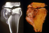
What characteristic feature does osteosarcoma show on imaging?

A

Codman’s triangle –> periosteol reaction to tumor destroying new bone before it ossifies

Indicates aggresive tumor

18
Q

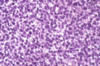
What does osteosarcoma look like microscopically?

A

Invades normal bone producing poorly formed bony spicules in a hypercellular matrix of osteoid and numerous pleomorphic malignant cells

Described as “lace-like” –> dainty little tumors

19
Q

What is seen in chondroblastic osteosarcoma?

A

Malignant cartilage formation

20
Q

What is the pathogenesis of osteosarcoma?

A

70% have acquired genetic mutation

Rb –> germline Rb 1000x increased risk

TP53 –> DNA repair and apoptosis

INK4a –> encodes tumor suppressor

MDM2 and CDK4 –> cell cycle regulators that inhibit p53 and Rb

21
Q

What are the cartilage-forming tumors?

A

1) Chondroma
2) Osteochondroma
3) Chondrosarcoma

22
Q

What is a chondroma?

What are the types of chondroma?

A

Benign cartilaginous tumor

1) Enchondroma –> arises from diaphyseal medullary cavity
2) Subperiosteal/juxtacortical
3) Soft tissue chondroma

23
Q

What genetic mutations are associated with enchondromas?

A

IDH1 and IDH2 (isocitrate dehydrogenase)

24
Q

What does a chondroma look like grossly?

A

Grey-white mass with color consistent with cartilage –> usually well circumscribed

25
What does chondroma look like microscopically?
Numerous chondrocytes closely packed near periphery in a pink ground substance that resembles normal cartilage
26
What is an osteochondroma?
Tumor of bone with an overlying cartilage cap --\> mushroom cap appearance on X-ray is pathognomonic
27
What is the epidemiology of osteochondroma?
Mostly in males 10-20 years old
28
What is the epidemiology and location of enchondroma?
Age 20-49, no gender preference Usually found in small bones of hands and feet
29
What is Maffuci syndrome?
Multiple enchondromas and soft tissue hemangiomas; also ovarian carcinoma and brain glioma
30
What is chondrosarcoma and where is it typically found?
Malignant cartilage forming tumor (no osteoid) Arises in medulla of pelvis or central/axial skeleton
31
What is the epidemiology of chondrosarcoma?
Men in their 40s
32
What is the gross appearance of chondrosarcoma?
Invasive cartilage growth in bone This example shows cartilage growing across joint space
33
What is the microscopic appearance of chondrosarcoma?
Cells mimic normal chondrocytes --\> appear to float in chondroid-like matrix
34
What are the fibrous and fibroosseous tumors?
1) Fibrous cortical defect 2) Fibrous dysplasia
35
What is a fibrous cortical defect (nonossifying fibroma)?
Benign fibrous lesion of bone
36
What is the epidemiology of fibrous cortical defect?
Children and adolescents
37
What is the microscopic appearance of fibrous cortical defect?
Fibrous spindle cells with pinwheel appearance Scattered giant cells, histiocytes, cholesterol clefts, and hemosiderin
38
What does fibrous cortical defect look like grossly?
Lesion \>5cm with intermedullary component
39
What is fibrous dysplasia?
A problem with osteoblastic maturation that causes fibrous tissue to form instead of medullary bone
40
How does fibrous dysplasia present and what are the most common sites?
Presents with painful swollen bones prone to fracture Ribs and femur are most commonly affected
41
What is the gross appearance of fibrous dysplasia?
Variable size, well circumscribed, _medullary_, tan-white, gritty mass lesions
42
What is the microscopic appearance of fibrous dysplasia?
Poorly formed islets of trabeculae of woven bone without a rim of osteoblasts --\> resemble chinese characters Fibroblastic activity surrounding trabeculae with lots of collagen
43
What is McCune-Albright syndrome?
Fibrous dysplasia plus cafe-au-laite spots and endocrine dysfuntion (especially early onset puberty)
44
What is a giant cell tumor of bone?
Tumor composed of multinucleated giant cells and stromal cells
45
What is the epidemiology of giant cell tumor of bone and where does it typically arise?
Young adults (20-40) Epiphysis of long bones, typically in the knee
46
What is the characterstic appearance of giant cell tumor on X-ray?
"Soap bubble" appearance --\> dude is fucked
47
What feature of giant cell tumor cells makes them a good candidate for targeted therapy?
High level of RANK-L expression
48
What is the microscopic appearance of giant cell tumor?
Regular and uniform distribution of stromal and giant cells --\> nuclei of mononuclear cells and osteoclasts are ovoid with prominent nucleoli (top pic) Fibroblastic activity and formation of reactive bone (bottom pic, pink shit)
49
What is Ewing sarcoma (primitive neuroectodermal tumor)?
Malignant proliferation of poorly-differentiated cells derived from neuroectoderm
50
What is the epidemiology and typical location of Ewing sarcoma?
Usually in males \<15 years old Typically found in diaphysis of long bones
51
What is the gross appearance of Ewing sarcoma?
White, fleshy, ill-defined tumor with involvement of medulla and cortex with periosteal elevation
52
What is the microscopic appearance of Ewing sarcoma?
Sheets of small, round, uniform cells (resemble lymphocytes) Indistinct cell membranes
53
What is the characteristic appearance of Ewing sarcoma on X-ray?
Onion skin appearance of periosteum - layered periosteum
54
What translocation is seen in Ewing sarcoma?
t(11,22) EWS-FLI1 fusion
55
What cellular organization pattern is sometimes seen in Ewing sarcoma?
Homer Wright rosettes --\> dark blue tumor cells surrounding pink fibrillar material resembling neuropil
56
What is a solitary bone cyst?
Benign lytic lesion, fluid-filled cyst with a thin wall
57
What is the epidemiology and typical location of solitary bone cysts?
Males \<20 years old Medullary in metaphysis of proximal humerus or femur --\> typicaly presents as a fracture
58
What is an anuerysmal bone cyst?
Exactly what is sounds like asshole Benign, but grows rapidly Looks like a sponge filled with blood
59
What is the epidemiology of anuerysmal bone cysts?
No gender preference; ages 1-20 Removed by curettage
60
What is the microscopic appearance of anuerysmal bone cysts?
Richly vascularized bone tissue --\> differentiating feature is rich capillary bed
61
What do metastatic bone tumors do to bone? What is the exception?
Produce punched out lesions radiographically **Prostatic carcinoma** produces osteoblastic (sclerotic) lesions
62
What is the mnemonic to remember sites of metastasis for bone shit
**BLT** with a **K**osher **P**ickle Breast Lung Thyroid Kidney Prostate
63
Whats does metastatic bone tumor from prostatic carcinoma look like radiographically?
Osteoblastic lesions --\> highly sclerotic bone