Thorax 1 Flashcards
What does the base of the heart consist of?
- the left atrium
- a small portion of the right atrium
- the proximal parts of the great veins (superior and inferior venae cavae and the pulmonary veins)

Where is the base of the heart directed?
It is quadrilateral and directed posteriorly + fixed posteriorly to the pericardial wall
Oesophagus lies immediately posterior to the base
What is the apex of the heart formed by and where is it positioned?

Positioned deep to the left fifth intercostal space, 8-9 cam from midsternal line
Formed by the inferolateral part of the left ventricle
What does the anterior surface face and consist of?

- Faces anteriorly
- Consists mostly of right ventricle
- Has some of right atrium on right
Has some of left ventricle on the left

In the anatomical position, what does the heart rest on?
- The diphragmatic surface
What does the diphragmatic surface consist of?
- Consists of left ventricle + small portion of right ventricle
- Rests on the diaphragm
What does the left pulmonary surface face + what does it consist of?
- Faces the left lung
- Broad + convex
- Consists of left ventricle + portion of left atrium
What does the right pulmonary surface face + consist of?
- Faces the righ lung
- Is broad + convex
- Consists of right atrium
What are the right and left margins the same as?
- The right and left pulmonary surfaces of the heart
What is the interior margin defined as?
- The sharp edge between the anterior and diaphragmatic surface of the heart
- formed mostly by right ventricle + small portion of left ventricle near apex
What is the obtuse margin?
- Separates anterior and left pulmonary surfaces
- It’s round + extends from left auricle to cardiac apex
- Formed mostly by left ventricle + superiorly by small portion of left auricle
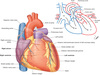
What does the right border in a standard posteroanterior view consist of?
- The superior vena
- Right atrium
- Inferior vena cava

What does the left border consist of?
- arch of aorta
- pulmonary trunk
- left auricle
- left ventricle
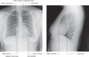
What does the inferior border consist of?
= Right ventricle and left ventricle at the apex

Label the image


What is the coronary sulcus and what does it contain?
- Circles the heart, separating the atria from the ventricles
- Contans the right coronary artery, the small cardiac vein, the coronary sinus + circumflex branch of left coronary artery
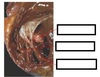
Label the image of coronary sulcus


What sulci separates the 2 ventricles + what does it contain?
- Anterior and posterior interventricular sulci
- The anterior interventricular sulcus is on anterior surface of the heart -> contains the anterior interventricular artery and great cardiac vein
- Posterior interventricular sulcus is on diaphragmatic surface of heart -> contains posterior interventricular artery + middle cardiac vein
Why does the left ventricule have a thicker muscular wall than the right?
More force is required to pump blood through the bdy than through the lungs
How does the blood return to the right atrium?
Enters through 1 of 3 vessels:
- Superior + inferior venae cavae -> together deliver blood to heart from body =
- coronary sinus -> returns blood from heart walls
Where does the superior and inferior vena cava enter the right atrium?

- Superior vena cava -> enters upper posterior portion of right atrium
- Inferior vena cava + coronary sinus enter lower posterior portion of right atrium
What is the heart housed inside of?
- Located within the mediastinum (the middle mediastinum)
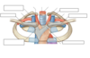
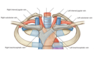
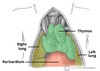
Label this image of the mediastinum and what type of image is it

- Cross section through the mediastinum

Label this image

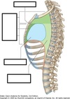
- Saggital view of mediastinum
Inside the mediastinum, what is the heart contained in?
- Contained in the pericardial sac
What is the pericardial sac?
- Fibroserous sac surrounding heart and great vessels
- Consists of 2 layers: fibrous and serous
Describe the fibrous layer of the pericardium
- Strong connective tissue
Describe the serous layer of the pericardium
- Inner layer
- Encloses pericardial cavity which encloses pericardial fluid
- Divided into 2 layers:
1) Parietal (lines fibrous pericardium) -> most superficial layer of pericardium
Made up of dense + loose connective tissue, protects the heart + anchors it to surrounding walls
2) Visceral part -> adheres to heart
What are the 2 separate pumps of the heart?
- Pulmonary pump -> delivers poorly oxygenated blood to the lungs where it is oxygenated and returned to the heart
- Systemic pump -> delivers highly oxygenated blood to rest of body
Describe the pathway at which blood moves through the heart
- Blood leaves the left ventricle via aorta (head, neck to rest of body)
- Returns to right atrium through the superior vena cava or inferior vena cava (deoxygenated)
- Goes to right ventricle where it exits the heart into the pulmonary trunk and into either right or left pulmonary artery.
- Dexoygenated blood travels to lungs
- Blood returns from the lungs via pulmonary veins into left atrium
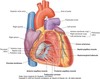
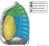
What do the different surfaces consist of?
- Anterior or sternocostal surface, formed mainly by the right ventricle
- Inferior or diaphragmatic surface, formed mainly by the left ventricle
- Posterior surface, formed mainly by left atrium
- apex formed entirely by left ventricle
What is the line of demarcation between the right atrium and right ventricle?
- The atrioventricular groove
- The right coronary artery lies in this groove

What groove is inferior to the root of the pulmonary trunk?
- Anterior interventricular groove -> inferior to the root of the pulmonary trunk
- Allows distinction between the right and left ventricles on surface of heart

Label this digram


How are the brachiocephalic veins formed? What do they form once they join together?
- Brachiocephalic veins are formed from the union of an internal jugular vein in the neck and a subclavian vein
- Left brachiocephalic vein and right brachiocephalic vein join together to form superior vena cava (empties into right atrium)
Label this diagram


What is the name for the right atrioventricular valve and what is its function?
- Tricuspid valve
- Allows blood from right atrium to right ventricle
- Prevents blood back-filling from ventricle to atrium

What is the name of the right semilunar valve and what does it do?
- Pulmonary valve
- Allows blood from right ventricle into pulmonary trunk

Label this image


What are the small attachments of tendons that anchor the tricuspid cusps into the ventricle wall?
Chordae tendinae

What do the chordae tendinae insert into?
Insert into ventricle wall (into the papillary muscle)
How many cusps do the tricuspid valve have and what are they?
- 3 cusps
- Anterior
- Septal towards the septum
- Posterior cusp

Label the image

Label this image


Where does the right coronary artery emerge from and where does it travel?
- Emerges from right aortic sinus
- Travels along margin of right atrium and right ventricle
- A marginal branch travels on inferior surface of heart towards apex

What is the name of a main branch of left coronary artery that descends between L and R ventricle?
- Anterior interventricular branch
- Circumflex branch that goes behind + down posterior surface of the heart

Label the image


Where is the electrical impulse generated in the heart?
- Impulse is generated in the SAN

From the SAN, where does the impulse travel?
- Transmitted from SA node to AV node along bundle of His + into ventricular wall

Label the image


What is the mediastinum + how is it divided?
- Central component of the thoracic cavity, located between the pleural sacs
- contains most of the thoracic organs
- divided into two parts by imaginary line from sternal angle to T4 vertebrae

What is the superior mediastinum?
Extends upwards
terminates at superior thoracic aperture

Vessel structures contained within the superior mediastinum?
- 3 major branches of aortic arch (braciocephalic, left common carotid + left subclavian artery)
- tributaries of superior vena cava (brachiocephalic veins, left superior intercostal vein, supreme intercostal vein + azygos vein)

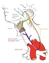
Nerves in the superor mediastinum?
- Vagus nerve (right vagus nerve runs parallel to trachea + passes posteriorly to SVC and right primary bronchus)
(left vagus nerve enters superior mediastinum between left common carotid + left subclavian arteries. descends anteriorly to aortic arch + travels posterior to left bronchus)
- phrenic nerves enter superior mediastinum from anterior scalene muscle + descend anteriorly into middle mediastinum, passing anteriorly to lung hilum.
Other structures in superior mediastinum?
- Thymus gland (most anterior structure in superior mediastinum + sits aganst posterior surface of sternum)
- Trachea (birfurcates into primary bronchi posterior to ascending aorta)
- Oesophagus (join with pharynx at C8)
- Throacic duct (passes to left of oesophagus on its path to the junction of the left internal jugular and subclavian veins.)

What are the borders of the anterior mediastinum?
Lateral borders: Mediastinal pleura (part of the parietal pleural membrane).
Anterior border: Body of the sternum and the transversus thoracis muscles.
Posterior border: Pericardium.
Roof: Continuous with the superior mediastinum at the level of the sternal angle.
Floor: Diaphragm.

What resides in anterior mediastinum?
- No major structure: some lymphatic vessels + nodes etc
- In infants, thymus extends inferiorly into anterior mediastinum BUT recedes during puberty + replaced mostly by adipose tissue in adults.
What are the borders of the middle mediastinum?
Anterior: Anterior margin of the pericardium.
Posterior: Posterior border of the pericardium.
Laterally: Mediastinal pleura of the lungs.
Superiorly: Imaginary line extending between the sternal angle (the angle formed by the junction of the sternal body and manubrium) and the T4 vertebrae.
Inferiorly: Superior surface of the diaphragm.
What organs does the middle mediastinum contain?
- the heart + its protective sheath (pericardium)
- tracheal bifurcation + left and right main bronchi

What nerves are located in the middle mediastinum?
- Cardiac plexus -> network of nerves located at the base of the heart (containing sympathetic, derived from T1-T4 segments of spine + parasympathetic fibres -> supplied by vagus nerve)
- Phrenic nerves (motor innervation to diaphragm -> arise in neck + descend through middle mediastinum to diaphragm)

What lympatics are located in middle mediastinum?

- Tracheobronchial lymph nodes
- Group of nodes associated with trachea + bronchia -> form from the gathering of bronchial nodes within the hila of the lungs
What are the borders of the posterior mediastinum?
Lateral: Mediastinal pleura (part of the parietal pleural membrane).
Anterior: Pericardium.
Posterior: T5-T12 vertebrae.
Roof: Imaginary line extending between the sternal angle (the angle formed by the junction of the sternal body and manubrium) and the T4 vertebrae.
Floor: Diaphragm.

What vessels are contained in the posterior mediastinum?
- The thoracic aorta (continuation of the arch of the aorta beginning at lower edge of T4 vertebra -> descends through posterior mediastinum. At inferior border of T12, thoracic aorta becomes abdominal aorta, passing through aortic hiatus of the diaphragm)
- Thoracic duct -> largest lymphatic vessel in the body (allowing lymph return from rest of body into venous system -> enters mediastinum via aortic hiatus)
ascends to lie directly anterior to T6-T12 vertebrae

