Thoracic Cavity Development Flashcards
(25 cards)
Pleural Cavities
Space in which lungs develop and persist. Develops from caudal limbs

Pericardial cavity
Space in which heart develops and persists. Develops from the most cranial end of the intraembryonic coelom. Embryo undergoes the head-fold, bringing the pericardium and heart ventrocaudally to bring its position anterior to the foregut.
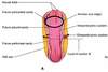
Peritoneal Cavity
Space in which abdominal viscera develops and persists. Herniation of small intestine and portion of large intestine. Develops from caudal limbs

Septum Transversum
Develops into the center tendon of the thoracic diaphragm
Intraembryonic Coelom
Space in the embryo. Primordium of embryonic body cavities. Develops in 4th week, ends by 8th. During the 4th week, a horseshoe cavity develops in the lateral mesoderm via apoptosis. Most cranial aspect gives rise to pericardial cavity/voelom.

Caudal limbs
Future pleural and peritoneal cavities. Open to the extraembryonic coelom to accommodate the growth and movement of the developing organs. Lose connection with the extraembryonic coelom during 10th week of development as developing intestines return to the body cavity.
Head Fold
Cranial end undergoes head fold. Brings pericardium and heart ventrocaudally. Developing heart now anterior to foregut. Brings pericardial coelom/angioblastic cord (earliest sign of heart) at ~4 weeks. Inflow to heart will come in from inferior aspect, outflow from heart will come out of superior aspect.

Lateral fold
Caudal limbs undergo caudal and lateral (horizontal) folds. Lateral folds bring the caudal limbs together which fuse to form the peritoneal cavity. Results in a gut tube suspended between two layers of membrane (mesentery) suspended from the body wall of the embryo.

Ectopia Cordis
congenital malformation in which the heart is abnormally located either partially or totally outside of the thorax. The ectopic heart can be found along a spectrum of anatomical locations, including the neck, chest, or abdomen. In most cases, the heart protrudes outside the chest through a split sternum.
Mesentery
Double layer of peritoneum extending from the abdominal wall. Conveys blood vessels, nerves, and lymphatics to the organs. Divide the peritoneal cavity into left and right halves. In each half, both parietal and visceral peritoneum found
Dorsal Mesentery
Permanent structure, provides the route for the vasculature, nerves and lymphatics to get to developing organs
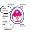
Ventral Mesentery
Not permanent. Remains attached to caudal portion of foregut (primordial stomach and proximal aspect of duodenum). Becomes lesser omentum, falciform ligament, and visceral peritoneum surrounding the liver. Any of the mesentaries attached to liver are Ventral mesentary.

Parietal Peritoneum
Serous membrane attached to and covering the abdominal wall
Visceral peritoneum
Serous membrane attached to/associated with internal organs
Pericardioperitoneal Canals
Term used for the intraembryonic coelom after the folding of the embryo. Tubes! Lies lateral to the foregut (future esophagus) and dorsal to the septum transversum (future central tendon of the diaphragm). Partitions form in each canal due to developing bronchial buds which divide developing lungs from pericardial space. The phrenic nerve can get caught up in these folds.
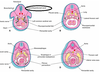
Cranial ridges of pericardioperitoneal canals
Goal is to divide pericardial space and pleura (lungs). Pleuropericardial folds. Located superior to developing lungs. Separate pleural cavities from pericardial cavities. Develop first

Caudal ridges of pericardioperitoneal canals
Goal is to divide pleural space and peritoneum. Pleuroperitoneal folds. Develop after the cranial ridges. Located inferior to developing lungs. Separate pleural cavities from peritoneal cavities. Do not fuse in center like pleuropericardial membrane; right one closes first, left closes later.

Divisions of the Embryonic Body Cavity
Pericardioperitoneal canals, pleuroperitoneal membranes, and pleuroperitoneal membranes
Pleuropericardial membranes
Separate pleura from pericardium. Enlargement of these pleuropericardial folds becomes the partition that separates the pleural and pericardial cavities. Contain the common cardinal veins which drain the primordial venous system into the snus venosus of the primordial heart. Contains the phrenic nerve. By 7th week, pleuropericardial membranes have fused with ventral mesenchyme, forming primordial mediastinum which contains all organs of thoracic cavity except lung and pleura

Pleural cavities
As bronchial buds mature into lungs, they grow into the pleural cavity. Pleural cavities expand ventrally around the heart which splits mesenchyme into outer layer (thoracic wall) and inner layer (fibrous pericardium/pleuropericardial membrane, which contains the phrenic nerve)
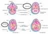
Pleuroperitoneal Membranes
Sperates pleural spaces from peritoneum. Right space closes first, LEFT closes LATER. Enlargement of pleuroperitoneal folds becomes partition separating pleural from peritoneal cavities. During week 6, these membranes extend ventromedially and fuse with dorsal mesentary of esophagus and septum transversum which separates pleural cavity from peritoneal cavity. This closure is assited by migration of mytoplasts into pleuroperitoneal cavity
Diaphragm
Develops from Septum transversum (from cranial fold), pleuroperitoneal membranes, dorsal mesentary of esophagus, and muscular ingrowth from lateral body walls.

Innervation of diaphragm
Myoblasts migrating into pleuroperitoneal membranes from 3-5 cervical myotomes carry their nerves with them. These nerves pierce the pleuropericardial membranes (in adults, located on fibrous pericardium) –> C3,4,5.
Posterolateral defect of diaphragm
Most common (95%). Diaphragm abnormality is characterized by a hole in the postero-lateral corner of the diaphragm which allows passage of the abdominal viscera into the chest cavity. Usually resultant of a failure of the diaphragm to close completely during development.
