Deep Back Flashcards
Development of Muscular System
Each somite differentiates into a sclerotome and a myo - dermatome. The myotome region of myo - dermatome gives rise to myoblast cells (embryonic muscle cells) that form the muscles of the trunk. Myotomes further divide into epaxial and hypaxial divisions. Even though most of the myoblasts migrate away from their specific myotome level of origin, they always maintain their original nerve supply from that segmen
Epaxial Division
Dorsal division. Supplied by dorsal primary rami of spinal nerves. Muscles derived of the epaxial division are the deep extensor muscles of the back
Hypaxial division
Ventral division. Supplied by ventral primary rami of spinal nerves. Muscles of the hypaxial division are the infrahyoid, flexor muscles of the vertebral column, and quadratus lumborum.
Original arrangement of deep back muscles
muscles have a segmental arrangement and only extend from one vertebra (segment) to the next. muscles extend from the skull (cranium) to the pelvis covered by the deep fascia
Developmental fusion of segmented muscles
During development fusion between adjacent segments takes place to form larger muscle masses covering more than one segment. Sequential splitting of muscles takes place later to form different superimposed layers
Action and movement of deep back muscles
Act to maintain posture and balance and also produce various movements (flexsion, extension, rotation) of the back. Since most of the body weight is anterior to the vertebral column, many back muscles function to support the body weight by extending the vertebral column. entire vertebral column moves smoothly during flexion, extension and rotation
Innervation of deep back (intrinsic) muscles
The intrinsic back muscles are the only muscles of the body innervated by the dorsal primary rami of spinal nerves. Many of the deep back muscle are very long they cross more than one segment of the vertebral column and are innervated by more than one spinal cord level
Back strain
Results from extensive extension and rotation – results in microscopic tearing of muscle cells or ligaments
Intrinsic Muscles of the back
Mainly involved with extension of the spine. Span the entire back from the skull to the sacrum. Are often grouped as follows, according to the direction of muscle fibers and length of muscle bundles: Splenius group, erector spinae group, transversospinalis group, and suboccipital muscles
Layers of intrinsic muscles
The intrinsic back muscles can also be divided into three layers: superficial, intermediate, and deep layers
Superficial layer of intrinsic muscles
all three subgroups of erector spinae and splenius muscles
Intermediate layer
two parts of Transversospinalis (Semispinalis and multifidus)
Deep layer
Segmental muscles and suboccipital muscles
Splenius group
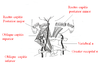
arise from the posterior midline of the back and proceed laterally and superiorly to their insertion. Most superficial intrinsic muscles of the posterior part of the neck. The word splenius refers to a “bandage-like” structure: muscle represents a bandage wrapped around the neck. Muscle fibers arise from the midline and run superiorly and laterally. Two muscles in group: splenius capitus and splenius cervicus
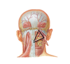
Splenius Capitus
Broad flat muscle of the neck. Partly covered by the trapezius and the sternocleidomastoid. Upper and larger of the two splenius muscles. Fibers proceed superiolaterally from their origin (ligamentum nuchae, spines of upper thoracic vertebrae) to attach to the skull (occipital bone, mastoid process)

Splenius Cervicis
Narrow muscle located below and parallel to the splenius capitis. Fibers extend superiorly and laterally - from spinous processes of upper thoracic vertebrae (T3-T6) and insert at the transverse processes of upper cervical vertebrae (C2-C4). No fibers attach to the skull.

Erector Spinae Group
Arise from the posterior midline of the back, or more laterally, and run up longitudinally. Largest muscle mass in the back. Forms a prominent bulge on each side of the lower part of the vertebral column between the spinous process and the angle of ribs. Origin is from a very extensive common tendon of origin. Chief extensor of the vertebral column. Ascending from the common tendon of origin, divides into three vertical columns: iliocostalis, longissimus, and spinalis. Each column is further divided into three parts according to its superior attachments. The muscle of each part is made up of overlapping short muscle fibers to provide smooth controlled action

Erector spinae muscles’ action
Unilaterally: Flex the head/neck & vertebral column laterally to the same side. Bilaterally: Extend the head/neck & vertebral column. Erector spinae is the chief extensor of the vertebral column
Common tendon of origin attachments
Sacrum, illiac crests, spinous process of lumbar and last two thoracic vertebrae (T11/T12)

Iliocostalis
Associated with the ribs. It has the following three parts: iliocostalis lumborum, iliocostalis thoracis, and iliocostalis cervicis. The most lateral column of the erector spinae group. All three parts of this muscle have their origin and or insertion on the ribs

Iliocostalis lumborum
Component of iliocostalis from common tendon to ribs 6-12

Iliocostalis Thoracis
Component of iliocostalis from lower 6 ribs to upper 6 ribs

Iliocostalis cervicis
Component of iliocostalis from from ribs 3-6 to transverse process of C4 – C6

Longissimus
associated with the transverse processes of vertebrae. Divided into three parts: Longissimus thoracis, Longissimus cervicis, and Longissimus capitus. Intermediate division of the deep back muscles. Its lowest section is the longissimus thoracis (there is no lumborum part like the iliocostalis muscle). Lower part of this muscle blends with iliocostalis lumborum

Longissimus thoracis
largest part of longissimus (common tendon to lower 9 ribs)

Longissimus cervicis
Runs from transverse process of T1-T5 to Transverse process of C2-C6

Longissimus capitus
Runs from the tendon of insertion of longissimus cervicis to the mastoid process
Spinalis muscles
Most medial and smallest division of erector spinae. Poorly developed in most individuals. In well developed individual it extends from the thorax to the skull and contains a thoracis, cervical and a lumbar segment. Usually only the thoracic part is present. Lies medial to longissimus thoracis. It extends from the spinous processes of lumbar vertebrae to the spinous processes of upper thoracic vertebrae.

Transversospinalis Group
arise laterally but travel toward the midline to their insertion. Lie deeper to the erector spinae group. Shorter than erector spinae. Slant inward from their origin to their insertion. Consist of three muscle masses: semispinalis, multifidus, and segmental muscles. Originate from transverse processes and proceed to spinous processes of more superiorly located vertebrae

Semispinalis muscles
Divided into three parts: Semispinalis thoracis, Semispinalis cervicis , and Semispinalis capitis. Muscle fibers arranged longitudinally like the erector spinae group. Muscle fibers span over about 4-6 vertebrae. Lacking in the lumbar region. Lie superficial to the multifidus.
Semispinalis thoracis
Spans thoraic vertebrae from transverse processes of T6-T12 to spinous processes of T1-T4 & C6/C7.

Semispinalis cervicis
Origin: Transverse processes of T1-T6
Insertion: Spinous proceses C2-C5

Semispinalis capitis
Origin: T1-T5
Insertion: on the occipital bone

Multifidus
Deeper to semispinalis. Thickest in lumbar area. Present throughout the back. Muscle fibers go FROM Transverse TO spinous processes. Individual segments are not known by separate names; the different parts are not usually distinct. Ends in the cervical region. In sacral and lumbar areas the muscle is covered by erector spinae; in the thoracic and cervical regions it is covered by semispinalis. Muscle fibers cover 2 - 4 vertebrae

Segmental muscles
Deepest part of transversospinalis group. Better developed in cervical and lumbar regions. Lie deep to the multifidus. Three sets of segmental muscles: interspinalis, intertransversarii, and rotators

Interspinales Muscles
Muscle running from spinous process to spinous process in the cervical, thoracic, and lumbar regions. Very small. Well developed in cervical and lumbar regions. The muscles are separated by interspinous ligaments. Lacking in thoracic region

Intertransversarii Muscles
Run vertically between adjacent transverse processes. Well developed in cervical and lumbar regions. Lacking in thoracic region

Rotatores Muscles
Have a single origin and a single insertion unlike most other back muscles. Span one or two vertebrae. Present in cervical, thoracic, and lumbar regions but most present in thoracic region. Two sets: long rotatores, and and short rotatores

Long rotatores
Aka Longus. Found in the lumbar, thoracic and cervical areas. Skip a vertebra.

Short rotatores
Found in the lumbar, thoracic and cervical areas. Attach to the vertebra above (do not skip a vertebrae)

Suboccipital Muscles
They are responsible for movement of the head and neck only. This group connects the skull to the atlas and axis. 2 obliquely oriented and 2 straight muscles: oblique capitis inferior, oblique capitis superior, rectus capitis posterior major, rectus capitis posterior minor. The oblique capitis inferior, oblique capitis superior and the rectus capitis posterior major form the suboccipital triangle.

Suboccipital triangle
Triangle formed by oblique capitis inferior, oblique capitis superior and the rectus capitis posterior major. Rectus capitis posterior minor is in the triangle, but is not a boundary. Vertebral artery originates at the base of the neck from the sunclavian artery and passes superiorly through the transversae foramina of cervical vertebrae. It enters the suboccipital triangle and by coursing across the floor of the triangle enters the skull through the foramen magnum.

Nerve supply of suboccipital muscles
Nerve supply to all four suboccipital muscles is via the dorsal ramus of C1 (suboccipital) nerve. Greater occipital nerve (dorsal ramus of C2) extends below the oblique capitus inferior, but DOES NOT PROVIDE INNERVATION TO ANY OF THE MUSCLES. It crosses the muscle and travels obliquely across the triangle to the back of the skull. This is a sensory nerve to the posterior part of the scalp
Oblique Capitis Inferior
Origin:Spinous Process of Axis
Insertion: Transverse Process of Atlas
Fxn: Aids in rotating the atlantooccipital joint
Innervation: Dorsal Ramus of C1 (Suboccipital Nerve)

Oblique Capitis Superior
Origin:Transverse Process of atlas
Insertion: Occipital Bone
Fxn: Aids in rotating the atlantooccipital joint
Innervation: Dorsal Ramus of C1 (Suboccipital Nerve)

Rectus capitis posterior minor
Origin:Posterior Tubercle of Atlas
Insertion:Occipital Bone
Fxn: Aids in rotating the atlantooccipital joint
Innervation: Dorsal Ramus of C1 (Suboccipital Nerve)

Rectus capitis posterior major
Origin: spine of axis
Insertion: Occipital bone
Fxn: Aids in rotating the atlantooccipital joint
Innervation: Dorsal Ramus of C1 (Suboccipital Nerve)

Actions of deep back muscles
No individual discrete action for any of the deep back muscles. Muscles function together as large groups because the origins and insertions of the muscles fibers overlap
Flexion of vertebral column
Anterior trunk muscles; gravity. Back muscles act as antagonistic – relax gradually to produce a smooth movement
Extension of vertebral column
All deep back muscles except intertransversarii. Erector spinae acts as the chief extensor muscle
Lateral Flexion
Intertransversarii. Splenius capitis and cervicis rotate the head to the same side and laterally flex the neck. Erector spinae muscles initiates lateral flexion unilaterally. All muscles on the opposite side control flexion by relaxing gradually
Rotation of vertebral column: Trunk
Multifidi, Rotatores and Semispinalis muscles rotate the trunk to the opposite side. Splenius muscles and the Erector spinae extend the trunk and help to rotate it to the same side
Rotation of vertebral column: Head
Oblique capitis inferior and rectus capitis posterior major and minor rotates the atlas and turns the face to the same side. The oblique capitis superior and the rectus capitis posterior major and minor extend the head bilaterally and flex it to the same side unilaterally