Heart 2: Development of Heart Flashcards
(45 cards)
Cardiovascular system development timingAttach Images
First system to functionally develop. Begins to develop during the middle of the 3rd week. Blood circulates at beginning of 4th week. Done development by 8th week.
Early development
Angioblastic cords are originally found in the cardiogeic area (cranial end of the embryo). These cords canalize (hollow out) to form the endocardial heart tubes at the late 3rd week. Heart begins to beat at 22-23 days

Head fold effect on cardiac development
The head-folding of the embryo brings the developing heart and pericardial cavity ventral to the foregut and caudal to the oropharyngeal membrane

Lateral fold effect on cardiac development
the lateral folding brings the endocardial heart tubes together, fusing to form a single heart tube with three layers.

Three layers of the heart tube
Visceral pericardium cardiac jelly (connective tissue that will become the AV valves), and the wall of the heart tube

Sinus venosus
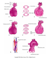
“Main drain vein” of the fetal heart. Receives venous blood flow from umbilical vein (chorion), vitelline vein (yolk sac) and cardinal vein (embryo). Develops right and left horns due to embryo shunting blood to the right side of the primitive heart.

Primordial Atrium
Site of collection of fetal blood. Not differentiated into left and right. Receives blood from sinus venosus

Primordial Ventricle
Develops into blood inflow portion of adult ventricle. Site of inflow from primordial atrium. Pumps blood out. Largest part of fetal heart

Bulbis cordis
Develops into conus arteriosus and aortic vestibule. Outflow portion of developing heart ventricle. Proximal to truncus arteriosus. Receives blood from primordial vestibule and sends blood to truncus arteriosus.

Truncus arteriosus
Outflow portion of developing heart. Continuous with aortic sac from which aortic arches develop

Aortic Sac
Develops into aortic arches. Primordial vascular channel anterior to the truncus arteriosus. Outflow of fetal heart

Cause for heart outflow in front and inflow in back
Bulbus cordis and ventricle (largest aspects) grow faster than other regions of the developing heart, causing the heart to bend upon itself. The bend is down and to the right side of the embryo. As the primordial heart bends at the bulboventricular loop, the atrium and sinus venosus come to lie dorsal to the truncus arteriosus, bulbus cordis, and ventricle. BENDING OCCURS AT INFERIOR BORDER OF HEART. Results in adult positioning of heart in mediastinum: inflow of blood on posterior aspect and outflow of blood on anterior aspect.
Bulboventricular Loop
U shaped junction between bulbus cordis outflow and primordial ventricle inflow. Location of bending of heart at inferior border of heart.
Blood flow of primitive heart prior to folding
Sinus venosus –> primordial atrium –> (AV Canal) –> primordial ventricle –> bulboventricular jxn –> bulbus cordis –> truncus arteriosus –> aortic sac –> aortic arches

Development of Semilunar valves
Develop from three swellings of subendocardial tissue via blood flowing through and hollowing out/remodeling the endocardium. Swellings are restructured via this blood flow to form thin-walled cusps. The position of the cusps after rotation is the normal anatomical way of naming cusps

Development of AV valves
Proliferation of endocardial cushions (cardiac jelly!) around the AV canals. Endocardium at the atrioventricular junctions undergo remodeling TOWARD THE VENTRICLE –> chordae tendinae, papillary muscle. This development toward the ventricle is due to the DIRECTION OF BLOOD FLOW (from atrium to ventricle) pushing the valves toward the ventricle. Three cusps on right (Tricuspid valve) and two cusps on left (mitral valve)

Partitioning of primordial atrium
Two Walls and Two Holes (phrasing!). Goal: primordial atrium into left and right atrium.
Septum Primum
“First wall”. The first septum. Grows inferiorly to the atrioventricular junctions. Its job is to close the opening between the primordial atrium known as the Foramen Primum (first hole). The Septum Primum divides the atrium into the right and left atria.

Foramen Primum
First hole. Closed by foramen primum. Aka Ostium primum. The original opening between the right and left atria before they are separated by the septum primum.

Foramen Secundum
Before the foramen primum is closed, a second hole forms in the septum primum. This hole is the foramen secundum. This hole allows for blood flow between the two atria as the fetal lungs are not yet sufficiently developed, so a bypass is necessary.

Septum Secundum
On the RIGHT side of the septum primum, the septum secundum grows inferiorly ONLY TO OVERLAP THE NEWLY FORMED FORAMEN SECUNDUM. This overlapping forms a flap-valve over the foramen (Foramen ovale)

Foramen ovale
Allows blood to flow between from right to left atria. USUALLY closes within minutes at birth, forming the fossa ovalis.

Formation of adult right atrium
Sinus venosus and primitive atrium. Right horn of sinus venosus becomes the posterior wall of right atrium –> sinus venarum (smooth portion of adult right atrium). Left horn of sinus venosus becomes coronary sinus. Primitve atrium becomes the rough anterior wall of adult right atrium (pectinate muscles)

Formation of adult left atrium
Smooth walls of left atrium formed by incorporation of primordial pulmonary vein IN NORMAL HEART.


