what are the lines of normal skin development called
Blaschko’s lines
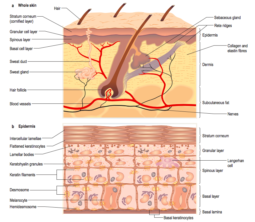
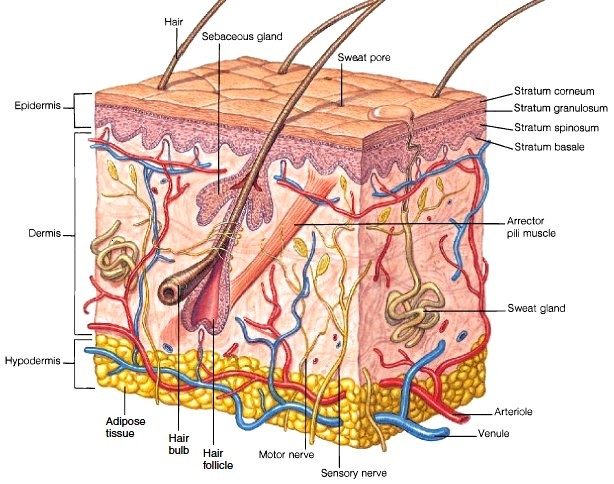
epidermis layer
- what does it arise from
stratified squamous that arises from dividing basal keratinocytes
varies at different body sites
rete ridges
downward projections of the epidermis into the dermis
name the layers of the epidermis
keratin
granular
prickle
basal

keratin layer of epidermis
- also called stratum corneum
- Composed of layers of flattened, scale-like cells. Thousands of these dead cells shed from the skin surface every day and are replaced by new ones from deeper layers.
- Corneocytes – terminally differentiated keratinocyte, are toughened and provide protection to the skin. Compose most of the stratum corneum
- contains filaggrin, involucrin and keratin (80% keratin and filaggrin)

where is teh keratin layer of epidermis thicker
areas of increased mechanical pressure eg sole of foot
what does the keratin layer of epidermis provide
a tight waterproof barrier
granular layer of epidermis
- Consists of 3/4 flattened layers of cells
- Keratohyalin granules that are the precursor to keratin, producing the keratin layer above.
- Odland bodies

prickle layer of epidermis
contains polyhedral cells with lots of desmosomes

basal layer of epidermis
- single layer of cells in conact with dermis
- cells in the epidermis proliferate from the basal layer - most metabolically active during foetal development
- contains melanocytes, merkel cells, keratinocytes and langerhans cells
keratinocytes
- can secrete a variety of cytokines in response to tissue injury or certain skin disease. play a role in immune function (innate and adaptive), cutaneous inflammation and tissue repair
- Produce keratin, which toughens and waterproofs the skin
what can keratinocytes be activated by
UV or sensitizers (eg allergic contact dermatitis)
which cell carry out vitamin D metabolism
kertatinocytes
what does HPV infection of keratinocytes cause
warts
melanocytes
dendritic cells formed in the basal layer and secrete the pigment melanin
where do melanocytes migrate from
neural crest to the epidermis in the first 3 months of foetal development
found above basal layer
what is the function of melanocytes
provide DNA protection from UV radiation
- convert tyrosine to melanin which absorbs light
- full melanosomes transferred to adjacent keratinocyte via dendrites and form protective cap over nucleus.
what are racial differences in skin pigmentation due to
- Found in equal numbers in black and white skin, but vary in melanin production
vitilgo
autoimmune disease with loss of melanocytes
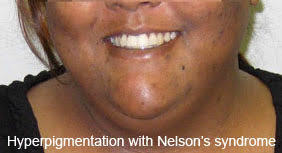
nelson’s syndrome
excess of melanin stimulating hormone
merkel cells
found in the basal layer of the epidermis between keratinocytes and nerve fibres
act as mechanoreceptors - numerous on finger tips and oral cavity
basement membrane zone
- A structure consisting of collagen, hemidesmosomal proteins, integrins and laminin
- collectively, they hold the skin together, keeping the epidermis attached to the dermis

dermis
contains blood and lymphatic vessels, nerves, muscles, appendages and a variety of immune cells (mast cells and lymphocytes etc)
also contains, type I and III collagen, elastic fibres, ground substaces, fibroblasts
what cells are involved in collagen synthesis
fibroblasts


-
Viral Skin Infections86
-
Bacterial Virulence63
-
Pruritis12
-
Psoriasis44
-
Lichen Planus13
-
Vesiculobullous blistering25
-
Acne and Rosacea35
-
Genetics and Skin Disease33
-
Photodermatology20
-
Pigmented Skin Lesions67
-
Non-Melanocytic Tumours51
-
Photocarcinogenesis23
-
Leg Ulcers34
-
Dermatitis34
-
ITS 2 - Steroids13
-
Anatomy29
-
Structure and Function of the Skin62
-
Drug Reactions41
-
Extra Derm18
-
Immunology27
-
Melanoma39
-
Dermatitis 2.062
