Streptococci and Enterococci Flashcards
(48 cards)
Streptococci (Genus) Characteristics
- Gram +
- cocci in chains
- catalase -
- grow on blood agar
-hemolysis patterns
•the presence of certain carbohydrate antigens in cell-wall extracts, or a polysaccharide capsule, generally separates the high from the low virulence streptococci

Hemolysis Patterns
- β-hemolysis: Colonies are surrounded by a clear zone where the erythrocytes have been completely lysed.
- α-hemolysis: Colonies show hazy (incomplete hemolysis) with a green discoloration of the agar
- gamma hemolysis = none

Pyogenic Streptococci
- pyogenic: involving or realting to the production of pus
- presence of a carbohydrate in the cell wall defines the pyogenic streptococci which are then classified by the antigenic specificity (A, B, C, etc.) of that antigen (Lancefield carbohydrate)
- β-hemolysis strongly suggests that the strain has one of the Lancefield group antigens, but some Lancefield positive strains may be α-hemolytic or even nonhemolytic
- Of the groups most frequently isolated from humans (A, B, C, F, and G) groups A and B are the most frequent causes of disease.
- The Lancefield group D carbohydrate is found in the genus Enterococcus which used to be classified as streptococci.
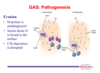
S. pyogenes - aka Group A streptococcus (GAS) Diseases
- Pharyngitis, scarlet fever, impetigo, erysipelas, toxic shock syndrome, rheumatic fever, glomerulonephritis
- Strep throat!
Group A streptococcus (GAS) - Growth
- Colonies are usually compact, small, and surrounded by a zone of β hemolysis that is easily seen and sharply demarcated.
- β-hemolysis is caused by hemolysins called streptolysin S and streptolysin O (oxygenlabile)
Group A streptococcus (GAS) - Structure
- The GAS cell wall is built upon a peptidoglycan matrix within which lies the group carbohydrate antigen.
- A number of other molecules such as M protein, and lipoteichoic acid are imbedded in the cell wall and may extend beyond often in association with the hairlike pili.
- GAS are divided into more than 100 serotypes based on antigenic differences in the M protein.

Group A streptococcus (GAS) - M Protein
•M protein is a fibrillar coiled-coil molecule with structural homology to myosin. Its carboxy terminus is rooted in the peptidoglycan of the cell wall and the amino-terminal regions extend out from the surface.
- Specificity of the more than 100 serotypes of M protein is determined by variations in the surface portions of the molecule.
- The middle part of the molecule is conserved across many M types.
- The biologic functions of M protein can be assigned to specific domains. This includes both antigenicity and the capacity to bind other molecules like fibrinogen, serum factor H and immunoglobulins.
- These actions make M protein the major factor in both the initiation of acute infection and the development of rheumatic fever

Group A streptococcus (GAS) - Other Surface Molecules
- A fibronectin binding protein F and lipoteichoic acid (LTA) are both exposed on the streptococcal surface and play a role in pathogenesis.
- A hyaluronic acid capsule is present in some strains but has no proven unique role in disease.
Group A streptococcus (GAS) - Toxins: Streptolysin O
•Streptolysin O
- Streptolysin O (SLO) is a pore-forming exotoxin similar to complement and staphylococcal α-toxin
- SLO is antigenic and the quantitation of antibodies against it is the basis of a serologic test called antistreptolysin O (ASO).
- Cytokines released through the superantigen mechanism.

Group A streptococcus (GAS) - Toxins: Streptococcal superantigen toxins (StrepSAgs)
•Pyrogenic Exotoxins GAS produce a family of nine superantigen (SAg) toxins called streptococcal superantigen toxins or StrepSAgs produced by roughly 10% of GAS.
- Similar in structure and biological activity to the StaphSAgs of Staphylococcus aureus.
- Multiple effects including fever, rash (scarlet fever), T-cell proliferation, B-lymphocyte suppression

Group A streptococcus (GAS) - Pharyngitis
- Most common bacterial cause of pharyngitis in school-age children
- Transmission is person-to-person from the large droplets produced during coughing, sneezing, or conversation.
- GAS + StrepSAg à scarlet fever which for unknown reasons is less common now that in the past.
- Unless treated, the organism will persist for 1 to 4 weeks.

Group A streptococcus (GAS) - Impetigo
- Impetigo occurs when transient skin colonization with GAS is combined with minor trauma such as insect bites.
- Spread locally by scratching and to others by direct contact

Group A streptococcus (GAS) - Wound and Puerperal Infections
•As with staphylococci, transmission to patients is on the hands of people, including physicians who fail to follow recommended hand washing practices.
Group A streptococcus (GAS) - Cellulitis (erysipelas)
- Deep cellulitis
- Spread
- Bacteremia
Group A streptococcus (GAS) - Streptococcal Toxic Shock Syndrome
- A severe invasive and toxic form of GAS soft tissue infection
- Rapid progression to death in previously healthy persons
- Multiorgan involvement with frequent spread to the blood stream and distant organs.
- As with staphylococcal TSS the findings of shock, renal impairment, and diarrhea are related to massive cytokine release stimulated by the superantigenicity of the StrepSAgs.
- Enhanced invasiveness of group A streptococci is an added feature of STSS compared to its staphylococcal counterpart.
- Systemic spread of the GAS is common as are satellite infections.

Group A streptococcus (GAS) - Poststreptococcal Sequelae: Acute Rheumatic Fever
- The association between GAS and acute rheumatic fever (ARF) is based on epidemiologic studies linking it to cases of GAS pharyngitis.
- Follows weeks after GAS pharyngitis
- ARF does not follow skin or non-respiratory infection with GAS.
- Recurrences of ARF can be triggered by infection with any GAS serotype.
- Injury to the heart caused by recurrences of ARF leads to rheumatic heart disease
- Host factors - Hyper-reactors to streptococcal products (genetic)
-Anti SLO, M protein, other
•Molecular mimicry
- Type II hypersensitivity
- Anti-M protein antibody cross-reacts with host
*Connective and neural tissue
*Sarcolemmal membranes (heart)
Group A streptococcus (GAS) - Poststreptococcal Sequelae: Poststreptococcal Glomerulonephritis
- Poststreptococcal glomerulonephritis may follow either respiratory or cutaneous group A streptococcal infection.
- It is caused only by certain “nephritogenic” GAS strains.
- Type III hypersensitivity – antigen-antibody complexes
Group A streptococcus (GAS) - Acute Infections
- Most important adhesins are M protein, LTA, and protein F.
- In the nasopharynx (NP) all three are involved in mediating attachment to the glycoprotein fibronectin covering epithelial surfaces.
- In NP, M protein provides an essential scaffold for LTA to reach its binding site
- In skin, M protein binds directly to keratinocytes,
- Protein F is involved primarily in adherence to antigen-presenting Langerhans cells.
- SLO mediates direct injury to host cells by membrane insertion.

Group A streptococcus (GAS) - Acute Infections M Protein Resistance to Phagocytosis
- GAS have the capacity to be highly invasive. M protein also plays an essential role in GAS resistance to phagocytosis.
- Related to the ability of domains of M protein to bind fibrinogen and serum factor H leading to a diminished availability of alternative pathway generated complement component C3b
- In the presence of M protein type-specific antibody, classical pathway opsonophagocytosis proceeds. The end result is immunity to subsequent infection with the same M protein serotype.
Group A streptococcus (GAS) - Immunity
- Antibody directed against M protein is protective for subsequent group A streptococcal infections.
- Protection is only for subsequent infection with strains of the same M type. This is called type-specific immunity.
- This protective IgG reverses the antiphagocytic effect of M protein.
- Streptococci opsonized with type-specific antibody bind complement C3b by the classical mechanism facilitating phagocyte recognition
- Because there are over 100 M types, subsequent infections with other M types can occur.
- In ARF patients it is the hyperreaction to self in each episode that produces the chronic cardiac lesions of rheumatic heart disease.
Group A streptococcus (GAS) - Diagnosis
- Blood agar plates demonstrate β-hemolysis
- Susceptibility to the antibiotic bacitracin is an indirect but simple way of separating GAS from the other pyogenic streptococci. GAS are sensitive. The others are resistant.
- Colonies are definitively identified by serologic grouping identifying the group A antigen
- Rapid Strep Test - Detection of group A antigen extracted directly from throat swabs is rapid and specific, but only 90% sensitive compared to culture. Therefore, a positive test is considered diagnostic, but negative results must be confirmed by culture.
- High titers of antistreptolysin O (ASO) are usually found in sera of patients with rheumatic fever
Group A streptococcus (GAS) - Treatment
- Group A streptococci are highly susceptible to penicillin G, the antibiotic of choice. Penicillin resistance is so far unknown.
- Adequate treatment of streptococcal pharyngitis within 10 days of onset prevents rheumatic fever
Group A streptococcus (GAS) - Prevention
- Penicillin prophylaxis is used to prevent recurrences of ARF during the most susceptible ages (5 to 15 years).
- Throughout life persons with a history of rheumatic fever or known heart disease receive prophylaxis while undergoing procedures known to cause transient bacteremia, such as dental extraction.
- Treatment of the acute infection may not prevent poststreptococcal glomerulonephritis.
Streptococcus agalactiae - aka Group B streptococcus (GBS) Diseases
- Neonatal sepsis and meningitis
- Group B streptococci (GBS) have all the characteristics of the pyogenic streptococci with the addition of a polysaccharide capsule composed primarily of sialic acid. The typical GBS case is a newborn in the first few days of life who is not doing well.
- Fever, lethargy, poor feeding, and respiratory distress are the most common features. Localizing findings are usually lacking and the diagnosis is revealed only by isolation of GBS from blood or cerebrospinal fluid. The mortality rate is high even when appropriate antibiotics are used.

