Stomach & SI Flashcards

See clinical correlates - Anatomy 4

1
Q

What does the celiac trunk supply

A

Foregut & spleen

2
Q

What does the celiac trunk give rise to (3)

A
  1. Common hepatic artery
  2. Splenic artery
  3. Left gastric artery
3
Q

What is the parasympathetic innervation of the celiac trunk

A

Vagus nerve - CN X

4
Q

What is the sympathetic innervation of the celiac trunk

A

PRE-GANGLIONIC

Thoracic splanchnic nerves (T5 - T9)

POST-GANGLIONIC CELL BODIES

Celiac ganglion

5
Q

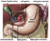
Name the 2 main branches of the common hepatic artery

A

Gastroduodenal

Proper hepatic artery

6
Q

3 branches of the gastroduodenal artery

A
  1. R gastro-omental
  2. Supraduodenal
  3. Superior pancreaticoduodenal - ant & post
7
Q

3 branches of the proper hepatic artery

Where does it run

A
  1. R gastric
  2. L hepatic
  3. R hepatic -> Cystic

Proper hepatic artery runs through the hepatoduodenal ligament - free edge of lesser omentum

8
Q

3 branches of the splenic artery

Where does it run

A
  1. Short gastric
  2. Left gastric omental
  3. Dorsal, greater and inferior pancreatic

Runs along superior border of pancreas to spleen

9
Q

Branch of the left gastric artery

Where does it run

A

Oesophageal branches

Descends along lesser curvature of stomach

10
Q

What does the superior mesenteric artery supply

A

Midgut

11
Q

What is the parasympathetic innervation of the sup mesenteric a.

A

Vagus nerve (CN X)

12
Q

What is the sympathetic innervation of the sup mesenteric a.

A

PRE-GANGLIONIC

Thoracic splanchnic nerves (T9 - T12)

POST-GANGLIONIC CELL BODIES

Sup mesenteric ganglion

13
Q

5 branches of the sup mesenteric a.

A
  1. Inferior pancreaticoduodenal
  2. Intestinal
  3. Middle colic
  4. Right colic
  5. Ileocolic
14
Q

Inf pancreaticoduodenal supplies

A

Duodenum distal to papillae

15
Q

Intestinal supplies

A

Vasa rectae (15-18 branches)

16
Q
  1. What does the middle colic form part of
  2. clinical anastomosis with
A
  1. Marginal artery of Drummond
  2. Inferior mesenteric a.
17
Q

Right colic supplies

A

Ascending colon

18
Q

Ileocolic supplies

A

Appendicular artery

19
Q

3 branches of the inferior mesenteric a.

A
  1. Left colic
  2. Sigmoid
  3. Superior rectal
20
Q

Parasympathetic innervation of the inferior mesenteric a.

A

Pelvic splanchnic nerves

21
Q

Sympathetic innervation of the inferior mesenteric a.

A

PRE-GANGLIONIC

Lumbar splanchnic nerves (L1-L2)

POST-GANGLIONIC CELL BODIES

Inferior mesenteric ganglion

22
Q

What does the left colic form part of

Clinical anastomosis with

A

Marginal artery of Drummond

Superior mesenteric a.

23
Q

Superior rectal supplies

A

Proximal 2/3rds of rectum

(terminal branch)

24
Q

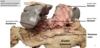
Anterior view of the supracolic viscera ex situ, with the lesser omentum removed

A
25
Where does the oesophagus travel through the oesophageal hiatus of the diaphragm
T10
26
How is the oesophagus attached to the diaphragm
By the phrenico-oesophageal ligament
27
Where does the oesophagus terminate
Cardial orifice of the stomach * Inferior oesophageal sphincter - PHYSIOLOGICAL
28
Arterial supply to the oesophagus
Oesophageal branches from the left gastric artery
29
Venous drainage of the oesophagus
LEFT GASTRIC VEIN Portal venous system OESOPHAGEAL VEINS Systemic venous system via azygos vein
30
Lymphatics of the oesophagus
Left gastric lymph nodes -\> celiac lymph nodes
31
Parasympathetic innervation of oesophagus
Vagus (CN X)
32
Sympathetic innervation of oesophagus
Greater splanchnic nerve (T5-T9)
33
Where is the cardia (1) when supine
T11
34
What is the fundus (2)
Dilated part - gas or sometimes fluid
35
What does the body have (3)
2 curvatures
36
What controls the outflow of chyme What is it divided into
Pylorus PYLORIC ANTRUM - Chamber PYLORIC CANAL - outflow region
37
Gastric rugae temporary vs permanent
Longitudinal folds of mucosa Can be temporary - diminish as stomach distends Can be permanent - form gastric canal
38
Gastric canal
Groove that appears along the lesser curvature during swallowing
39
Pyloric sphincter
Circular band of SM Junction between stomach and SI - Embryological clinical correlate - congenital hypertrophic pyloric stenosis
40
Arterial supply of stomach via
CELIAC TRUNK
41
Arterial supply of lesser curvature (2)
1. Left gastric ( 2. Right gastric (
42
Arterial supply of greater curvature (2)
1. Left gastro-omental ( 2. Right gastro-omental (
43
Arterial supply of fundus
Short/posterior gastric arteries (
44
Venous drainage of right gastric v.
Hepatic portal vein
45
Venous drainage of left gastric v.
Hepatic portal vein
46
Venous drainage of right gastro-omental v.
Superior mesenteric vein
47
Venous drainage of left gastro-omental v.
Splenic vein
48
Venous drainage of short gastric v.
Splenic vein
49
Drainage of prepyloric vein
Right gastric vein
50
Summary of Stomach
51
Infracolic viscera - below transverse colon
SMALL COLON * Duodenum - part * Jejunum * Ileum LARGE COLON * Ascending * Transverse * Descending * Sigmoid CECUM * Appendix RECTUM * Anal canal
52
Abdominal viscera - anterior view ex situ
53
What is the duodenum in relation to the small colon
First section Shortest widest most fixed
54
Superior part of duodenum - intraperitoneal (mobile)
* Duodenal cap/ampulla is continuous with the pylorus of the stomach (approx L1) * Hepatoduodenal ligament connects superiorly * Greater omentum connects inferiorly * Supplied by celiac trunk
55
2. Descending part - Retroperitoneal (immobile and covered by transverse mesocolon)
* Relationship to HEAD of pancreas & gallbladder * Lies right of midline (approx L2) & is anterior to the hilum of the right kidney * Supplied by the coeliac trunk and superior mesenteric artery * Major & minor duodenal papilla
56
Major duodenal papilla
* Elevation of mucosa protruding into duodenum and is larger due to the hepatopancreatic ampulla and sphincter internally within the head of the pancreas * Opening of **common bile duct** and **main pancreatic duct** * Releases pancreatic juice and bile
57
Minor duodenal papilla
* Where the accessory pancreatic duct opens * Releases pancreatic juices only * Located 2cm superior to the major duodenal papilla
58
3. Transvers/inferior/horizontal part
* Secondarily retroperitoneal * Crosses posteriorly under superior mesenteric artery and vein * Passes anteriorly over the IVC * Closely related to the uncinate process of the pancreas and the root of the mesentery * Lies at the level of L3 and crosses from right to left * Longest section of the duodenum * supplied by superior mesenteric artery
59
4. Ascending
* Mostly retroperitoneal * Terminates at the duodenojejunal flexure * Closely related to abdominal aorta * Ascends back up to L2 * Supplied by superior mesenteric artery * Supported by LIGAMENT OF TREITZ
60
Histologically, what does the duodenum contain
Submucosal Brunner's glands
61
What does the ligament of Treitz (suspensory ligament of the duodenum) do
* Connects the duodenum to the diaphragm and the posterior abdominal wall * Facilitates with the movement of the intestinal content * Marks the duodenojejunal flexure - junction between duodenum and jejunum * clinically divides GIT - Upper GIT bleed is PROXIMAL - blood appearing in vomit, stool would be dark and tarry - Lower GIT bleed is DISTAL - blood would only appear in stool
62
What do circular folds create
A large internal SA - Plicae circulares
63
Dual arterial supply of the duodenum
1. From the CELIAC TRUNK - Ant & post - Superior to pancreaticoduodenal arteries (yellow) 2. From the GASTRODUODENAL ARTERY - Supraduodenal artery (blue) 3. From the SUPERIOR MESENTERIC ARTERY - Ant & post - inferior pancreaticoduodenal arteries
64
Summary of duodenum
65
What section of the SI is the jejunum
2nd
66
What section of the SI is the ileum
3rd/last
67
Where is the jejunum mostly found
Left upper quadrant (LUQ)
68
What does the jejunum account for length wise
Accounts for proximal 2/5ths - 2.5m
69
Is the jejunum intraperitoneal or extraperitoneal
INTRAPERITONEAL - highly mobile and attached to the posterior abdominal wall by the root of the mesentery
70
What about the jejunum helps it to absorb nutrients
Specialised epithelial lining for absorbing nutrients that have been digested Gradual change - no clear delineation of when ileum begins
71
Where is the ileum mostly found
Right lower quadrant (RLQ)
72
How much of the SI does the ileum account for
Distal 3/5ths - 3.5m
73
Is the ileum intraperitoneal or extraperitoneal
INTRAPERITONEAL - mesentery proper acts as a conduit for neurovasculature
74
Function of ileum
Absorb any products of digestion that the jejunum missed
75
What is prominent in the ileum
Peyers patches
76
Embryological clinical correlate of ileum
Meckel's diverticulum
77
Jejunum vs Ileum - COLOUR
J - deeper red I - pale pink
78
Jejunum vs Ileum - DIAMETER
J - 2-4cm I - 2-3cm
79
Jejunum vs Ileum - WALL
J - thick and heavy I - thin and light
80
Jejunum vs Ileum - VASCULARITY
JEJUNUM * greater density * long vasa recta * few large arterial arcades ILEUM * lesser density * short vasa recta * many short arterial arcades
81
Jejunum vs Ileum - MESENTERIC FAT
J - less I - plenty
82
Jejunum vs Ileum - CIRCULAR FOLDS
J - large, tall, closely packed I - low and sparse, possibly absent
83
Jejunum vs Ileum - PEYER'S PATCHES
J - few I - many
84
Summary of jejunum and ileum