Clinical correlates Flashcards
Marginal artery of Drummond
- Describes the arteries supplying the midgut and hindgut when collectively they form a continuous circle along the inner border of the large colon
- During development, the middle colic artery may not meet with the left colic artery - this is why the most common area of ischemia is at the splenic flexure (SUDECK’S POINT)

Cirrhosis of the liver
Progressive destruction of hepatocytes
Liver cells are replaced by fibrous (scar) tissue and regenerative nodules (lumps)
- Liver becomes firm
- Circulation becomes inhibited
Common causes of liver cirrhosis
- Chronic alcoholism
- Hepatitis B and C
- Fatty liver disease
Treatment of liver cirrhosis
May involve a shunt of venous blood from the portal system to the caval (systemic) system, or if the cirrhosis is very advanced then a liver transplant may be necessary
Portal hypertension
An increase in pressure of the blood travelling in the veins of the portal system
How does portal hypertension occur
- Venous blood draining away from GIT usually drains to the liver before draining into the IVC so if the route to the liver is obstructed then the reverse (collateral) flow from the portal system veins through to the caval system veins instead can divert blood to the heart instead of the liver
- The small caliber veins of both the portal and caval system are not suited to handle this reversal of blood for an extended period of time, as these collateral veins are forcing through a very large vol of blood
Suprahepatic causes of portal hypertension
- Cardiac diseases
- Hepatic vein thrombosis
Hepatic causes of portal hypertension
- Cirrhosis and acute liver failure
- Hepatocellular cancer
- Schistosomiasis
Infrahepatic causes of portal hypertension
- Arteriovenous malformation
- Tumour in head of pancreas
- Splenomegaly
- Portal vein thrombosis
Varices
Increased portal blood pressure can result in potentially fatal abnormally dilated veins
Portacaval anastomoses
- The hepatic portal vein and its tributaries have no valves
- Therefore if the venous drainage of GIT gets blocked at the hepatic portal vein, blood can bypass the liver by flowing in a REVERSE DIRECTION and drain to the IVC through an alternative route
GASTROESOPHAGEAL
- Left gastric -> oesophageal
PARAUMBILICAL
- Paraumbilical -> epigastric
ANORECTAL
- Superior rectal -> middle/inferior rectal

Where is there anastomosis between (in the portal and caval systems)
PORTAL - left and right gastric veins
CAVAL - oesophageal
Oesophageal varices
- In severe cases of portal hypertension the blood is unable to effectively flow through the liver which causes retroflow in the gastric veins which change to drain into the OESOPHAGEAL veins instead
- Potentially fatal if one of these fragile, dilated oesophageal veins gets damaged and excessive bleeding occurs

Treatment of oesophageal varices
Can be treated using an endoscope to directly inject the varices with clotting medicine or by placing a band to cut off circulation
Symptoms of oesophageal varices
- Black, tarry stool
- Paleness
- Light headed
- Vomiting - emesis
- Symptoms of chronic liver disease
Caput Medusa
- In severe cases blood is unable to effectively flow through the liver which can cuase retroflow in PARAUMBILICAL VEINS
- The superficial veins of ant abdominal wall (superficial epigastric and thoracoepigastric) then become extremely dilated and varicose

Treatment of caput medusa
Divert portal blood by creating a shunt between larger veins of the caval system in order to relieve pressure
- Hepatic portal vein -> IVC
- Splenic vein -> left renal vein
CIPS
Hemorroids anastomoses
Portal system - superior rectal veins
Caval system - inferior rectal veins
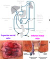
Internal hemorrhoids
- Found above pectinate line
- Will not be painful - VISCERAL INNERVATION
- If damaged, bright red blood in stool - lower GIT bleed
External hemorrhoids
- Found below pectinate line
- Painful - SOMATIC INNERVATION
- Develop from varicose perianal veins that are part of the caval system

Peritonitis
Infection can occur if gas, fecal matter or bacteria enter the peritoneal cavity which would result in inflammation of the peritoneum
Exudate
A fluid rich in cellular elements - serum, fibrin, acid or pus that has seeped out and been discharged from an inflamed organ or vessel
Ascitic fluid
Excess fluid in the peritoneal cavity = ASCITES
Paracentesis
Surgical puncture of the peritoneal cavity for the aspiration/drainage of the ascitic fluid
Inflammation of parietal peritoneum
- Sharp, well-localised pain
- Tenderness on palpation
Inflammation of visceral peritoneum
- Generalised, referred pain that is felt in the associated dermatome of the organ
Direction of peritoneal fluid flow when supine
- Inflammatory exudate tends to collect
1. Hepatorenal recess
2. Right posterior subphrenic recess
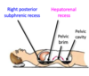
Direction of peritoneal fluid flow when inclined
Inflammatory exudate collects in the pelvic cavity where there is a slow absorption of toxins

Normal vs abnormal flow of peritoneal fluid

What is dialysis
The separation of particles in aliquid on the basis of differences in their ability to pass through a membrane
What sort of membrane is the peritoneum
A semi-permeable membrane which permits relatively rapid absorption of solutions
Peritoneal dialysis in the case of renal failure
- A dilute, sterile solution can be introduced into peritoneal cavity on 1 side of the patient
- Excess water and soluble waste products (e.g. urea) can be transferred from the BVs
- The dilute solution and waste products can be drained out of the peritoneal cavity from the other side
Pringle manoeuvre
- The portal triad travels within the hepatoduodenal ligament and this can be clamped to control bleeding
- If a patient is still bleeding internally after clamping, there must be a haemorrhage elsewhere from:
- R or L hepatic veins
- Retrohepatic IVC

Omental bursa herniation
- Part of GIT (usually a loop of SI) can pass through the omental foramen and become twisted inside the lesser sac
- Relatively rare
PRE-DISPOSING FACTORS
- Large omental foramen
- Redundant/mobile mesentery
- Elongated right liver
- Defect in lesser omentum
Boundaries of omental foramen
ANTERIOR - Hepatoduodenal lig. (containing portal triad)
POSTERIOR - IVC and right crus of diaphragm
SUPERIOR - caudate lobe of the liver
INFERIOR - 1st part of duodenum
How do peritoneal adhesions form
As a result of damage to the peritoneal surface when sticky fibrin appears in order to assist with the healing process
VIsceral -> adjacent organ
Visceral -> parietal peritoneum
What do adhesions limit
The normal movement of viscera and could lead to complications
- Intestinal obstruction (volvulus)
- Chronic pain
Laparotomy
Surgical incision into abdominal cavity prior to major surgery
Adhesiotomy
Surgical separation of adhesions
Nerves most at high risk of damage during appendectomy
- Iliohypogastric nerve (L1)
- Ilioinguinal nerve (L1)
(either through transverse/grid iron incision at McBurney’s point or laproscopic surgery)
NB Correct ligation of appendicular artery
Initial appendicitis
General visceral afferent (GVA) - T10 dermatome
Acute appendicitis
General somatic afferent (GSA) - localised at McBurney’s point
Splenic rupture
Most commonly injured abdominal organ
If the spleen ruptures, this will lead to:
- shock
- intraperitoneal hemorrhage (profuse internal bleeding)

Splenomegaly
Pathological enlargement of the spleen (10x normal size) accompanied by high BP
Cholelithiasis/gallstones
- How do they form
- Common in…
- Symptoms
- Crystals form in gallbladder when there are high concentrations of cholesterol and can be associated with individuals who are regularly dehydrated
- Relatively common in females and often ASYMPTOMATIC
- pain in RUQ
- Pain may be referred to right neck/shoulder region
- Nausea
- Cholecystitis
- Jaundice - due to obstruction of major duodenal papilla/common bile duct
Common constriction site of cholelithiasis
The hepatopancreatic ampulla is a common constriction site where cholelithiasis often become painfully lodged
Cholecystectomy
Surgical procedure to remove gallbladder
Gallbladder is not a vital organ so if gallstones have a high risk of reoccurence and regularly cause severe biliary colic then an individual may elect to undergo a cholecystectomy to remove gallbladder
Cystohepatic triangle
Borders
Must be identified in a cholecystectomy to determine if there is a variation in the cystic artery or biliary apparatus
SUPERIOR BORDER
Inferior border of the liver
MEDIAL BORDER
Common hepatic duct
LATERAL BORDER
Cystic duct

Pancreatic cancer (head)
What does it obstruct
What does it lead to
- Head is most common type of pancreatic cancer
- Tumour could obstruct the common bile duct or the hepatopancreatic ampulla
- Retention of bile will lead to jaundice (yellowing of skin and sclera of eyes if bile is unable to be released into the duodenum)
- Can lead to faeces become ACHOLIC -light/grey coloured
Cancer of the neck and body of the pancreas
Tumour could obstruct:
- Hepatic portal vein
- IVC
Pyrosis/heartburn
What is it associated with
Most common type of oesophageal discomfort and substernal pain
- burning sensation in the abdominal part of the oesophagus which is perceived in the chest
- Gastro-oesophageal Reflux Disorfer (GERD)
- Inferior oesophageal sphincter prevents acid reflux
- May be associated with a hiatal hernia - when the proximal part of the stomach protrudes through the oesophageal opening in the diaphragm into the mediastinum

Peptic ulcers
A distinct lesion or necrosis of the mucosa in either the stomach, pyloric canal or duodenum as a result of acid erosion
Gastric ulcers and gastritis are the 2 most common (men are affected more)
STRONGLY ASSOCIATED WITH
- Mucosal exposure to gastric acid and pepsin
- Helicobacter pylori bacterial infections
- Non-steroidal anti-inflammatory drugs
- Aspirin
Symptoms of peptic ulcers
HEMATESMESIS
Vomiting “coffee ground” blood
MELENA
Black. foul-smelling faeces
What are the majority of gastric cancers
Troiser’s Sign
Adenocarcinomas - originate in glandular tissue
TROISER’S SIGN - Hard, palpable, enlarged left supraclavicular lymph nodes indicate metastatic cancer in the abdomen
Crohn’s Disease/Colitis
Similar symptoms to ulcerative colitis but colitis is limited to colon
- Chronic inflammation of GIT
- Most common affects ileum and beginning of LI but can occur anywhere from mouth the anus
- Treatment is designed to suppress immune system’s abnormal inflammatory response
Symptoms of Crohn’s Disease
- Persistent diarrhea
- Rectal bleeding
- Urgency when time to defecate
- Abdominal cramps and pain
- Constipation - bowel obstruction
Invasive treatment of Crohn’s Disease
COLECTOMY
Terminal ileum, colon and rectum are removed
ILEOSTOMY
Artificial opening (stoma) of the healthy ileum is created through abdominal wall
COLOSTOMY
Opening is created to drain faeces
Colonic diverticulosis
When multiple false diverticulae (out-pocketings of the mucosa of the colon) develop along the LI
Acquired mucosal herniations which protude through weak areas of the muscular wall
MOST COMMONLY:
- Occur on the mesenteric side of 2 bands of taenia coli (omental and free) due to perforating nutrient arteries
- Affect middle-aged and beyond
- Found on the sigmoid colon
- RISK FACTORS = low fibre, high meat, BMI > 25
Volvulus of the sigmoid colon
- Condition involving the twisting and rotation of the mobile loops of the intestinal tract - SIGMOID COLON
- CONSTIPATION
- ISCHEMIA OF INTESTINE
- If left untreated, necrosis could occur and an immovable collection of compressed feces may develop
Referred pain from abdominal organs

