Abdomen Flashcards
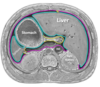
When the anterior abdominal wall is reflected to expose the contents of the abdominal cavity, what is everything that can be seen covered in
Peritoneum
What is the peritoneal cavity
“space” between the parietal & visceral layers of peritoneum where no structures are found - no fluid occupies this potential space to allow movement & mobility of certain organs
What is the peritoneal cavity divided into
* greater sac = main compartment * lesser sac = hidden from view
What does the peritoneum viscera have a relationship with
All abdominal viscera - INTRAPERITONEAL = within - EXTRAPERITONEAL = outside
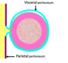
Parietal layer of peritoneum
- Covers & lines the internal walls of the abdomen
- Pain is well-localised => sensitive
- served by same neurovasculature as adjacent wall/structure
(pink)

Visceral layer of peritoneum
- Covers & invests abdominal organs that protude into peritoneal cavity
- Pain is generalised/REFERRED
- Served by same neurovasculature as organ it is covering
(blue)

What is the peritoneal cavity
“Potential” fluid-filled space between parietal & visceral peritoneum
What are peritoneal structures formed from
More than 1 layer of peritoneum
- Omenta
- Mesentery
- Peritoneal ligament
(yellow)
What is the peritoneal cavity divided into
The greater & lesser sac
- What is the greater sac
- Where does it extend from
- Are there organs in this potential space
- Main component of peritoneal cavity
- Extends from diaphragm -> pelvis
- NO
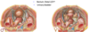
Peritoneal cavity in males
Completely closed
Peritoneal cavity in females
Opening in peritoneum through the uterine tubes
- What is the lesser sac also known as
- Where is it positioned
- Omental bursa - no organs here
- Small compartment that is lined with peritoneum & is positioned:
- BEHIND stomach and greater omentum
- IN FRONT OF peritoneum & pancreas
how is the lesser sac/omental bursa formed
Twisting & rotation of gut
- What does the omental foramen provide
- What is it also known as
- Direct communication between the greater & lesser sacs
- Epiploic foramen/foramen of Winslow

Mesentery
Defined as a double layer of peritoneum resulting from invagination of peritoneum by organs
Greater & lesser omenta
Extensions of peritoneum that form ligamentous “aprons” extending from stomach & proximal duodenum -> adjacent organs
Peritoneal ligaments
Membranous fold (double layer of peritoneum) that supports an organ by helping to keep it in anatomical position
Peritoneal recesses
Potential spaces in abdominal cavity where excess fluid may collect
Peritoneal fossae
Depressions in anterior abdominal wall that are lateral to the 3 respective umbilical folds
What do mesenteries provide
Pathways for vessels, nerves and lymphatics to pass between abdominal organs

What does the mesentery contain
Fat
Arteries
Veins
Lymphatic vessels
Nerves supplying abdominal organs
Function of mesentery
Connect organs to posterior abdominal wall
- Mesooesophagus (embryonic)
- Mesogastrium (embryonic)
- Mesentery proper of small bowel
- Mesoappendix
- Transverse mesocolon
- Sigmoid mesocolon
What is the lesser omentum
DOUBLE LAYERED PERITONEAL FOLD
- Connects the liver to the lesser curvature of the stomach and the first part of the duodenum
What are the 2 ligaments of the lesser omentum
- HEPATOGASTRIC - thin and membranous portion
- HEPATODUODENAL - thick free edge containing the portal triad

What is the greater omentum
- FOUR LAYERED PERITONEAL FOLD
- Connects transverse colon to greater curvature of stomach
- hangs down and is able to move within the peritoneal cavity
- Can wrap around organs to localise inflammation & prevent peritoneal adhesions (abdominal policeman)
Name the 5 hepatic peritoneal ligaments
- Coronary ligament - limited by left/right triangular ligaments
- falciform ligament
- Round ligament of liver - ligamentum teres hepatis
- hepatogastric ligament - membranous lesser omentum
- hepatoduodenal ligament - thick edge lesser omentum

Name the 5 gastric peritoneal ligaments
- Hepatogastric
- gastrophrenic
- gastrosplenic/gastrolienal
- gastrocolic
- (phrenicocolic)

Name the 2 splenic peritoneal ligaments
- Gastrosplenic ligament
- Splenorenal ligament

What is the paracolic gutter
- Groove between abdominal wall and lateral aspect of the ascending or descending colon
- Allows communication between the supracolic & infracolic regions of the greater sac
- Important for peritoneal flow

What is the hepatorenal recess
- Extension of the subhepatic space between the visceral surface of liver and right kidney
- potential site for fluid collection

What are the subphrenic recesses
- Located below the diaphragm
- Separated into left & right by falciform ligament
- Potential space for fluid collection

What is the subhepatic space
Immediately inferior to liver

- Are the intraperitoneal organs mobile
- Which ones are they
- YES
- Liver
- Stomach
- Transverse colon
- SI

Please Don’t Act Rash
- Are retroperitoneal organs mobile
- Which ones are they
- NO
- Pancreas
- Duodenum (part is intraperitoneal)
- Aorta
- Rectum
- What are the intraperitoneal organs defined as
- How are they suspended
- Being almost entirely wrapped in visceral peritoneum
- Suspended by mesentery in the abdominal cavity
List the intra-peritoneal organs
- Stomach
- Liver
- Spleen
- tail of pancreas
- duodenum - 1st part
- jejunum
- ileum
- cecum & appendix
- transverse colon
- sigmoid colon
- uterus
- uterine tubes
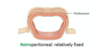
What are the retroperitoneal organs defined as
Lying behind the peritoneal cavity with only part of its surface covered by peritoneum
What are primary retroperitoneal structures
Retroperitoneal from the start of their development
- Suprarenal/adrenal glands
- Ureters
- Kidneys
- Aorta & IVC
- Esophagus
- Rectum - proximal 1/3rd
SAD PUCKER
What are secondary retroperitoneal structures
Were once suspended within the abdominal cavity by mesentery but migrated posteriorly to end up behind the peritoneum
- Duodenum - 2nd, 3rd, 4th parts
- Pancreas - head, neck and body
- Ascending & descending colon
What happens to the kidneys if someone loses a lot of weight drastically
Kidneys become mobile because they’re supported by fascia
What are infra/extra/subperitoneal organs
Defined as being BENEATH the peritoneal cavity
only part of its surface is covered by peritoneum
- Rectum - Distal 2/3rds
- Urinary bladder
What delineates the boundary between the supracolic and infracolic abdominal compartments
The transverse mesocolon
SUPRACOLIC:
Visceral structures below the diaphragm and above the transverse mesocolon
What are the 7 supracolic viscera
- Distal esophagus
- Stomach
- Duodenum - 1st and part of 2nd
- Pancreas
- Spleen
- Liver
- Gallbladder
(unpaired digestive glands/organs)
What are the 6 infracolic viscera
- SI - 2nd, 3rd, 4th parts; jejenum; ileum
- Cecum
- Appendix
- Large colon
- Rectum
- Anus

- Where does the celiac trunk branch from
- What does it supply
- Branches from the abdominal aorta at the level of T12
- Foregut
- Distal oesophagus
- Stomach
- Spleen
- Pancreas
- Liver
- Gallbladder
- Duodenum - 1st & proximal 2nd part

- Where does the superior mesenteric artery branch from
- What does it supply
- Abdominal aorta at the level of L1
- MIDGUT
- Duodenum (distal) - 2nd, 3rd, 4th parts
- Jejunum
- Ileum
- Cecum
- Appendix
- Ascending colon
- Transverse colon - proximal 2/3rds

- Where does the inferior mesenteric artery branch from
- What does it supply
- Abdominal aorta at the level of L3
- HINDGUT
- Transverse colon - distal 1/3rd
- Descending colon
- Sigmoid colon
- Rectum and anal canal (upper half only)

