Skin Infections Flashcards

1
Q

How is the skin the first line of defense against microbial pathogens ?

A
  1. It is a physical barrier 2. Secrets a low pH sebacous fluid and fatty acids as well as antimicrobial peptides to inhibit growth of pathogens. 3.By possessing its own normal flora, it prevents the colonization of other flora
2
Q

What are the steps to bacterial colonization ?

A
  1. Invasion- normally through a break in the skin 2. Interaction between the bacteria and the host tissue causes the clinical effects.
3
Q

What are the three steps of disease pathogenesis once the bacteria has invaded the skin ?

A
  1. Bacterial adherence to host cells 2. Invasion of tissue with evasion of host defenses 3. Elaboration of toxins
4
Q

What are the two main classes of toxins and how do they cause damage ?

A

Exotoxins are actively secreted proteins that cause tissue damage or dysfunction through various mechanisms. Endotoins

5
Q

What are super antigens ?

A

Usually made by S. Areus and S. Pyrogenes. These are a group of exotoxins that cause a massive release of cytokines that grossly exaggerates inflammatory responses.

6
Q

What is Impetigo ?

A

Superficial crusting epidermal skin infection that presents in bullous and nonbullous forms.

7
Q

Who usually gets impetigo ?

A

children and it usually infects the face. Caused by both S. Areus and S. Pyogenes. The characteristic feature is a honey colored crust.

8
Q

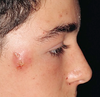
What is Erysipelas ?

A

It is a streptococcal infection of the superficial dermal lymphatics that demonstrates sharply demarcated and raised borders. **Picture was inflammation with above described manifestation over the entire orbital bone

9
Q

What is cellulitis ?

A

It is an infection of the deeper dermis and sub-q with poorly demarcated borders. The vast majority are streptococcal in origin.

10
Q

What is a cutaneous abscess ?

A

A collection of pus in the dermis and sub-q tissue

11
Q

What is folliculitis ?

A

A superficial infection of the hair follicle with a collection of pus in the epidermis.

12
Q

What is a furuncle ?

A

A boil with a deeper involvement of the particle. The infection extends into the sub-q tissue

13
Q

What is a carbuncle ?

A

Numerous furuncles coalesce to form a single inflamed area.

14
Q

Diffuse generalized arrhythmia and superficial desquamation with flexural accentuation. Mucous membranes are uninvolved. There is perioral and periocular crusting and radial fissuring with mild facial swelling

A

Staph scalded skin syndrome. IT will present primarily in infants or adults with renal failure. There will be granular splits in the dermis that lacks inflammatory infiltrate.

15
Q

What is necrotizing fasciitis ?

A

NF is an insidious and deadly soft tissue infection characterized by widespread tissue necrosis. MUCH DEEPER PLANE THAN CELLULITIS

16
Q

Type 1 Necrotizing Fasci

A

Polymicrobial

17
Q

Type 2 NF

A

Strep flesh eating

18
Q

Type 3 NF

A

Clostridial Myonecrosis- Gas Gangrene

19
Q

How could you know you have NF ?

A

Pain that is much worse than the clinical findings. Exquisite tenderness and erythema, warmth and swelling. -Skin changes from red and purple to gray blue within 36 hours.

20
Q

What happens if you delay NF diagnosis-

A

Fatal

21
Q

What causes Toxic shock syndrome ?

A

TSST-1 Toxin which is released by S. Areus **Surgery Deep abscesses and Tampons

22
Q

What are the clinical features of toxic shock syndrome ?

A

Fever, strawberry tongue, sunburn like erythema and sandpaper papules. Later causes desquamation. You must remove the focus of infection and give ABX

23
Q

What produces periorfical abscesses ?

A

Anarobic organisms

24
Q

What organism produces pus ?

A

Staph Areus

25
How do you treat a skin bacterial infection ?
Usually a culture to determine abx susceptibility and then appropriate ABX treatment
26
Where are superficial fungal infections localized to ? What usually causes them ?
They are confined to dead keratinous tissue, the epidermis and hair follicles. They are usually caused by dermophytes, Nondermatophyte mold or yeasts
27
How do deep fungal infections form ?
They involve all skin layers and often extend into the subcutis tissue. They usually occure through direct inoculation of the skin. ( Sporotrichoses Mycetoma and chromomucosis )
28
What are the most common fungal infections ?
Dermatophyte cutaneous infections.
29
How do dermatophytes infect skin ? What are the 3 genra
They adhere to kerratin and invade by secreting enzymes. Trichophyton- Tri Rubryn is the most common. Micosporum Epidermophyton-
30
Tinea Pedis
Athletes Foot- 95% Dermatophytes. Superficial infection
31
Tinea Unguium or Onchomycosis
Infection of the nail bed or plate- leads to nail bed deformity ( Onchydystrophy with thickening and hyperkeritosis with discoloration )
32
Tinea Corpis
Superficial fungal infection of the skin commonly on the trunks and limbs. Ring Worm
33
Tinea Faci
Ring worm on the face. Superficial fungal infection.
34
Tinea Curis
Jock itch- infection in the groin region that occurs exclusively in adult men.
35
How does tinea curis present ?
Erythematous patch involving the inner thigh and inguinal fold. Sparing the scrotum and penis. (Insert Dick Pic Here )
36
Tinea Capitis - What is the bug that causes this?
Ring worm in the hair. 95% are T. Tonsurans
37
What causes Candidiasis ? Who usually presents with it ?
Candida albicans affects the skin and mucous membranes. It presents most frequently in women with vulvovaginal candidiasis.
38
What is Tinea Versicolor ?
Harmless skin disease most frequently in the tropical region. It is characterized by overgrowth of the malassezia species leading to the formation of hypo-pigmented patches with associated fine scale.
39
Where do HHV-1, HHV-2 and VZV remain latent ?
In the sensory ganglia.
40
What is the most common form of herpes infection ? How is HSV-1 spread.
Herpes Labialis- the most common form of herpes infection and presents as ulcurative lesions of the oral cavity or perioral skin and mucosa. Caused by HSV-1 contact with oral secretions
41
What is genital herpes caused by ?
HSV-2 but has increasing HSV-1 coverage.
42
What determines the clinical presentation of HSV and what is predictive of the onset ?
The immune response of the host and is preceded by tingling pain.
43
What are the symptoms of HSV-1 and HSV-2 ?
HSC-2 is much more severe and has constitutional symptoms such as fever and lethargy. Usually a painful group of vesicles on a red base that ulcerate and heal over in 4-5 days.
44
How is HSV diagnosed ?
Usually by clinical evaluation alone, TSANK smear viral culture and direct fluorescence Ab structure.
45
What is HSV superinfection with atopic dermititis ?
Eczema Herpeticum- Remember disgusting looking herpes finger.
46
Describe the pathogenesis of this disease
Impetigo- Is characterized by the accumulation of neutrophils in the stratum corneum that often produces a pustule. Usually by staph areus or S. Pyrogenes. This primarily affects children and characteristly has a honey colored crust.
47
What is this disease ?
Bulous presentation of Impetigo
48
What disease is this ? What is the pathogenesis ?
Erysipelas - A strep infection of the superficial dermal lymphatics that is characterized by the sharply defined borders
49
What disease is this and what bacteria cause this?
Impetigo- S. Areus and S. Pyrogenes. Honey colored crust. Affects children
50
What is the pathogenesis of this disease ?
Cellulitis is an infection of the deeper dermal tissue with poorly demarcated borders. The vast majority of cases it is Streptococcal in origin
51
What are these ?
Abscesses - A collection of pus in the dermis and deeper dermal tissues
52
What is this disease ?
Folliculitis is the superficial infection of hair follicles with the accumulation of pus in the epidermis where the infection spreads in the subdermal tissue
53
How do you treat an uncomplicated soft tissue infection you know is NOT MARSA (No recent hospitalizations, no known colonizations of MARSA, not from a region where MARSA is prevalent) ? (Swepman Style) If it progresses and you are seeing them in the hospital ? What if they are allergic to B lactams ?
An oral anti - Staph penicillin sich as Dicloxacillin or a first gen cephalosporin such as Cephalexin. If they are hospitalized give IV Nafcillin, oxacillin or Cefazolin. If they are allergic to B lactams give IV Nafcillin, Oxacillin or cefazolin.
54
What causes this disease ?
This is a furuncle which is an follicular infection that often involves
55
What happens when a bunch of these form together ?
A Carbuncle forms
56
What is the pathogenesis of this disease ? Who and where does it normally attack ?
This is Staph Scalded Skin Syndrome- An infection with exotoxin - producing S. Areus. Binding to cell adhesion molecule DESMOGELIN-1 and cleaving leaving a loss of cell cell adhesion. Primarily affects young infants or adults with renal failure. ERYTHMA AND SUPERFICIAL DESQUAMATION
57
A patients presents with a lesion that has exquisit tenderness, erythema, warmth and swelling. There is a skin change from red and purple to a characteristic gray-blue colir in ill-defined patcheswithin 36 hours of onset THe pathent tells you with a cut that hurts MUCH worse than the clinical presentation. What do you think it is?
Necrotizing Fasciitis- Spreads like cellulitis but the infection is on a much deeper plain. NF is an insidious and deadly soft tissue infection characterized by widespread necrosis. This is a surgical emergency needing widespread debridment. A ciolaceous and / or hemorrhagic bulla may develop
58
What are all of these examples of ?
Superficial Tinea infections.
59
What is the pathogenesis of this disease ?
This is Tinea Pedis- it is a superficial dermatophyte infection. Remember dermatophytes have enzymes for attachment, invasion, and digestion of keratin. They rarely invade viable tissue.
60
What is this ? What will it lead to if it is not treated ?
Tinea Unguinum- Dermatophyte infection of the toes. It can lead to onchodystrophy which is nail deformity or thickening which is hyperkeratosis and discoloration.
61
What is this ?
Tinea Corporis which is a superfical fingal infection of glabrous skin occuring mos commonly on the trunk and limbs, whereas tinea facei affects the face. Both of these are considered ring worm given their tendency to produce annular plaques or patches with a red raised, scaling border.
62
Insert picture of... {an erythematous patch involving the upper inner thigh and inguinal folds of an elderly man's crotch but the patch does not spread onto his testicles. }
**Tinea Cruris** or jock itch describes infection of the groin region that occurs almost exclusively in adult men.
63
What is this ? What causes this ?
Tinea capitus which is a superficial fungal infection of the scalp and hair. T. tonsurans
64
What is the histopathology of superficial fungal infections ?
**Neutrophils** in the stratum corneum Fungal **hyphae** in the stratum corneum as seen with **PAS Stain ** Culture and material scraping usually shows the infecting agent.
65
What infection is this.
Canadia Albicans- it is a yeast and infects the skin mucus membranes, nales and UG tract. It is common in women as a vulvovaginal infection.
66
What bug causes this and what disease is it ?
This is Tinea Versicolor and is a harmless skin disease most common in the tropical regions. It is characterized by an overgrowth of Malassezia species leading to the fomation of hypopigmented or hyperpigmented patches with an associated fine scale.
67
What is this ? What virus causes this
Herles Labialis HSV-1 which is spread through oral secretions
68
What is this ?
Herpes Labialis- HSV-1 primary infection
69
What is this ? If shows up on your genitals. What will this person feel usually before this lesion shows up ?
This is Herpes Genitalis- caused by HSV-2 and some HSV-1. These lesions are usually preceeded with tingling pain
70
What does HSV-1 usually present as ?
HSV-1 recurrences typically present as painful, grouped vesicles on an erythematous base on the vermillion border of the lip, lasting 2 to 3 days. ## Footnote • The lesions subsequently develop ulceration with crusting, and ultimately heal in 4 to 5 days. • HSV-2 recurrences present with similar skin findings, but tend to be less sev
71
How do you diagnose herpes ?
Usually by clinical presentation alone. Tests are available : Biopsy T-Snack Smear Viral Culture Serological testing Direct Fluorescence
72
What is this ?
Herpatic whitlow- A digital HSV infection
73
What is this ?
Eczema Herpeticum- HSV superinfection of atopic cermatitis.
74
What is the pathogenesis of this ?
VZV- Chichen Pox- Dew drop on a rose petal- Transmitted by airborn droplets or by direct contact. first degree infection begins with fever, malaise, mylagia followed by pruritic eruption spreading fro face / scalp to trunk
75
What is this ?
This is VZV shingles. IT is reactivation from the HSV that is dormant in the dorsal root ganglia. Notice it is in the V1 distribution over the face
76
What is the yellow arrow pointing to and what disease does this indicate ?
Multinicleated acantholytic keratinocytes This indicates a herpetic viral infection histology
77
What is the pathogenesis of this disease ?
This disease is Molluscum contagiosum- this is characterized by smooth, umbilicated papules Histologic exam revelas cytoplasmic inclusions called HENDERSON PATTERSON BODIES
78
What is this disease ?
Molluscum contageosum
79
What are the viral characteristics of HPV ?
Icosahedral, naked dsDNA virus
80