Shoulder Joint Conditions Flashcards

1
Q

How prevalent is a dislocated shoulder? In what population is it most common?

A
  • Approximately 1.7% of the population will experience a dislocated shoulder.
  • Bimodal distribution
    • 20-30yrs (Males:Females 9:1) - Sports injuries
    • 61-80 (Males:Females 1:3) - Falls
  • It is less common in children as thier epiphyseal plate is weaker and tends to fracture before dislocating.
  • More common in elderly: reduced collagen crosslinking - weaker capsule / tendons / ligaments.
2
Q

How is the glenohemeral joint stable?

A
  • Congruency of humeral head in glenoid fossa and glenoid labrum
  • Negative intra-articular pressure (‘suction cup’ effect)
  • Static stabilisers e.g. labrum, joint capsule, extra-capsular ligaments
  • Dynamic stabilisers e.g. rotator cuff muscles, long head of biceps brachii
  • BUT.. weak inferiorly as there isnt much support.
3
Q

What are the two types of anterior dislocation of the shoulder?

A

90% of dislocations are anterior; two subtypes:

  • Subcaracoid (60%): Head of humerus lies anterior to glenoid fissa, inferior to caracoid
  • Subglenoid (30%): Head of humeus lies antero-inferior to glenoid

Humeral head dislocates antero-inferiorly

Disruption of glenohumeral ligaments

Head pulled anteriorly by muscles (subscapularis, pectoralis major)

4
Q

How do you anteriorly dislocate your shoulder?

A
  • Hand behind head - Abduction and external rotation
  • Trauma pushes arm posteriorly
  • Humeral head dislocates antero-inferiorly

OR

  • ​Direct blow to posterior shoudler
5
Q

What is a Bankart lesion?

A

A tead of glenoid labrum +/- glenoid fracture

6
Q

What is a Hill-Sachs lesion?

A
  • Anterior dislocation
  • Posterior aspect of jumeral head jammed agaisnt anterior lip of glenoid - Held tightly by mucles e.g. infraspinatus
  • Indentation fracture = ‘dent’
  • 50% of patients aged under 40 with an anterior dislocation have this lesion
  • 80% of patients have a recurrent dislocation have this lesion
7
Q

How does a posterior shoulder dislocation occur (mechanism)?

A
  • Violent muscle contraction
    • Epilectic seizure
    • Electrocution
    • Lightning strike
  • Blow to the anterior shoudler
  • Arm flexed across body and pushed posteriorly
8
Q

How does a patient with a posterior dislocation present?

A
  • Squaring of shoudler (loss of normal contour)
  • Arm adducted and internally rotated
  • Prominent caracoid process anteriorly
  • Humeral head may be prominent posteriorly.
  • Arm cannot be externally rotated into anatomical position
9
Q

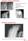
How can you use an x-ray to see if a posterior dislocation has occured?

A

..

10
Q

What causes inferior dislocation?

A

Rare = 0.5% cases

Hyperabduction injury - e.g. grasping an object from above to break the fall.

High rate of associated injuries

  • Nerves 60%
  • Rotator cuff 80%
  • Vascular 3%
11
Q

What are complications of shoulder dislocation?

A
  • Recurrent dislocation
    • 60% overall risk
    • Risk decreased with age because there is decreased elasticity of the capsule and ligaments.
  • Axillary artery damage
    • More common in the elderly
  • Nerve injury
    • Axillary nerve most common
    • Cords of brachial plexus
    • Other branches e.g. musculocutaneous
  • Fractures
    • Most common during first-time dislocation
    • Humeral head, greater tubercle, clavicle, acromion
  • Rotator cuff tears
    • More common in elderly and after inferior dislocations
12
Q

Anatomy of the clavicle

A
  • S shaped bone
  • Acts as strut between sternum and glenohumeral joint
  • Lateral 1/3 is flat - insertion of trapezius and origin of the deltoid
  • Middle 1/3 is tubular - weak to axial load
  • Medial 1/3 is quadrangular - insertion of sternocleidomastoid and origin of pectoralis major (clavicular head)
  • Protects apex of lung, subclavian vessels and trunks and divisions of brachial plexus.
13
Q

How do you treat a clavicle fracture?

A

Most treated conservatively in a sling but sometimes indications for surgical fixations.

14
Q

When is surgical fixation needed in clavicle fractures?

A
  • Complete displacement
  • Severe displacement with tenting of skin (risk of conversion to open fracture)
  • Open fracture
  • Neurovascular compromise
  • Interposed muscle
  • Floating shoulder (clavicle and glenoid neck fracture)
15
Q

What are some complications of clavicle fractures?

A
  • Non-union
  • Malunion
  • Infection (if open fracture)
  • Damage to nerves
    • Subrascapular nerve
    • Supraclavicular nerves (3 of these)
    • trunks and divisions of brachial plexus
  • Vascular - Subclavian vein and artery
  • Apex of lung - Pneumothorax (air in chest cavity).
16
Q

What are the four rotator cuff muscles and what is the most common site for a tear?

A
  • Supraspinatus
  • Infraspinatus
  • Teres minor
  • Subscapularis

The most common site for a tear is the supraspinatus under the coraco-acromial arch

17
Q

What is the pathology of rotator cuff tears?

A

Although acute tears of the rotator cuff can occur (e.g. following shoulder dislocation), most rotator cuff tears are chronic, resulting from extended use in combination with other factors such as poor biomechanics or muscular imbalance.

The most common cause is age-related degeneration. With age, the blood supply to the rotator cuff tendons decreases, impairing the body’s ability to repair minor injuries.

The principal theory is a degenerative-microtrauma model, which supposes that age-related tendon damage, compounded by chronic microtrauma, results in partial tendon tears that then develop into full rotator cuff tears. Inflammatory cells are recruited and oxidative stress leads to tenocyte (tendon cell) apoptosis, leading to further degeneration, thus a ‘vicious circle’ is created

18
Q

What are risk factors for rotator cuff muscles?

A
  • Age
  • Recurrent overhead activity
    • Painters
    • Carpenters
    • Swimming
    • Volleyball
    • Tennis
    • Weightlifting
  • Recurrent heavy lifting
  • OA shoulder with osteophytes
  • Abnormally-shaped acromion
19
Q

What is the clinical presentation of a supraspinatus tear?

A
  • Can be asymptomatic
  • Most commonly anterolateral shoulder pain radiating down arm
  • Precipitated by activity (can be present at rest)
    • Above shoulder activity (abduction and external rotation)
    • Learning down on elbow (displaces humeral head superiorly)
    • Pushing self out of a chain (displaces humeral head superiorly)
    • Reaching forward (flexing shoulder and lifting e.g. milk from fridge.)
  • May also complain of weakness of shoudler abduction but, this is often only found on examination because they don’t do these activites as too much pain.
20
Q

What is impingement syndrome?

A
  • Impinge = to rub or catch
  • Supraspinatus tendon impinges on coraco-acromial arch
  • Caused by anything that narrows subacromial space
    • Thickening of coraco-acromial ligament
    • Inflammation of supraspinatus tendon
    • Subacromial osteophytes in OA
  • Pain, weakness and reduced range of motion
  • Pain eacerbated by overhead movement
21
Q

How does impingement syndrome present?

A
  • Painful arc when patient lifts arm between 60°-120°. This is caused by impingement of the supraspinous tendon under the acromion during abduction of the shoulder.
  • This is because, when the shoudler is abducted or flexed this space is narrowed further resulting in pain, weakness and reduced range of motion.
  • Pain is dull rather than sharp
  • Other symtoms can include grinding or popping sensation upon movement of the shoulder.
  • Worse when completing overhead shoulder movements and at night.
    *
22
Q

Whar is cacific supraspinatus tendonitis?

A
  • Hydroxyapetite crystals in supraspinatus tendon
  • Pain on abduction or flexion of shoulder
    • reduced coraco-acromial space
  • Mechanical ‘block’ to movement
    • Stiffness
    • Snapping sensation
    • Catching
    • Reduced range of movement
  • Treatment is initially conservative with rest and analgesia but surgical treatment is often required for persistant symptoms.
23
Q

What is the theory of the pathology of calcific supraspinatus tendonitis?

A
  • Multifactorial
  • Theory 1:
    • Regional hypoxia
    • Tenocytes - chondrocytes
    • Endochrondrial ossification
  • Theory 2:
    • Metaplasia of mesenchymal stem cells onto osteogenic cells
    • Ectopic bone formation
  • Crystaline in resting phase then reabsorbed by phagocytes (In this phase they look like “toothpaste”). Reabsorbtion phase is most painful.
24
Q

What is adhesive capsulitis?

A
  • When glenohumeral joint is inflammed and stiff
  • Pain is constant, worse at night
  • Exacerbated by movement and cold weather
  • Possible autoimmune component, often triggered by trauma to shoulder.
25
Q

What are the risk factors for frozen shoulder?

A
  • Femal gender
  • Trauma to shoulder
    • Acute
    • Calcific tendonitis
    • Rotator cuff tear
  • Epilepsy with tonic seizures (sudden muscle contractions)
  • Diabetes mellitus (glucose binds to capsular collagen)
  • Connective tissue disease
  • Thyroid disease (both hypo and hyper)
  • Inactivity
    • Stroke
    • Trauma
    • Illness
26
Q

How do you treat a frozen shoulder?

A
  • Physiotherapy
  • Analgesia
  • Anti-inflammatory medication
  • Sometimes manipulation under anaesthesia - Just move it to break adhesions.

Usually resolves with time

90% of shoulder motion regained

6-17% risk of other shoudler within 5 years.

27
Q

Where in the shoulder is osteoarthritis most common?

A

Acromioclavicualr joint is more common that Glenohuemral joint.

28
Q

What is the treatment of osteoarthrits?

A