Sensory organs Flashcards
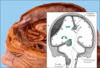
How many ventricles have the brain?
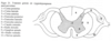
The brain has four fluid-filled chambers, the ventricles. They are:
- Lateral ventricle (2)
- Third ventricle
- Fourth ventricle

Explain the Cerebrospinal fluid.
Cerebrospinal fluid
CSF is a clear and colorless liquid produced in the ventricles and circulating around the CNS. Total volume is 140 ml. 500 ml is produced every day.
Functions:
- Protection against physical injury.
- The volume can be regulated.
- Transport of substances.
Explain Hydrocephalus
Hydrocephalus is a condition in which there is an abnormal accumulation of cerebrospinal fluid (CSF) within the brain.
It can be:
- Congenital
- Aquired
- Obstructive, non-communicating
- Communicating (the ventricles ”communicate”)
The skull is protective but cannot expand

Explain Craniosynostosis.
Craniosynostosis
Craniosynostosis is a condition in which one or more of the fibrous sutures in an infant skull prematurely fuses by turning into bone (ossification), thereby changing the growth pattern of the skull. Because the skull cannot expand perpendicular to the fused suture, it compensates by growing more in the direction parallel to the closed sutures.

Explain the Meninges.
The Meninges

The brain and spinal cord is surrounded by the meninges (connective tissue).
- Pia mater – closest to the nervous tissue.
- Arachnoid mater (spider´s web) – membrane.
- Dura mater (tough)
Explain Dura Mater.
Dura Mater
- The tough mother.
- The periosteum of the skull, wall between the hemispheres and above the cerebellum.

Explain the blood supply to the brain
Blood supply to the brain
Two pair of arteries:
- Carotis interna – from aorta. Through the skull base. Is devided in a cerebri media and a cerebri anterior.
- Vertebralis – from the vessels to the arm. Passes through the cervical vertebraes and enters the cranium together with the brain stem.
- Basilaris.

Explain how blood drains from the brain.
Venous flow

The blood from the brain is drained by a superficial and a deep system.
Both system is drain in v. jugularis – the large vein of the neck.
Which parts are included in the CNS?
Central nervous system
- Brain
- Brainstem
- Spinal cord
What is the function of the PNS?
Peripheral nervous system
Connects the CNS to the limbs and organs
Explain the Peripheral nervous system.
Peripheral nervous system
Spinal nerves
31 pair of spinal nerves that exits from different levels of the spinal cord. They are mixed (motoric and sensoric).
- 8 cervical
- 12 thoracal
- 5 lumbar
- 5 sacral
- 1 coccygeal

Name two classifications of Spinal nerves.
Spinal nerves

Motoric – efferent
Sensoric - afferent
How many pair of cranial nerves exits from the brain?
Cranial nerves
12 pair of nerves exiting from the brain.

Explain Medulla spinalis.
Medulla spinalis
- Consists of grey and white matter.
- From skull base to 2nd lumbal vertebrae.
- Spinal nerve is divided in ventral and dorsal rote.
A pair of spinal nerves are connected to each segment of the medulla spinalis. The spinal nerve is consists of a ventral and a dorsal root.
The ventral root contains axons that send signal from the CNS (efferens).
The dorsal root contains axons that send signal to the CNS (afferens).

Describe Afferent signals.
Afferent signals:
- Reflex
- Continue upwards in the dorsal column, cross midline, via thalamus reaching the cerebral cortex – vibration and proprioception (muscles tendons).
- Spinothalamic tract, cross midline, via thalamus to cerebral cortex – pain and temperature.

Describe Efferent signals.
Efferent signals
- Pyramidal tract – from cerebral cortex via medulla spinalis to muscles. Cross midline in the brainstem.
- Non-voluntary autonomic signals to organs.

Cranial nerves
Cranial nerves
- Nervus Olfaktorius
- N. Opticus
- N. Oculomotorius
- N. Trochlearis
- N. Trigeminus
- N. Abducens
- N. Facialis
- N. Vestibulacochlearis
- N. Glossopharyngeus
- N. Vagus
- N. Accessorius
- N. Hypoglossus

Cranial nerves – smell, vision and eye movements
- N. olfactorius (n.I)
- N. opticus (n.II)
- N. trochlearis (n.IV) - m. obliq sup
- N. abducens (n.VI) - m. rectus lat
- N. oculomotorius (n.III) – all remaining eye muscles, m. sphincter pupillae and m. levator palpebrae

Cranial nerve V, VII och VIII

- N. trigeminus (n.V) – muscl of mastication and sensory to face.
- N. facialis (n.VII) – muscl to face, glands, sensory – tongue.
- N. vestibulocochlearis (n.VIII) – hearing and balance.
Cranial nerve IX-XII

- N. glosso-pharyngeus (n.IX) – sensoric info from the tongue, tonsil, pharynx, middle ear. Motor – pharynx.
- N. Vagus (n.X) – motor and sensoric – pharynx, thorax, abdomen.
- N. Accessorius (n. XI) – m. sternocleidomastoideus och trapezius
- N. Hypoglossus (n.XII) - tongue
Explain the ANS.
Autonomic nervous system - ANS
ANS is the part of the nervous system not controlled by the free will – e.g., heart, bowel and glands.
Hypothalamus is the boss of this system.
ANS is situated both in the CNS and the PNS.
ANS is divided in:
a. Sympathetic – fight-flight
b. Parasympathetic – energy saving and recharging, e.g., after a meal.
Different responses to the Sympatic and Parasympatic systems.
See picture.
Explain Sensations, Perception and Adaption.
SENSATIONS
Definition of sensation: the conscious or subcounscious awareness of external or internal conditions of the body
- Stimulus
- Sensory receptor
- Conduction along a neural pathway
- Integration in the brain
Perception – conscious sensation. How you perceive sensations is very individual, there is a strong psychological aspect.
Adaptation – decrease in the strength of a sensation during a prolonged stimulus (hot shower)
Which are Somatosensory (general senses)?
Somatosensory (general senses)
- Tactile
- Thermal
- Pain
- Proprioception
- Visceral
Which are the Special senses?
Special senses.
- Taste,
- Smell,
- Vision,
- Hearing,
- Balance
List and explain different types of sensory receptors (6)
Different sensory receptors
- Mechanoreceptors
- Mechanical pressure, vibration, proprioception
- Thermoreceptors
- Changes in temperature
- Nociceptors
- Painreceptors, respond to physical or chemical damage to tissue
- Photoreceptors
- Detect light that meets the retina of the eye
- Chemoreceptors
- Detect chemicals in the mouth (taste), nose (smell) and body fluids.
- Osmoreceptors
- Sense osmotic pressure in body fluids
Describe Tactile sensations.
Tactile sensations
- Touch
- Pressure
- Vibration
- Itch
- Tickle

List the anatomy of Sensory receptors
Sensory Receptors
- Free nerve endings – pain, touch pressure
- Meissner corpuscle – light touch, small receptor fields
- Merkel disc – touch and pressure, small receptive fields
- Ruffini end organs – touch, heavy pressure, large receptive fields
- Pacinian corpuscles – vibration
- Hair end organs - touch
Explain Thermal sensation.
Thermal sensation.
Thermoreceptors – free nerve endings
Temperatures between 10° and 40°C – cold receptors
Temperatures between 32° and 48°C – warm receptors
Temperatures >48° and <10°C – nociceptors
High degree of adaptation
Explain Pain sensation.
Pain sensation

Nociceptors (noci=harmful)
– free nerve endings
Respond to several types of stimuli
Only little adaptation
Fast pain (Aδ-fibers)– within 0,1 s, sharp
Slow pain (C-fibers) – after a second, chronic, burning
Potential causes of pain.
Potential causes of pain
- Tissue damage
- Tissue ischemia (acidosis, laktate)
- Thermal pain
- Spasm of muscle, artery or ”hollow viscus”
- Inflammation
- Neurogenic pain
Name three Pain syndromes.
Pain syndromes
- Hyperalgesia- increased sensitivity
- Allodynia- pain triggered by nonpainful
stimuli
• CRPS – complex regional pain syndrom
What is phantom pain?
Phantom pain
- The cerebral cortex interprets signals from the proximal portion of neurons (spinal cord) that used to carry impulses from the phantom limb
- Still active regions in the cortex that used to receive impulses

Explain Referred Pain.
Referred Pain

Visceral (internal organs, intestines) pain fibers synapse onto the same secondary neurons in the spinal cord
Explain the term Gate control.
Gate control.

Stimulation of large fibers (touch) prevents pain impulses.
Explain Proprioceptive sensations.
Proprioceptive sensations
1) Static position sense
Conscious perception of the orientation of the different parts of the body with respect to one another.
2) Dynamic proprioception
Rate of movement sense.
Knowledge of position, both static and dynamic, depends on knowing the degrees of angulation in all joints in all planes and their rate of change. Multiple types of receptors are involved and used together.
Adaptation is slow and only slight.
Proprioception is both conscious and subconscious
Name three Proprioceptors.
Proprioceptors

- Muscle spindles
- Golgi tendon organs
- Joint receptors
Explain Olfaction.
Olfaction – sense of smell

10-100 million receptors
Olfactory hairs
Odorants – 10.000 differents odors
Cribriform plate of the ethmoid bone
Olfactory nerv, bulb and tract (CN I)
Primary olfactory area in temporal lobe, frontal lobe, limbic system and hypothalamus
Explain Hyposmia.
Hyposmia = decreased sense of smell
Due to:
- Trauma
- Tumors
- Smoking
- Drugs
- Age
Explain Gustation.
Gustation - sense of taste
5 primary tastes: sour, sweet, bitter, salty, umami
All other tastes are combinations plus olfactory and tactile information
Explain the structure of taste buds.
Structure of taste buds
- 10 000
- Vallate papillae
- Fungiform papillae
- Filiform papillae
- Taste pore
- Gustatory hair

Explain Gustatory pathway.
Gustatory pathway
1) The Facial nerve (CN VII)
2) The Glossopharyngeal nerve (IX)
- serves the toungue
3) The Vagus nerve (CN X) – serves the throat and epiglottis, Parietal lobe, limbic system, hypothalamus
Explain accessory structures of the eye in terms of protection.
Protection of the eye
- Eyebrows and eylashes
- Eyelids
- Lacrimal apparatus
- -salt, mucus, lysozymes

Explain the layers of the eyeball (Fibrous tunic).
Layers of the Eyeball - Fibrous tunic
- Cornea
- Admits and refracts light
- Sclera
- Shape and protection
- Conjunctiva

Explain the layers of the eyeball (Vascular tunic).
Layers of the eyeball (vascular tunic).
- Choroid
- Blood supply, absorb scattered light
- Ciliary body
- Alters shape of lens
- Iris
- Regulates the amount of light
- Lens

Explain how the pupil reacts to different types of light.
See picture.

Explain the layers of the eyeball (Retina).
Layers of the eyeball (Retina)
- Macula lutea (yellow spot, central vision)
- Central fovea (sharp vision)
- Optic disc (blind spot)
- CN II
How are the layers of the eyeball categorized (3).
Layers of the eyeball
- Fibrous tunic
- Vascular tunic
- Retina

Which are the 3 neuronal layers of the Retina?
Retina

3 neuronal layers
- Photoreceptor layer
- Bipolar cell layer
- Ganglion cell layer
Which are the main photoreceptors in the eye?
Photoreceptors
- Rods – 120 million
- Cones – 6 million
Explain Image formation.
Image formation.

Refraction of light rays
The change in shape of the lens
-accomodation
Constriction of the pupil
Convergence
Explain Myopia.
Myopia - nearsightedness
The eye is too long or the lens is to strong

Explain Hyperopia.
Hyperopia - farsightedness

Eye is too short or lens too weak.
Explain Presbyopia.
Presbyopia
With age the lens become less elastic leading to lack of ackomodation - permanently focused at a certain distance.
Reason for Astigmatism.
Reason for Astigmatism
Asymmetrical cornea
Name three types of Refractive surgery.
Refractive surgery
- Flap procedures (eg LASIK)
- Surface procedures (PRK)
- Corneal incision procedures
Explain Stimulation of Photoreceptors
Stimulation of Photoreceptors
Photopigment
Rhodopsin - opsin+retinal
Cones - 3 different opsin proteins – red,green, blue
A photoreceptor cell is a specialized type of neuron found in the retina that is capable of phototransduction. The great biological importance of photoreceptors is that they convert light (visible electromagnetic radiation) into signals that can stimulate biological processes. To be more specific, photoreceptor proteins in the cell absorb photons, triggering a change in the cell’s membrane potential.
There are currently three known types of photoreceptor cells in mammalian eyes: rods, cones, and photosensitive retinal ganglion cells. The two classic photoreceptor cells are rods and cones, each contributing information used by the visual system to form a representation of the visual world, sight.
Test Color blindness.
See pic.

Explain the visual pathway.
See picture.

Which are the three main parts of the ear?
Main parts of the Ear
- Outer Ear
- Middle Ear
- Inner Ear

Describe the Outer Ear.
Outer Ear.
- Auricle
- External auditory canal
- Eardrum/tympanic membrane

Describe the Middle Ear.
Middle Ear.
- Air-filled cavity
- Auditory/eustachian tube
- Auditory ossicles: malleus (hammer), incus (anvil), stapes (stirup)
- Oval window

Explain the Inner Ear.
Inner Ear.
- Temporal bone
- Bony labyrinth
- Membranous labyrinth with endolymph
- Vestibule – utricle and saccule
- Semicircular canals
- Cochlea with the spiral organ (organ of Corti)
- Round window
- CN VIII

Explain Sound interpretation.
Sound interpretation
Loudness – higher amplitude
Frequency – 20-20.000 Hz

Describe the Hearing pathway.
Hearing pathway

- CN VIII
- Brainstem
- Auditary cortex
Explain different types of hearing loss.
Hearing loss
- Sensorineural – damage to organ of Corti, CNVIII, cortex, eg. presbyacusis
- Conductive – tympanic, membrane, ossicles, eg otosclerosis
Explain the functions of a Hearing Aid.
Hearing Aid.
Amplify and modify soundwaves
Explain CI – Cochlear inplant.
CI – Cochlear inplant

Severe hearing loss due to damage hair cells
- Microphone
- Transmitter
- Electrode
- CNVIII
Explain Physiology of Equilibrium.
Physiology of Equilibrium

Static equlibrium
Dynamic equlibrium -VESTIBULAR APPARATUS
Works together with vision and proprioception
Explain Static equlibrium.
Static equlibrium
- Maculae
- Otolithic membrane
- Hair cells
- CN VIII

Explain Dynamic equlibrium.
Dynamic equlibrium
- Ampulla
- Crista
- Hair cells
- Cupula
- Endolymph
- CN VIII
