Respiratory L5: Control of respiration Flashcards
What are 3 things that are involved in the control of respiration?
- Factors responsible for generating the alternating inspiration/expiration rhythm
- Factors that regulate the magnitude of ventilation to match body needs
- Factors that modify respiratory activity to serve other purposes such as speech, coughing, holding one’s breath
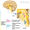
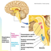
What are the control centres?
How much O2 in blood

What is the function of pace making cells?
On & off of breathing

What is the function of the pneumotaxis centre?
- Fine tune on/off rhythm (duration)
- Damage = abnormal breathing patterns

What is the function of the rostral ventromedial medulla?
On/off rhythm

What is the function of the dorsal respiratory group?
- Controls quiet breathing (only inspiration- eg. diaphragm)
- Expiration (passive)

What is the function of the ventral respiratory group?
- Controls increased ventilation (eg. during exercise)
- Both inspiration (diaphragm and external intercostals) and expiration (abdominal and internal intercostals)

What happens when the partial pressure of arterial O2 (PO2) is less than 60mmHG? (arterial PO2 < 60mm Hg)
1 pathways
- Emergency life-saving mechanism (hypoxic drive to breathe)
- Peripheral chemoreceptors
- Medullary respiratory centre
- Increased ventilation
- Increased arterial PO2
*central chemoreceptors are not detected = no effect on medullary respiratory centre

What happens when the partial pressure of arterial CO2 (PO2) is more than normal?
2 pathways
Pathway 1
- Increased brain ECF PCO2 (gets relieved)
- Breaks up into carbonic acid and bicarbonic ion + H+ ion
- Increase brain ECF H+ = more acidic (gets relieved)
- Central chemoreceptors stimulated
- Medullary respiratory centre
- Increased ventilation
- Decreased arterial PCO2
Pathway 2 (weakly)
- Peripheral chemoreceptors stimulated (blood pH becomes acidic)
- Medullary respiratory centre
- Increased ventilation
- Decreased arterial PCO2
- This increases diffusion co-efficient
- Hyperventilate = decreased CO2

Some patients with severe lung disease and chronic elevated arterial PCO2 do not show any increase in ventilation. Why?
In the presence of a prolonged increase of H+ in the brain ECF as a result of longstanding CO2 retention, and associated increase in arterial bicarbonate levels due to renal compensation of respiratory acidosis, enough bicarbonate may cross the blood-brain barrier to buffer the excess H+, and the brain ECF pH is restored to normal levels despite the fact that arterial PCO2 and ECF PCO2 remain high.
Oxygen therapy for such patients has to be carefully monitored. Why?
Because such patients may rely on the life saving mechanism of the peripheral chemoreceptors (PO2 under 60 mm HG) for ventilation. This is called ‘hypoxic drive to breathe’. O2 therapy will increase arterial PO2 above 60 mm Hg and thus decrease the patients drive to breathe. They may stop breathing all together and need to be mechanically ventilated.
- Not fully understood
What happens when arterial non CO2-H+is more than normal/increased?
Use respiratory system to compensate for metabolic issue Eg. increased acidic blood (increased lactic acid; ketoacidosis- diabete- not due to CO2)
Increased arterial non-CO2-H+ –> acidosis (gets relieved by a decreased arterial CO2-H+)
- Peripheral chemoreceptor (also weakly modulate blood pH)
- Medullary respiratory centre
- Increased ventilation
- Decreased arterial PCO2
- Decreased arterial CO2-H+
- Relieves acidosis

What are 3 three lines of defense against changes in non-CO2 induced [H+] to maintain the [H+] of body fluids at a nearly constant level (blood pH change)?
- Chemical buffer system
- immediate response
- Respiratory compensation
- few-minute delay
- Renal compensation
- hours to days delay
- excrete or hang on to bicarbonate
How long does it take for the chemical buffer system towork?
immediate response
How long does it take for the respiratory compensation to work?
few minute delay
How long does it take for the renal compensation to work? How does it work?
- hours to days delay
- excrete or hang on to bicarbonate
What are 3 factors that do not cause increased ventilation during exercise?
EXAM QUESTION
- Arterial PO2
- Arterial PCO2
- Arterial [H+]
What are 4 factors that do cause increased ventilation? EXAM QUESTION
- Joint and muscle receptors
- Body temperature
- Adrenaline
- Cerebral cortex
What are the 2 non-respiratory factors that influence ventilation?
- Involuntary control
- Voluntary control
How does involuntary control (non-respiratory factor) influence ventilation? List 3 factors.
- protective reflexes such as coughing and sneezing
- pain (take breathe in)
- emotions (laughing, crying, sighing, groaning)
How does voluntary control (non-respiratory factor) influence ventilation? List a factor
- speaking, singing, whistling
What is apnea?
During apnea, a person subconsciously “forgets to breathe”.
During sleep apnea, victims can stop breathing for seconds or even minutes for up to 500 times a night. True or false?
True
What can apnea lead to? In babies, what is a form of apnea?
respiratory arrest.
- SIDS (sudden infant death = “cot” death) may be a form of apnea.


