Resp. + body cavities embryology Flashcards
Explain the basic development of the respiratory system.
Acronym?
Gabriella Deluca Really Blows For Shrimps
- laryngeotracheal Groove forms in the floor of primordial pharynx caudal to 4th pair of pharyngeal pouches
- laryngeotracheal groove evaginates → laryngeotracheal Diverticulum
- elongates + distal ends enlarge → Respiratory Bud
- tracheoesophageal Folds develop lateral to laryngeotracheal groove
- approach each other → tracheoesophageal Septum: seperates
- ventrally: esophagus
- dorsally: laryngeotracheal tube
How does the larynx develop?
- arytenoid swellings at cranial end of laryngeotracheal tube
- grow toward tongue → laryngeal inlet
- temporary occlusion + recanalization
- epiglottis develops from hypopharyngeal eminence
additionally:
- 4th/6th pharyngeal arch → laryngeal mm.
- neural crest cells in 4th/6th pharyngeal arch → laryngeal cart.
- endoderm → endothelial lining
How does the trachea develop?
from larnygeotracheal tube
- endodermal lining distal to larynx → epithelium + glands of trachea + pulm. ep.
- splanchnic mesenchyme → cart., conn. tissue, tracheal mm.
What are “common” disorders resulting from faulty partioning of the laryngotracheal tube?
tracheoesophageal fistula (TEF):
passage btw trachea and esophagus due to incomplete division
⇒ often associated with a blind ending esophagus (= esophageal atresia)
How does the bronchial tree develop?
- respiratory bud seperates into 2 bronchial buds
- enlarge to main bronchi growing into pericardioperitoneal canals
- divide to secondary bronchi which undergo ramification
→ form lobar bronchi
→ form segmental bronchi + intersegmental branches - primordial bronchopulmonary segments are formed
- respiratory bronchioles develop
Which structures form parietal and visceral pleura?
- splanchnic mesoderm → visceral pleura
- somatic mesoderm → parietal pleura
NOTE analogy to peritoneum
What are the four stages of lung maturation?
- pseudoglandular stage (w6 - 16)
- canalicular stage (w16 - 26)
- terminal sac stage (w26 - birth)
- alveolar stage (w32 - 8y)
What happens in the pseudoglandular stage during the maturation of lungs?
Time period?
w6 - 16:
- developing lungs resemble exocrine glands histologically
- all major elements formed (→ terminal bronchi)
What happens in the canalicular stage during the maturation of lungs?
Time period?
w16 - 26:
- each terminal bronchiolus → 2+ resp. bronchioli w/ a few terminal sacs (type I + II pneumocytes)
- lung tissue vascularizes
- lumina of bronchi/terminal bronchioles become larger
⇒ respiration possible
canals (canalicular) are similar to vessels (vascularization), thus first contact with blood → gas exchange
What happens in the terminal sac stage during the maturation of lungs?
Time period?
w26 - birth:
- most terminal sacs (alveoli) develop
- vascularizations proceeds + lymphatic cap. form
⇒ survival of prematurely born infant
What is the function of pulmonary surfactant?
counteracts surface tension forces → facilitates expansion of alveolar sacs by preventing atelectasis (= collapse of sacs during inhalation)
What happens in the alveolar stage during the maturation of lungs?
Time period?
w32 - 8y:
increase of lung area mostly due to increased no. of resp. bronchioles/alveoli (by formation of sec. septa)
150 M in neonate → 300 M in adult
What are the conditions for the gas exchange btw the fetus and the placenta of the mother?
- transition of lungs from secr. to gas exchanging organs
- parallel pulmonary/systemic circulations
- surfactant
Why is the amniotic fluid so important for fetal lung development?
How is it cleared eventually?
How is the deficiency of amniotic fluid called?
fetal breathing moments force aspiration of some amniotic fluid into the lungs
→ condition resp. mm.
→ stimulate lung development by creating a pressure gradient btw lungs and amn. fluid
deficiency = oligohydraminos
routes of clearance:
- mouth/nose during vaginal delivery
- lymphatics + blood vessels
What are the parts of the embryonic body cavity?
- pericardial cavity
- 2 pericardioperitoneal canals
- peritoneal cavity
Explain the formation of body cavities.
Acronym?
-
intraembryonic Coelom = horseshoe-shaped cavity
- bend = future pericardial cavity
- limbs = future pleural/peritoneal cavity
- brought together during Folding
- limbs fuse ventrally → ventral mesentery degenerates → Peritoneal cavity
- growth of bronchial buds induces formation of
- cranially: pleuropericardial Folds
- caudal: pleuroperitoneal Folds
⇒ both become membraneous
-
pleuroperic. Memb. fuse → fibrous Pericardium/mediastinum
pleuroperit. Membr. → Diaphragm
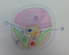
1 - 5

1) pleuropericardial membrane
2) n. phrenicus
3) common cardinal v.
4) lung
5) esophagus
6 - 8

6) pericardial cavity
7) fibrous pericardium
8) esophageal mesentery
What are the contents of the pleuropericardial folds?
common cardinal v. + n. phrenicus
How does the peritoneum form?
- somatic mesoderm → parietal peritoneum lining abdominal wall
- splanchnic mesoderm → visceral peritoneum covering organs
NOTE analogy to pleura
What is the peritoneal cavity initial continuous with?
When does it loose the connection?
extraembryonic coelom at umbilicus
looses connection as intestines return to the abdomen
How does the diaphragm develop?
4 parts fuse
- septum transversum → central tendon
- pleuroperitoneal membranes
- esophageal mesentery → median portion of diaphragm, myoblasts form crura
- muscular ingrowths of int. layer of post. body wall
1 - 5
purple layer is facing ventrally
1) IVC
2) aorta
3) esophagus
4) pericardioperitoneal canal
5) pleuroperitoneal membrane
6 - 8
purple layer is facing ventrally

6) muscular ingrowth of int. layer of post. body wall
7) septum transversum
8) esophageal mesentery
Explain the development nerve pathyway of the diaphragm.
What are consequences?
myoblasts from 3rd - 5th somite grow into diaphragm via septum transversum bringing their nerve fibers → fibers fuse: n. phrenicus
- nerves elongate during descent of diaphragm
- enter through pleuropericardial membranes (later fibrous pericardium)
